0842
A Pilot Evaluation of Amide Proton Transfer-Weighted (APTw) MR Imaging in Characteristics and Diagnosis of Premenopausal Breast Tumors1The First Affiliated Hospital of Dalian Medical University, Dalian, China, 2Philips Healthcare, Beijing, China
Synopsis
Amide proton transfer (APT) imaging is based on the chemical exchange between free bulk water protons and the amide protons (-NH) of endogenous mobile proteins and peptides in tissue. Previous studies have shown that APT-weighted (APTw) MRI could noninvasively identify and differentiate tumors in brain, head and neck etc. This study aims to explore the feasibility of APTw-MRI in characteristics and discrimination of premenopausal malignant and benign breast tumors.The results show that the APTw MR imaging efficiency of diagnosis of premenopausal breast tumors were 0.904.
Purpose
To explore the value of amide proton transfer-weighted (APTw) MR imaging for differential diagnoses of premenopausal malignant and benign breast tumors.Introduction
The sensitivity of APTw to these subtle chemical changes may provide complementary biomarkers of pathologic findings to conventional (T1- and T2-weighted) MR imaging while providing fundamentally different information than that provided with established quantitative techniques.Previous studies have shown that APT-weighted (APTw) MRI were feasible in human breast on 7T MRI1,2 and possible feasibility assessing chemotherapy response on 3T MRI3,4.Methods
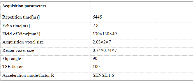
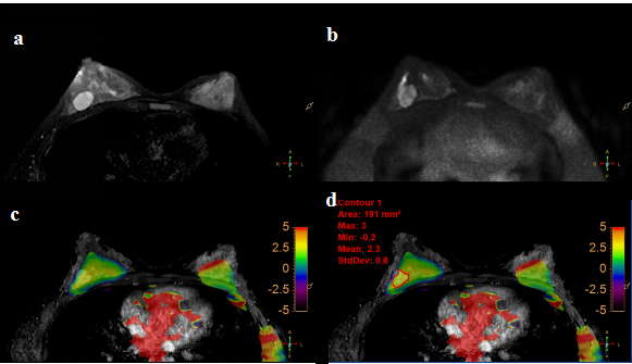
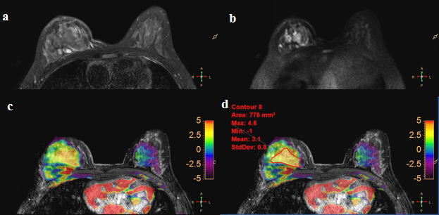
In this prospective study, a total of 18 premenopausal female patients (46.56±11.38,range:23-58years) were enrolled and informed consent was acquired from each patient. All patients were imaged using a 3.0 T whole-body MR scanner (Philips Ingenia CX, Philips Healthcare, the Netherlands) with a dedicated seven-channel bilateral phase-array breast coil. Specifically, group 1 included 12 patients with malignant breast tumors, and group 2 included 6 patients with benign breast tumors. APTw MR imaging (Table1) and conventional MR imaging (T1WI, T2WI, and DCE) were acquired. The region of interest (ROI) was obtained manually on the APTw MR images, DCE image was used to help drawing the ROIs, which corresponding with the most enhanced part of the lesion on DCE(Fig1 and Fig2). The mean value of ROI measurements was used as the final APTw values of lesions. The measured image parameters (APTw values, maximal lesion diameter) and age were compared between two groups, and the diagnostic performance based on parameters was quantified with ROC curve.Results
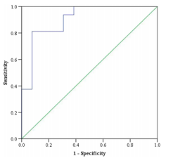
There was no significant difference in age and maximal lesion diameter between two groups (p = 0.985, 0.342). The APTw maps are shown as overlays on the anatomical images with the highest correspondence in 18 tumors . The intraclass correlation coefficients (ICC, 0.96 and 0.90 for group 1 and 2, respectively) indicate a good inter-observer agreement of the measured APTw values(Fig1&Fig2). APTw values of malignant lesions were significantly higher than those of benign lesions (3.26 ± 1.15 % &1.54±0.93 %, p <0.001). Area under the curve (AUC) acquired by APTw MRI was 0.904 with an optimal threshold of 2.62% (sensitivity of 81.3%, a specificity of 92.3%, and an accuracy of 86.2 % (Fig. 3).Discussion and Conclusion
APTw MRI detects the transfer of magnetization that is majorly contributed from amide protons (resonating at 3.5 ppm downfield from water). And it can be sensitive to the cellular mobile protein content as well as tissue pH[1-4]. In this study, we have established the inter-observer reproducibility of APTw imaging for detection breast tumors, and presented initial data to show that APTw imaging can also be used to assess effects of age and menstrual-cycle stages (as potential sources of changes in breast biochemistry) on diagnosis of breast tumors. High diagnostic performance can be achieved by APTw values for the differentiation of premenopausal malignant and benign breast tumors. These findings suggest that APTw MRI has good potential to discriminate malignant and benign breast tumors before surgery.Acknowledgements
This work was supported by the grant of the Basic Scientific Research Projects of the Universities in Liaoning Province (5061104) and The Provincical Natural Science Foundation of China (2019-ZD-0907) .References
[1] Zhou J, Heo HY, Knutsson L, et al. APT‐weighted MRI: techniques, current neuro applications, and challenging issues. Magn Reson Imaging. 2019,50(2) :347-364.
[2] By S, Barry RL, Smith AK, et al. Amide proton transfer CEST of the cervical spinal cord in multiple sclerosis patients at 3T. Magn Reson Med. 2018,79(2):806–814.
[3] Heo HY, Zhang Y, Jiang S,et al. Quantitative assessment of amide proton transfer (APT) and nuclear overhauser enhancement (NOE) imaging with extrapolated semisolid magnetization transfer reference (EMR) signals. II: Comparison of three EMR models and application to human brain glioma at 3 Tesla. Magn Reson Med. 2016,75(4):1630–1639.
[4] Jiang S, Eberhart CG, Zhang Y, et al. Amide proton transfer-weighted MR image-guided stereotactic biopsy in patients with newly diagnosed gliomas. Eur J Cancer. 2017,83(4):9–18
Figures