0837
CT and MRI based multi-modal and multi-parametric radiomics pre-operative prediction of perineural invasion in patients with rectal cancer1The First Hospital of Jilin University, changchun, China, 2GE Healthcare, beijing, China
Synopsis
Perineural invasion (PNI), defined by tumor invasion of nervous structures and nerve sheaths, which is thought an independent predictor of outcome in rectal cancer. However, for a radiologist, neither MRI nor CT can reliably evaluate PNI. Radiomics is an emerging and effective method for quantitative analysis and prediction using big data of medical imaging. Therefore, this study aims to develop and validate a radiomics prediction model based on MRI and CT for the preoperative prediction of PNI in rectal cancer. The results indicated that excellent diagnostic performance can be yielded with such multi-modal radiomics.
Synopsis
Perineural invasion (PNI), defined by tumor invasion of nervous structures and nerve sheaths, which is thought an independent predictor of outcome in rectal cancer. However, for a radiologist, neither MRI nor CT can reliably evaluate PNI. Radiomics is an emerging and effective method for quantitative analysis and prediction using big data of medical imaging. Therefore, this study aims to develop and validate a radiomics prediction model based on MRI and CT for the preoperative prediction of PNI in rectal cancer. The results indicated that excellent diagnostic performance can be yielded with such multi-modal radiomics.Introduction
Colorectal cancer (CRC) is the third most common malignant tumor and the second leading cause of cancer death in the world in 20181. Perineural invasion (PNI), defined by tumor invasion of nervous structures and nerve sheaths, which is thought an independent predictor of outcome in rectal cancer2, 3, and is also considered to be an important biomarker for personalized treatment therapies. 4, 5 However, for a radiologist, neither MRI nor CT can reliably evaluate PNI. Radiomics is an emerging and effective method for quantitative analysis and prediction using big data of medical imaging, and playing an important role in early diagnosis, treatment evaluation and prognosis prediction of tumors, ultimately achieving precision medicine.6, 7 However, to the best of our knowledge, hardly has the systematic integration of CT and MR multi-modality radiomics been applied for predicting PNI of rectal cancer. Hence, this study aimed to develop and validate a multimodal radiomics model based on MR and CT images for preoperative prediction of PNI in rectal cancer.Methods
A total of 94 consecutive patients who underwent radical surgical resection with pathological proven rectal cancer between Jan. 2016 and Jan. 2018 were included in this retrospectively study and were divided into a training cohort (n=65) and a validation cohort (n=29) randomly. All the patients underwent rectal MRI and CT scan within 2 weeks before surgery. The overall workflows chart is shown in Fig.1. The Volumes of Interests (VOIs) were outlined on T2WI, DWI images of MRI and Portal venous phase contrast-enhanced CT images on each slice of the tumor. A total of 1188 radiomics features were extracted from every patient. Subsequently, we used student t-test or Mann Whitney U test, Spearman correlation, and LASSO algorithms to select the strongest features to build single and multi-modal logistic models for predicting PNI. Receiver operating characteristic (ROC) curves and calibration curves were plotted to explore their predictive performance for PNI in the training and validation cohort, respectively.Results
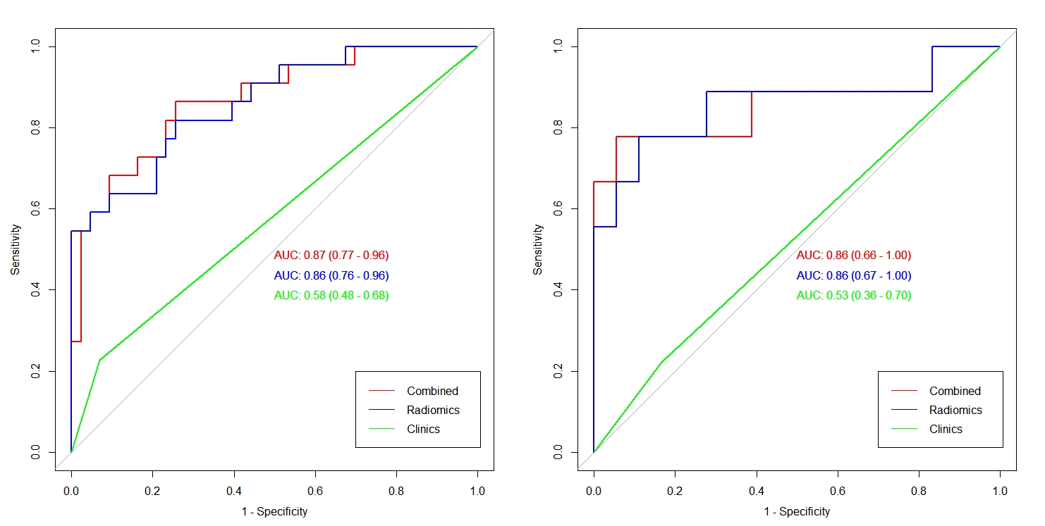
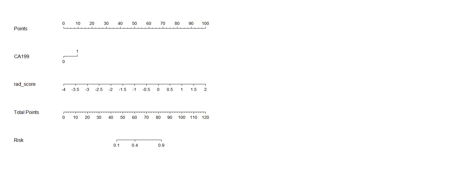
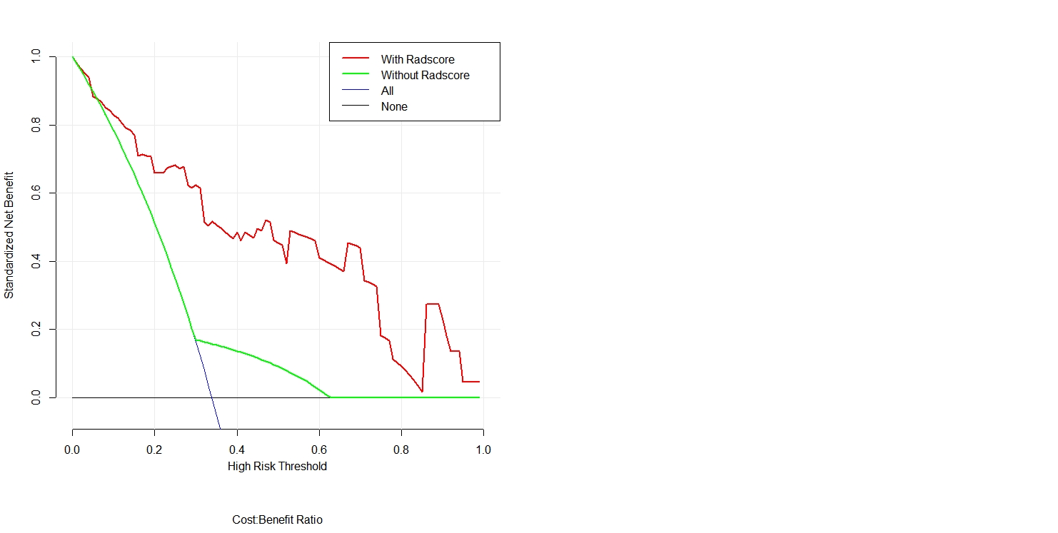
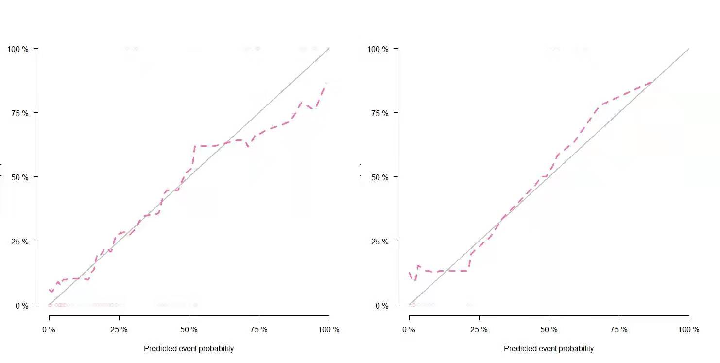
A total of 12 radiomics features were retained to construct a predictive model. As shown in Fig.2, The AUC of the clinical variable model in the training and the testing cohorts are 058(95% CI, 0.48-0.68) and 0.53(95% CI, 0.36-0.79). The AUC of the single radiomics model in the training and testing cohorts are0.87(95% CI, 0.77-0.96) and 0.86(95% CI, 0.66-1.00). The AUC of multi-model radiomics in the training and validation cohorts are 0.88(95% CI, 0.80–0.96) and 0.88(95% CI, 0.72–1.00). The optimal multimodal radiomics nomogram was developed for PNI estimation in each patient (Fig.3). Calibration curves (Fig.4) and decision curves analysis (Fig.5) indicated that the multimodal radiomics model provided greater clinical benefits.Discussion
The challenges of multi-modal fusion mainly include how to judge the confidence level of each mode and the correlation between modes, how to reduce the dimension of multi-modal characteristic information and how to register the multi-modal data collected asynchronously. We compared the advantages of two multimodal radiomics models for CT and MRI integration. The combined multimodal radiomics model was superior to the single model and the clinical model, indicating that the multimodal radiomics model approach may have a greater value in preoperative PNI prediction. The multimodal model can provide more abundant information than either model alone.Conclusion
Multiparametric radiomics features from multiparametric MRI of rectal cancer is a useful tool for predicting PNI preoperatively and has marked discrimination accuracy.Acknowledgements
No acknowledgment found.References
1. Bray F, Ferlay J, Soerjomataram I, Siegel RL, Torre LA, Jemal A (2018) Global cancer statistics 2018: GLOBOCAN estimates of incidence and mortality worldwide for 36 cancers in 185 countries. CA Cancer J Clin 68:394-424
2. Liebig C, Ayala G, Wilks J et al (2009) Perineural invasion is an independent predictor of outcome in colorectal cancer. J Clin Oncol 27:5131-5137
3. Betge J, Pollheimer MJ, Kornprat P, Rehak P, Vieth M, Langner C (2011) Perineural invasion is a strong and independent predictor of lymph node involvement in colorectal cancer. Dis Colon Rectum 54:e273
4. Suzuki T, Suwa K, Ogawa M et al (2015) Adjuvant chemotherapy for the perineural invasion of colorectal cancer. J Surg Res 199:84-89
5. Benson AB, 3rd, Venook AP, Al-Hawary MM et al (2018) Rectal Cancer, Version 2.2018, NCCN Clinical Practice Guidelines in Oncology. J Natl Compr Canc Netw 16:874-901
6. Lambin P, Leijenaar RTH, Deist TM et al (2017) Radiomics: the bridge between medical imaging and personalized medicine. Nat Rev Clin Oncol 14:749-762
7. Lambin P, Rios-Velazquez E, Leijenaar R et al (2012) Radiomics: extracting more information from medical images using advanced feature analysis. Eur J Cancer 48:441-446
Figures