0820
High Resolution T2W imaging using Deep Learning Reconstruction and Reduced Field-of-View PROPELLER1GE Healthcare, Houston, TX, United States, 2Global MR Applications & Workflow, GE Healthcare, New York, NY, United States, 3Global MR Applications & Workflow, GE Healthcare, Calgary, AB, Canada, 4Global MR Applications & Workflow, GE Healthcare, Waukesha, WI, United States, 5Department of Imaging Physics, MD Anderson Cancer Center, Houston, TX, United States, 6Global MR Applications & Workflow, GE Healthcare, Houston, TX, United States
Synopsis
T2W FSE-PROPELLER is robust to susceptibility artifacts and bulk motion, but requires longer acquisition times compared to conventional FSE methods. Recently, a reduced Field-Of-View PROPELLER sequence using rotating outer volume suppression method has been proposed and optimized to reduce the scan time for small FOV and high-resolution T2W imaging. However, image SNR is comparatively lower compared to the conventional PROPELLER with phase oversampling. In this work, a deep learning based PROPELLER reconstruction method was used to improve the SNR and image quality of the reduced Field-Of-View PROPELLER.
Introduction
T2W FSE-PROPELLER is commonly used due to its robustness to susceptibility artifacts and bulk motion. However, it requires longer acquisition times compared to conventional FSE methods, since the phase-encoding direction in PROPELLER is changed with each blade, and the oversampling is not confined to a single axis. Recently, a rotating outer volume suppression method (1,2) has been proposed and optimized to reduce the scan time for small FOV and high-resolution T2W imaging using PROPELLER. Compared to the other inner-volume imaging (3) or outer-volume suppression method (4), it also can support interleaved slice acquisition and variable refocusing flip angles (5). Notwithstanding these advantages, image SNR is comparatively lower compared to conventional PROPELLER with phase oversampling. Thus, the purpose of this work was to improve the SNR and image quality of the reduced Field-Of-View PROPELLER (rFOV PROPELLER) using a deep learning reconstruction method.Materials and Methods
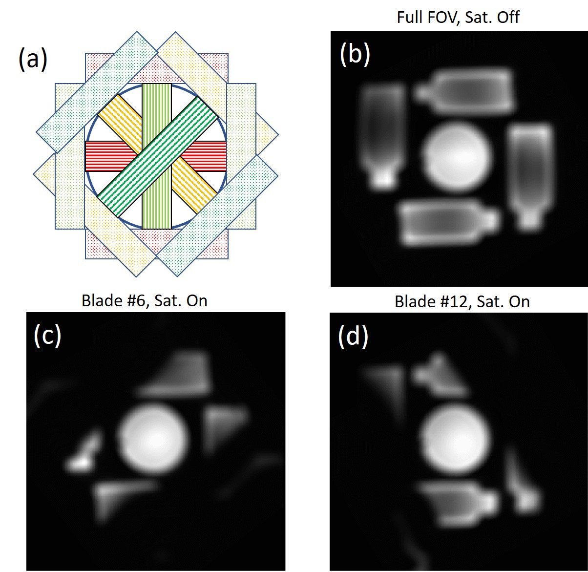
rFOV PROPELLER using rotating outer volume suppression (OVS) and variable refocusing flip (VRF) angles was implemented on a 3.0T MRI scanner (Discovery MR750, GE Healthcare, Waukesha, WI), as shown in Figure 1. Over 10,000 high quality images were used to train a convolutional neural network (CNN) to reconstruct T2W PROPELLER images with high SNR and high spatial resolution. A tunable noise reduction factor was offered to accommodate user preference. In this study, the noise reduction factor was 75%. In addition to this deep learning reconstruction (hereby named “DL Recon PROP”), a second set of images was reconstructed from the same raw data using conventional reconstruction (Conventional Recon). The saturation mechanism of rFOV PROPELLER was visualized with a large phantom. DL Recon PROP was first validated with an ACR phantom, then was evaluated in the prostate of 5 healthy volunteers with IRB approval and written informed consent. The prostate images were acquired in axial, sagittal and coronal planes. The typical in-vivo small FOV imaging parameters of the conventional PROPELLER included: axial orientation; FOV = 140 × 140 mm2; Resolution = 0.5 × 0.5 mm2; slice thickness = 4 mm; number of slices = 22; Oversampling Factor (OSF) = 3; FA = 120 degrees; ETL = 28; TE = 103 ms and auto TR. Same parameters were used in the rFOV PROPELLER, except ETL is 46 for VRF = 60 (minimum), 100 (middle),120 (last) degrees to match the TE, and the number of OVS saturation pulses was 2 for efficient saturation.Results and Discussion
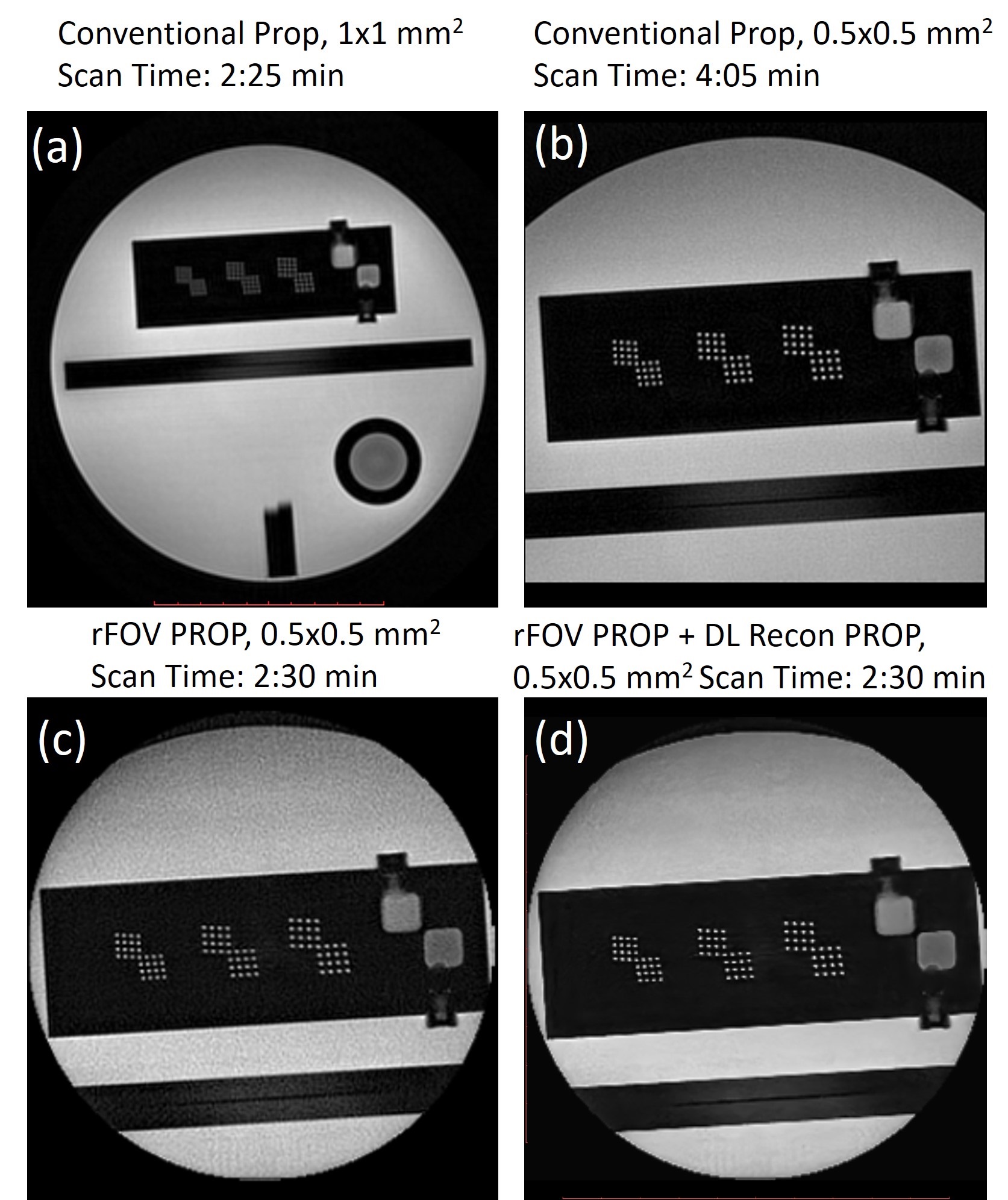
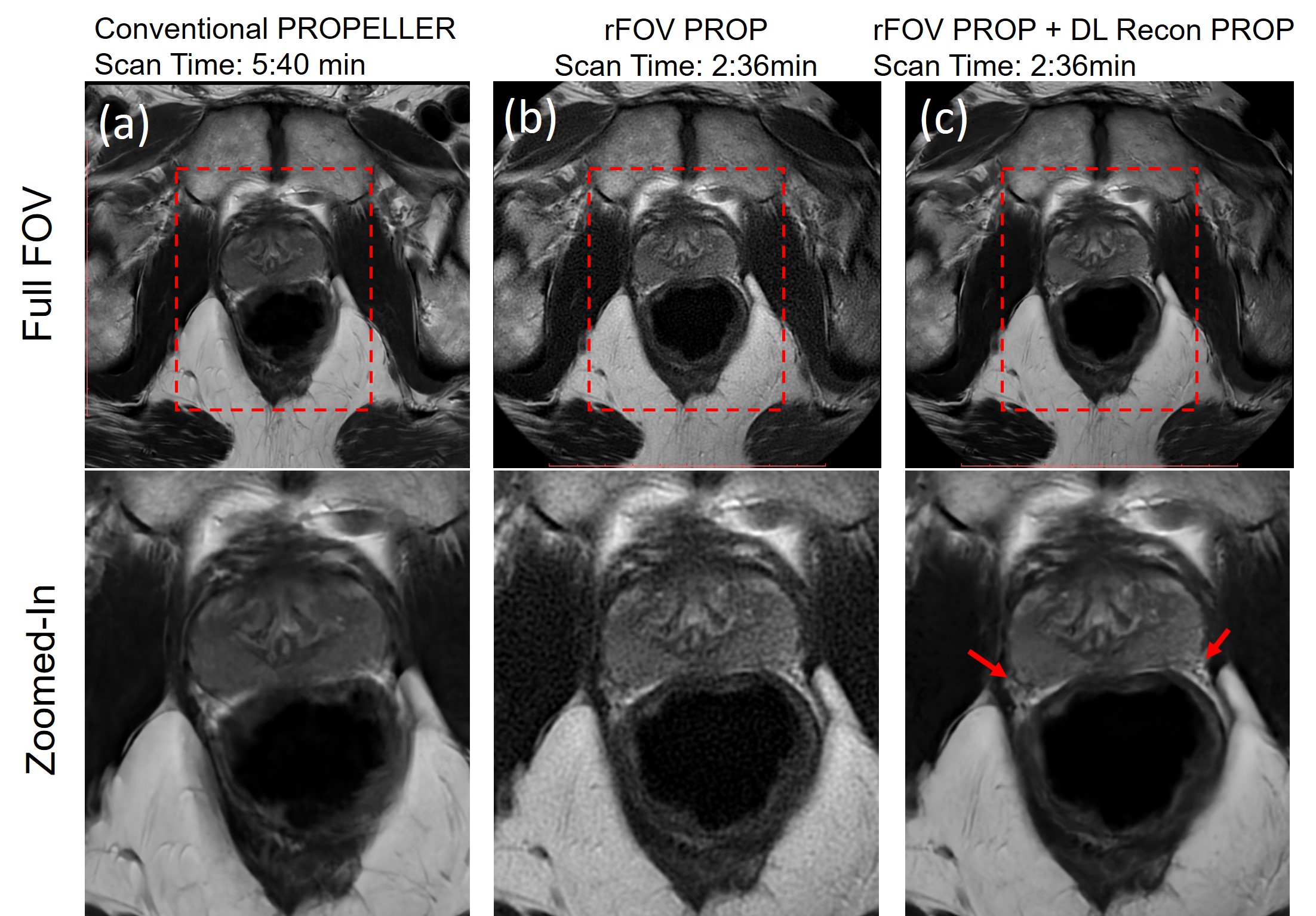
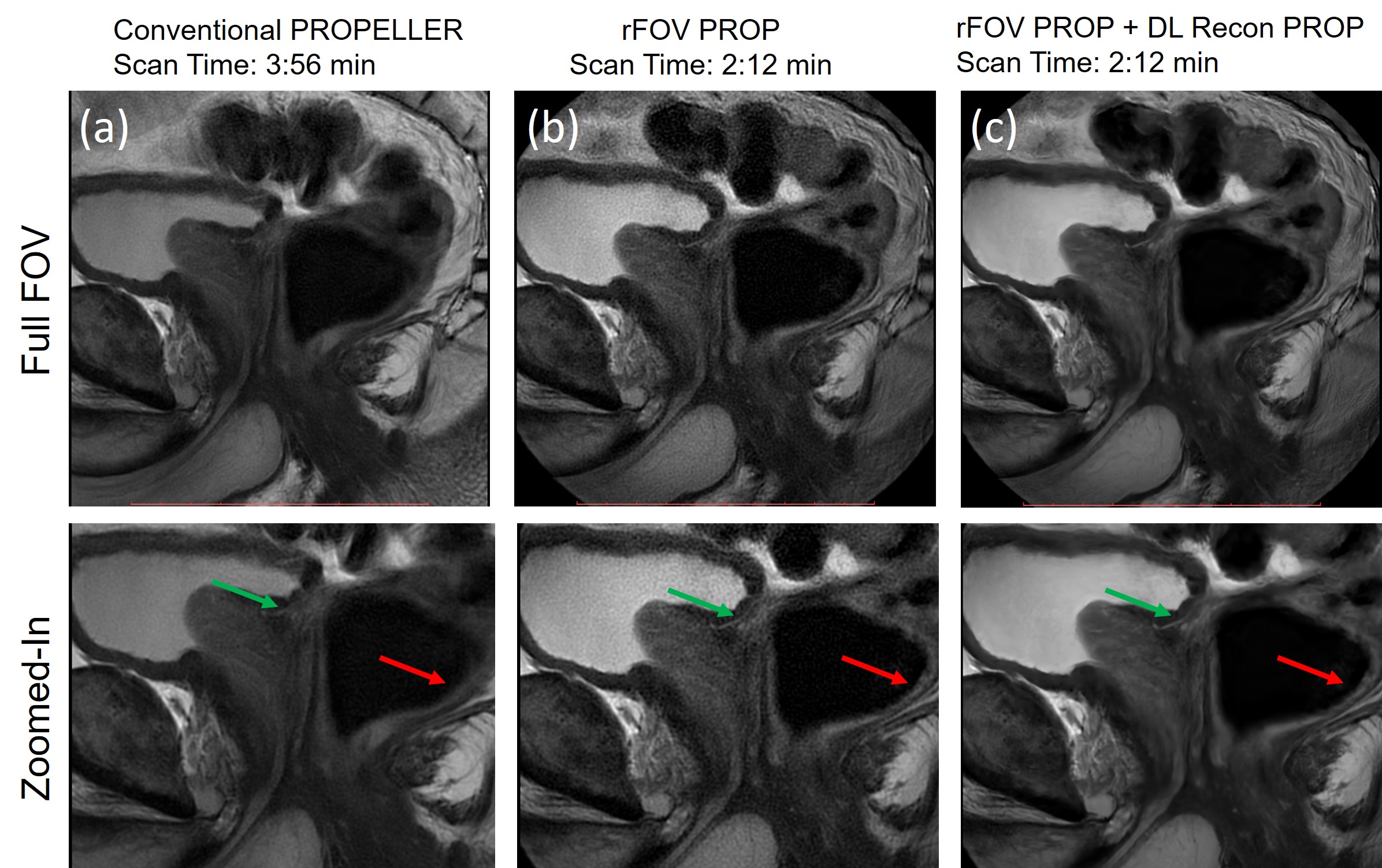
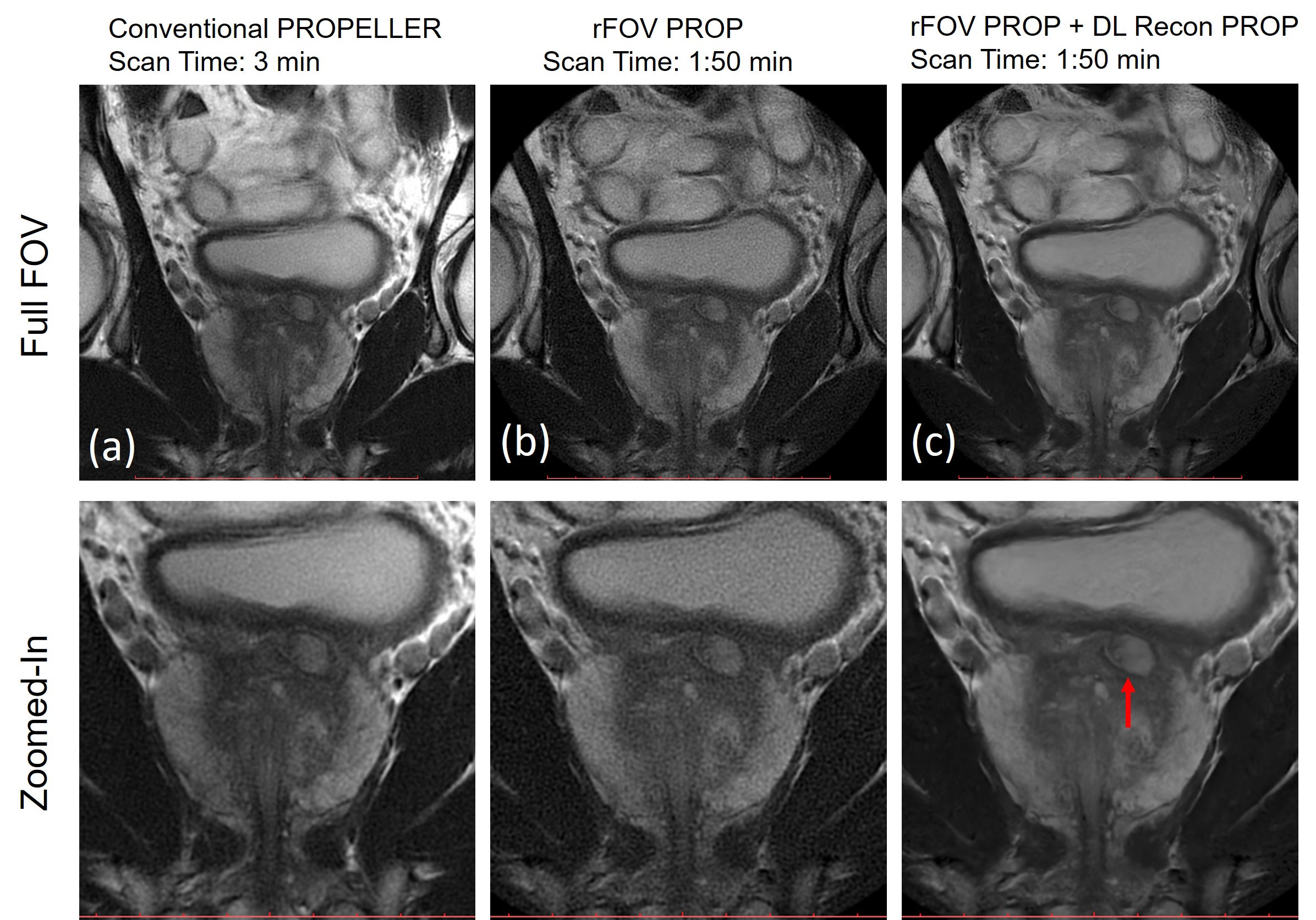
The spatial profile of saturation bands used in the rotating outer volume suppression is shown in Figure 1c, 1d. The saturation bands rotated with the blades, and efficiently saturated the signal from the phantoms out of the imaging volume. While the bright spots in the ACR phantom can be well separated in the high-resolution reduced FOV image (Fig. 2b, OSF = 1), the scan time is comparatively longer than the low-resolution image (Fig. 2a, OSF = 2) due to the increased phase oversampling factor. Combining rotating outer volume suppression, longer ETL and variable refocusing flip angles, rFOV PROPELLER (OSF = 1) reduced scan time (2:30 min vs 4:05 min) but resulting in low SNR, as shown in Fig. 2c. However, the SNR was improved using DL Recon PROP (Fig. 2d). Figure 2c and 2d were generated from the same raw data acquired using rFOV PROPELLER. Figure 3 shows high-resolution prostate images acquired in axial plane using conventional PROPELLER with an oversampling factor of 3 (Fig. 3a, 5:40 min) and rFOV PROPELLER (Fig. 3b, 2:36 min). While rFOV PROPELLER halves the acquisition time, the SNR is lower compared to the conventional PROPELLER with phase oversampling since SNR is proportional to the square root of the number of phase-encoding steps. However, DL Recon PROP can be used to improve the image quality of rFOV PROPELLER. DL Recon PROP improved the SNR and sharpness (red arrows) of images acquired using rFOV PROPELLER, as shown in Fig. 3c. Figure 4 and 5 show high-resolution sagittal and coronal prostate image acquired using conventional PROPELLER with phase oversampling (Fig. 4a, 5a; OSF = 3) and rFOV PROPELLER (Fig. 4b, 5b; OSF = 1). rFOV PROPELLER with VRF achieved ~50% acquisition time reduction compared with the corresponding high-resolution conventional PROPELLER, but with an expected reduction in SNR. DL Recon PROP improved the SNR and sharpness (Fig. 4c, red arrows) of images acquired using rFOV PROPELLER. rFOV PROPELLER images with DL Recon PROP were not visually inferior to conventional PROPELLER images with phase oversampling. In contrast, with DL Recon PROP and rFOV PROPELLER, it can depict fine anatomical details, such as vessels (Fig. 4c, green arrow) and the rectal wall (Fig. 4c, red arrow).Conclusion
Deep learning reconstruction method can improve the SNR and sharpness of high-resolution T2W images acquired using rFOV PROPELLER with rotating outer volume suppression and variable refocusing flip angles. Unlike conventional denoising techniques, deep learning reconstruction won’t smooth or blur the images. In contrast, deep learning reconstruction can improve the image sharpness.Acknowledgements
No acknowledgement found.References
1. Litwiller DV, Taviani V, Banerjee S, et al. Rotating Outer Volume Suppression for Reduced Field of View PROPELLER Imaging. ISMRM, 2018, 2685.
2. Wang X, Litwiller DV, Estkowski L, et al. High Resolution T2W imaging using Multi-shot Reduced Field-of-View PROPELLER. ISMRM, 2019, 0990
3. Deng J. and Larson A. Multishot targeted PROPELLER magnetic resonance imaging: description of the technique and initial applications. Invest Radiol. 2009;44(8): 454-462
4. Kojima S, Morita S, Ueno E, et al. Aliasing artifacts with the BLADE technique: Causes and effective suppression. J Magn Reson Imaging. 2011;33:432-440
5. Busse RF, Hari H, Anthony V, et al. Fast spin echosequences with very long echo trains: design of variable refocusing flip angle schedules and generation of clinical T2 contrast. Magn Reson Med. 2006;55(5):1030-7
Figures