0812
Unsupervised radial streak artifact reduction in time resolved MRI1Global MR Applications & Workflow, GE Healthcare, Tucson, AZ, United States, 2Global MR Applications & Workflow, GE Healthcare, Madison, WI, United States, 3Global MR Applications & Workflow, GE Healthcare, New York, NY, United States, 4Global MR Applications & Workflow, GE Healthcare, Hino, Japan, 5Global MR Applications & Workflow, GE Healthcare, Houston, TX, United States
Synopsis
Radial magnetic resonance imaging is attractive due to its inherently high motion robustness and its ability to support accelerated imaging but is plagued by streaking artifact. The problem is exacerbated in time resolved imaging, like DCE-MRI, which deal with higher levels of undersampling due to the need to jointly deliver high spatial and temporal resolution. While reconstructive methods typically based on sparse or low rank methods exist to minimize streak artifact, their use is currently limited due to their high computational complexity. As an alternative, we describe a temporal neural network to suppress streak artifact from a time-series of images.
Introduction
A consequence of undersampling in radial MRI is streaking artifact that tends to corrupt the entire imaging scene. While being mostly being at moderate undersampling rates, the artifact can become quite severe with increasing undersampling rates and degrades the diagnostic quality of images (poor SNR, high artifact, reduced resolution). Streaking artifact may occur even when the data is sampled fully due to other factors: gradient nonlinearity, motion, inadequate fat suppression.1 The artifact is even more problematic in higher dimensional imaging scenarios like time resolved imaging / contrast weighted imaging.2 Additionally, the long scan times here make them more susceptible to motion and associated artifacts (including streaking)A wide variety of methods have been developed to address streak artifacts in radial MRI. Methods based on parallel imaging and / or compressed sensing have been used with a fair amount of success to address the problem.2-3 However, all these methods are typically associated with large computational costs. Deep learning methods, usually supervised, offer the promise of delivering the high-quality performance of some of the more compute intensive methods but at a fraction of the compute cost.4-5 In contrast we propose an unsupervised, lightweight, patient specific temporal neural net to address the problem of streaking.
Unsupervised learning
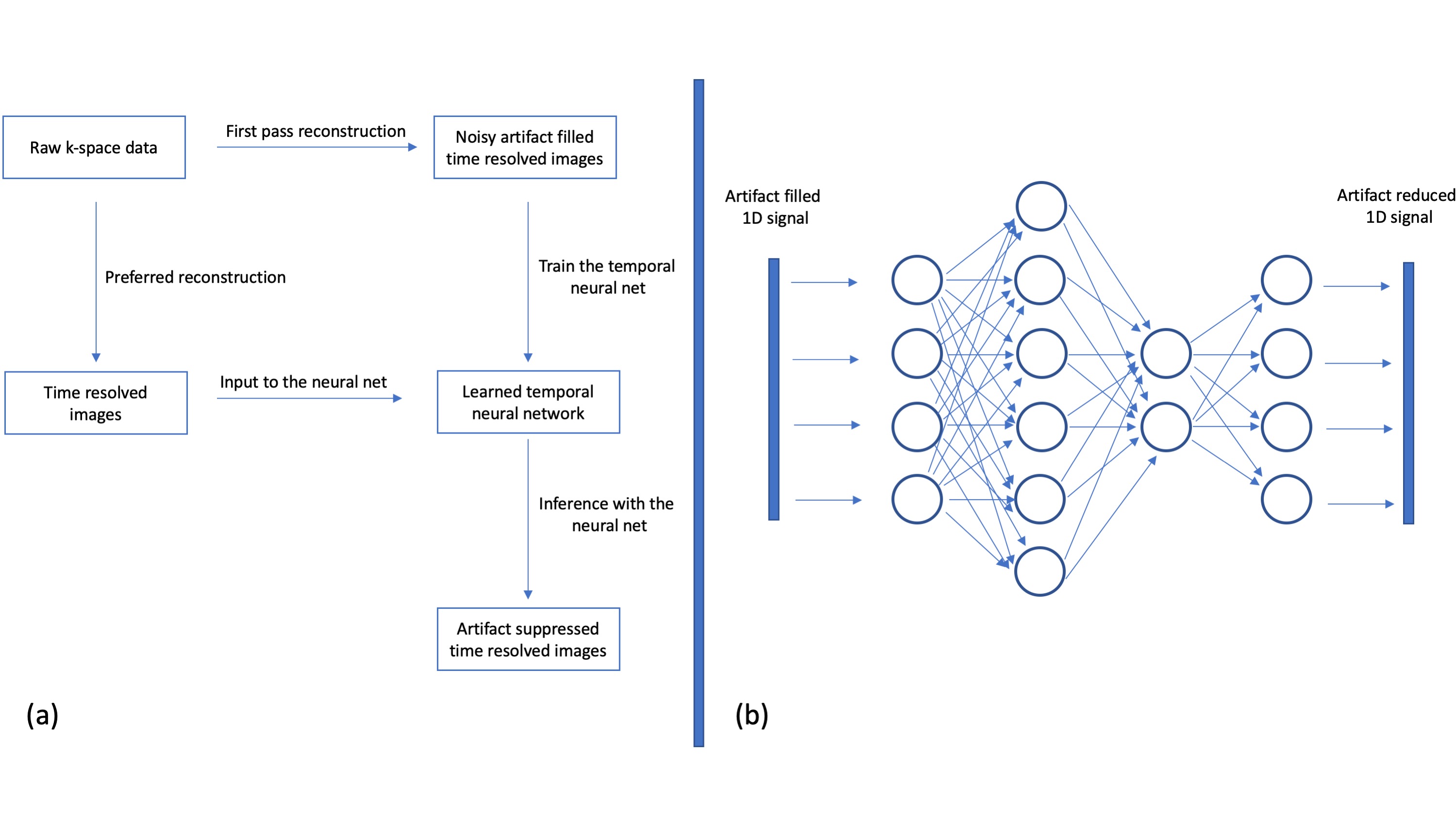
Time resolved imaging applications typically generate a time series of images and the time / contrast dimension tend to be compressible due to the significant amount of correlations in the data. Many low rank approaches exploit this fact to reconstruct images from undersampled data. While popularly used within a reconstructive setting, a rank operator can be built to act as a purely post-processing filter as well (analogous to using PCA for filtering noisy data). Such low rank filtering is one variant of an unsupervised method that generates a linear filter to reduce noise and artifact.6We extend this to the non-linear setting by using 1-D temporal neural nets that are trained in an unsupervised manner. The unsupervised method by-passes the need for labeled data and seeks to learn the underlying structure in the training data from the corrupted training samples themselves. The proposed method deals with 1D temporal or contrast signals and is trained on the fly. Fig 1 (a) shows one possibility for the training and inference pipelines, while Fig 1(b) shows an exemplar 1D signal being processed by the trained network. Note that as the inference using the trained model is spatially independent, it can be parallelized across that dimension leading to very low inference costs.
Methods and Results
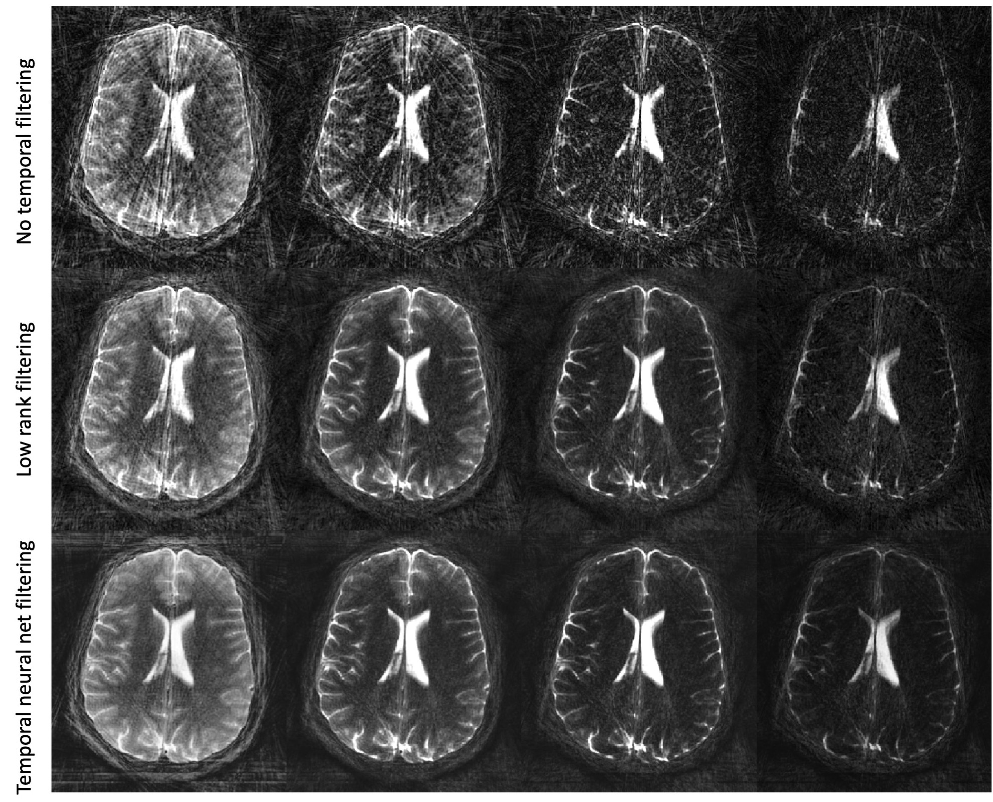
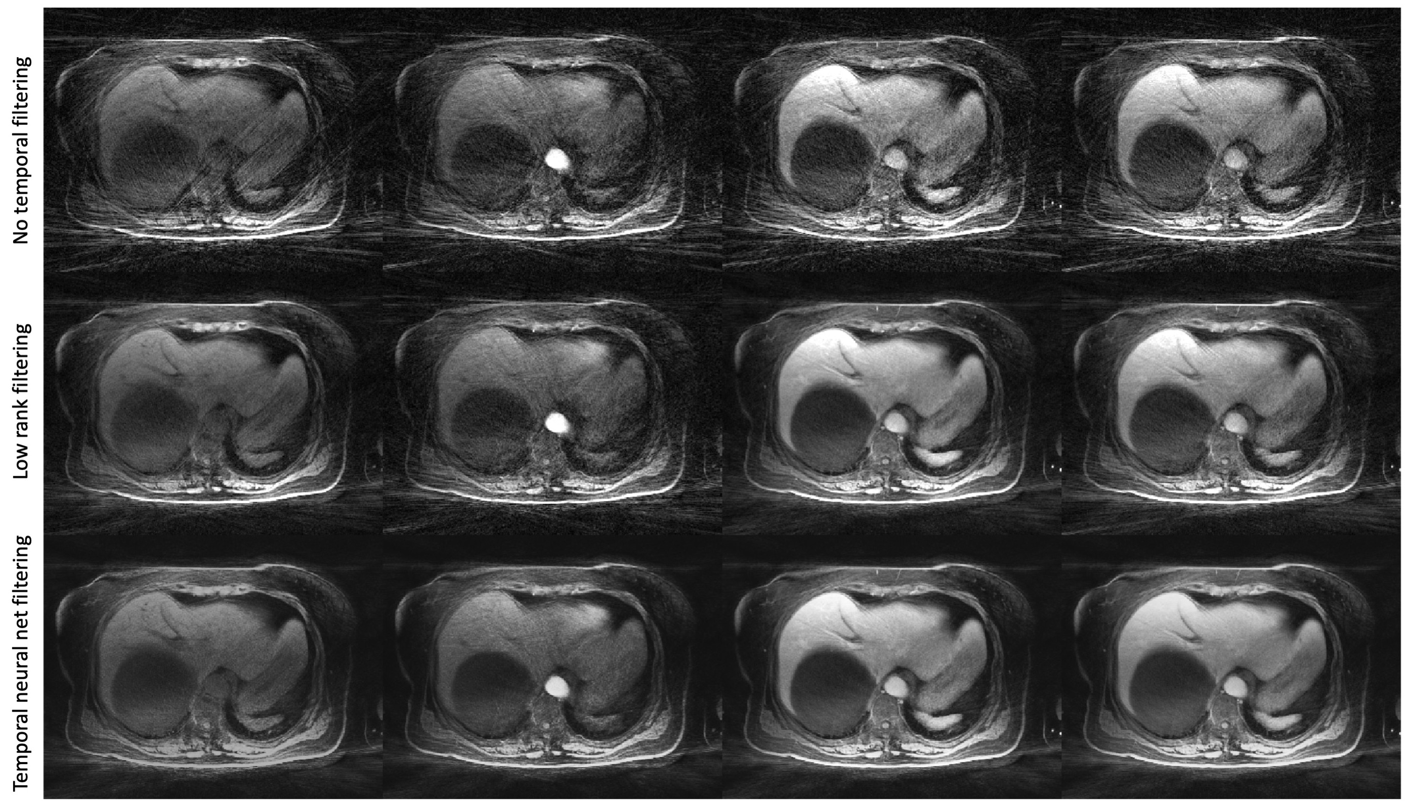
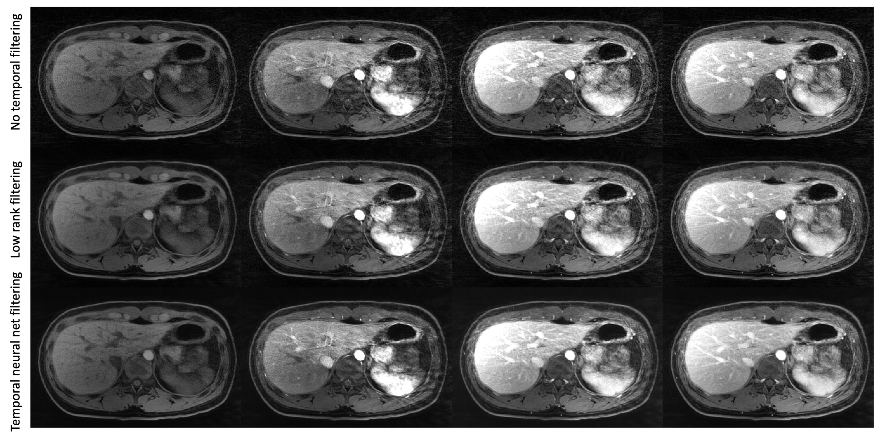
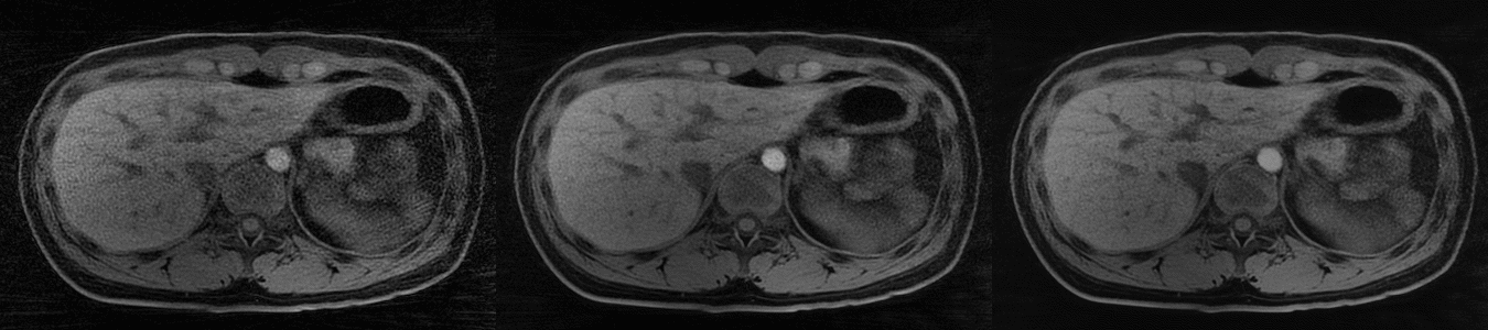
The proposed methods were tested at 3T on data generated from two sequences with a golden angle radial trajectory. The first sequence is a 2D radial single shot fast spin echo (radSSFSE) and uses a variable refocussing flip angle scheme with an echo train length of 264.7 The radSSFE data was retrospectively grouped at 22views/phase to generate 12 phases with different T2 weighting. The second sequence is the LAVA-STAR sequence with a stack-of-stars trajectory and intermittent fat suppression.8 The data from this scan was processed on a per slice basis at 100 views/phase and 20 phases. The time-series images were created using a gridding reconstruction and the resulting datasets were further processed with both the low rank and temporal neural-net based filters. Note that we refrained from the use of any spatial processing methods like parallel imaging or compressed sensing to enable a simpler evaluation of the performance of the proposed methods. The proposed methods are fully compatible with all other spatial processing methods as they operate purely along the time/contrast dimensions.The results of these additional filtering operations from the radSSFSE dataset are presented in Figure 2 where they are compared against the no filtering case which serves as a baseline for the artifact levels. Note the really strong reduction in streak artifact and the improved resolution and depiction of brain anatomy with the use of the temporal neural net. While the low rank filter also generates a strong reduction in artifact relative to the baseline, its performance is inferior to the temporal neural net. Figures 3 and 4 present results from DCE scans using the LAVA-STAR sequence. Note that the performance trends continue along the same lines. Figure 5 is a movie of the results in Fig 4 which enables a better visualization of the reduction in the flickering streak artifact with the baseline (no filtering), low rank and temporal neural nets in the first, second and third columns respectively.
Conclusion
We propose a simple method that can enable strong reduction in streak artifact in MR time series data with a very low processing overhead. The chief advantage of the method is the unsupervised nature of the network training. Additionally, as we deal with 1D signals, the training and inference is significantly cheaper than most deep learning approaches which typically seek to filter in 2D or 3D. While the streaking artifact reduction is strictly applicable to radial scans, the method is quite general and should be applicable to many applications that generate high-dimensional data.Acknowledgements
No acknowledgement found.References
1. Mandava et al., Radial streak artifact reduction using phased array beamforming. MRM, 2019; 81: 3915-3923
2. Feng et al., RACER-GRASP: Respiratory-weighted, aortic contrast enhancement-guided and coil-unstreaking golden angle radial sparse MRI. Magn Reson Med 2018;80: 77–89
3. Huang et al., T2 mapping from highly undersampled data by reconstruction of principal component coefficient maps using compressed sensing. MRM, 2012, 67, 1355–1366.
4. Han et al., Deep learning with domain adaptation for accelerated projection-reconstruction MR, MRM, 2018; 80: 1189-1205
5. Hauptmann et al., Real‐time cardiovascular MR with spatio‐temporal artifact suppression using deep learning–proof of concept in congenital heart disease. MRM, 2019; 81: 1143-1156
6. Mandava et al., A non-local low rank approach for high resolution parameter mapping. In Proceedings of the 26th Annual Meeting of ISMRM, Paris, 2018. Abstract No. 5611
7. Litwiller et al., Radial Single-Shot Fast Spin Echo: Toward Fast, Motion-Robust, Multi-Contrast Imaging. In Proceedings of the 27th Annual Meeting of ISMRM, Montreal, 2019. Abstract No. 4680
8. Zhang et al., Fast motion robust abdominal stack of stars imaging using coil compression and soft gating. In Proceedings of the 25th Annual Meetings of ISMRM, Honolulu, HI, 2017. Abstract No.1284
Figures