0783
Improved SMS Reconstruction using ReadOut-Concatenated K-space SPIRiT (ROCK-SPIRiT)1Electrical and Computer Engineering, University of Minnesota, Minneapolis, MN, United States, 2Center for Magnetic Resonance Research, University of Minnesota, Minneapolis, MN, United States, 3Department of Imaging Physics, Delft University of Technology, Delft, Netherlands
Synopsis
Simultaneous Multi-slice (SMS) imaging has great potential for high acceleration rates with minimal loss in SNR. However, due to the unfavorable coil geometry only moderate acceleration rates have been achieved in cardiac applications. Outer volume suppression (OVS) has been proposed to overcome this limitation by suppressing unwanted signal from extra-cardiac tissues such as chest and back. Despite OVS, existing reconstruction techniques may suffer from residual leakage artifacts or noise amplification. In this work, we sought to extend SPIRiT to a readout-concatenated space to improve image quality in highly accelerated perfusion and cine imaging while mitigating leakage and noise amplification.
Introduction
Simultaneous Multi-slice (SMS) imaging has gained interest in cardiac MRI (CMR) for improving coverage with minimal SNR loss1. However, due to unfavorable coil geometries, acceleration potential of SMS CMR remains limited compared to neuroimaging. Image quality with high acceleration rates is compromised due to leakage artifacts from extra-cardiac tissues1. To enable higher acceleration factors in SMS CMR, outer volume suppression (OVS) techniques have been proposed to suppress unwanted signals from surrounding tissues2,3. Even with OVS, the reconstruction method remains important for artifact-free SMS CMR. Numerous techniques have been proposed to reconstruct SMS accelerated images, providing different trade-offs between noise reduction and leakage-blocking4-6. In this work, we sought to develop a new SMS reconstruction technique called readout-concatenated k-space (ROCK)-SPIRiT to reduce leakage artifacts and noise amplification in highly-accelerated SMS imaging. ROCK-SPIRiT was evaluated on both perfusion and cine imaging and compared to SENSE/GRAPPA4, split slice(ss)-GRAPPA5 and SMS-SPIRiT6. It was shown to reduce leakage artifacts compared to SMS-SPIRiT and noise amplification compared to SENSE/GRAPPA and ss-GRAPPA.Methods
Reconstruction: ROCK-SPIRiT utilizes the idea of readout concatenation in image domain as in SENSE/GRAPPA4 to encode SMS acceleration. SMS-accelerated slices are inverse Fourier transformed along readout and concatenated, forming an extended k-space. SMS acceleration then corresponds to undersampling in this concatenated direction (Fig. 1). ROCK-SPIRiT solves the following objective function in the extended-kspace:$$arg\ \underset{\bf{\kappa_{ext}}}{min} ||\bf{P_{\Omega}}\bf{\kappa_{ext}}-\bf{\kappa_{SMS}}||_2^2 + \lambda||\bf{G_{ROCK}}\bf{\kappa_{ext}}-\bf{\kappa_{ext}}||^2_2 + \sigma\bf{\Psi}(\bf{E}\bf{\kappa_{ext}})$$
where $$$\bf{P_{\Omega}}$$$ is a subsampling operator in k-space, $$$\bf{\kappa_{ext}}$$$ is the extended k-space in readout direction, $$$\bf{\kappa_{SMS}}$$$ is the acquired SMS data across all coils in k-space, $$$\bf{G_{ROCK}}$$$ is the ROCK-SPIRiT self-consistency operator calibrated on low resolution extended k-space, $$$\bf{E}$$$ is SENSE-1 operator, $$$\bf{\Psi}$$$ is a regularizer, $$$\lambda$$$ and $$$\sigma$$$ are weight terms. Fig. 1 shows a schematic of data consistency (first term) and SPIRiT8 self-consistency (second term). The objective function is solved using ADMM.
Images were reconstructed with SENSE/GRAPPA4, ss-GRAPPA5, SMS-SPIRiT6 and ROCK-SPIRiT. SENSE/GRAPPA utilizes standard GRAPPA on readout-extended k-space, ss-GRAPPA works on regular k-space providing leakage-blocking as an extension to slice-GRAPPA, and SMS-SPIRiT employs individual SPIRiT kernels on SMS-accelerated k-space. For all methods, interpolation kernels were generated from calibration data.
ROCK-SPIRiT for cine imaging was implemented with $$$\lambda = 10^{-4}$$$ and $$$\sigma=1.2 \times 10^{-7}$$$ and the regularizer was based on locally low rank constraint9. For perfusion imaging, three-set of SMS-accelerated slices (for a total of 9 slices) were processed together for each dynamic. Thus, no temporal information was shared across dynamics. ROCK-SPIRiT for perfusion imaging was implemented with $$$\lambda = 10^{-4}$$$ and $$$\sigma=2.5 \times 10^{-8}$$$ and regularizer was based on low dimensional-structure self-learning and thresholding10.
In Vivo Imaging: Imaging was performed at 3T (Siemens Prisma) with institutional review board approval. Written informed consent was obtained. A multiband combination of two slab-selective single band saturation pulse along phase-encoding was utilized to perform OVS2 (Fig. 2), suppressing signals from chest and back simultaneously. First-pass perfusion imaging was performed on a healthy subject with injection of 0.05 mmol/kg gadabutrol at 4 mL/s followed by a 10-mL saline flush, followed by cine imaging2. Perfusion parameters were: SMS-factor=3, in-plane acceleration=4, partial Fourier=6/8, FOV/resolution=360×360/1.7x1.7mm2, slice-thickness=8mm, temporal resolution=110ms, saturation time=150ms. Cine parameters were: SMS-factor=5, FOV/resolution=320×320/1.7x1.7mm2, slice-thickness=6mm, temporal resolution=41ms. Cine data was further retrospectively undersampled to SMS=5 × in-plane acceleration=2 with 24 ACS lines. Slices were simultaneously excited with CAIPIRINHA shifts7. Both sequences used the aforementioned OVS module, which was interleaved within the imaging window between every 9 imaging pulses for perfusion and 8 for cine. OVS parameters were2: saturation slab=150mm (each side), 3.8ms asymmetric sinc, BWT=8 and RF peak shift = 15%. Calibration scans were acquired prior to imaging with FOV/resolution=360×360/1.7×5.6mm2 and FOV/resolution=320×320/1.7×4mm2 for perfusion and cine respectively.
Results
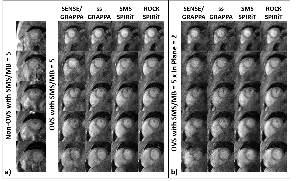
Fig. 3a shows cine results without and with OVS at SMS=5. Non-OVS images suffer from substantial artifacts. SENSE/GRAPPA with OVS shows leakage artifacts and suffers from noise amplification. SMS-SPIRiT with OVS shows less noise amplification, but residual leakage artifacts remain visible. ss-GRAPPA eliminates leakage artifacts albeit at increased noise amplification. ROCK-SPIRiT shows improved image quality compared to other methods with less noise and no leakage artifacts.Fig. 3b shows OVS-based cine with additional 2-fold in-plane acceleration. In this scenario, SENSE/GRAPPA has significant artifacts. SMS-SPIRiT shows more leakage artifacts with the higher acceleration rate. ss-GRAPPA eliminates the leakage artifacts but suffers from high noise amplification. ROCK-SPIRiT improves reconstruction by both eliminating leakage and reducing noise amplification, although some artifacts remain.
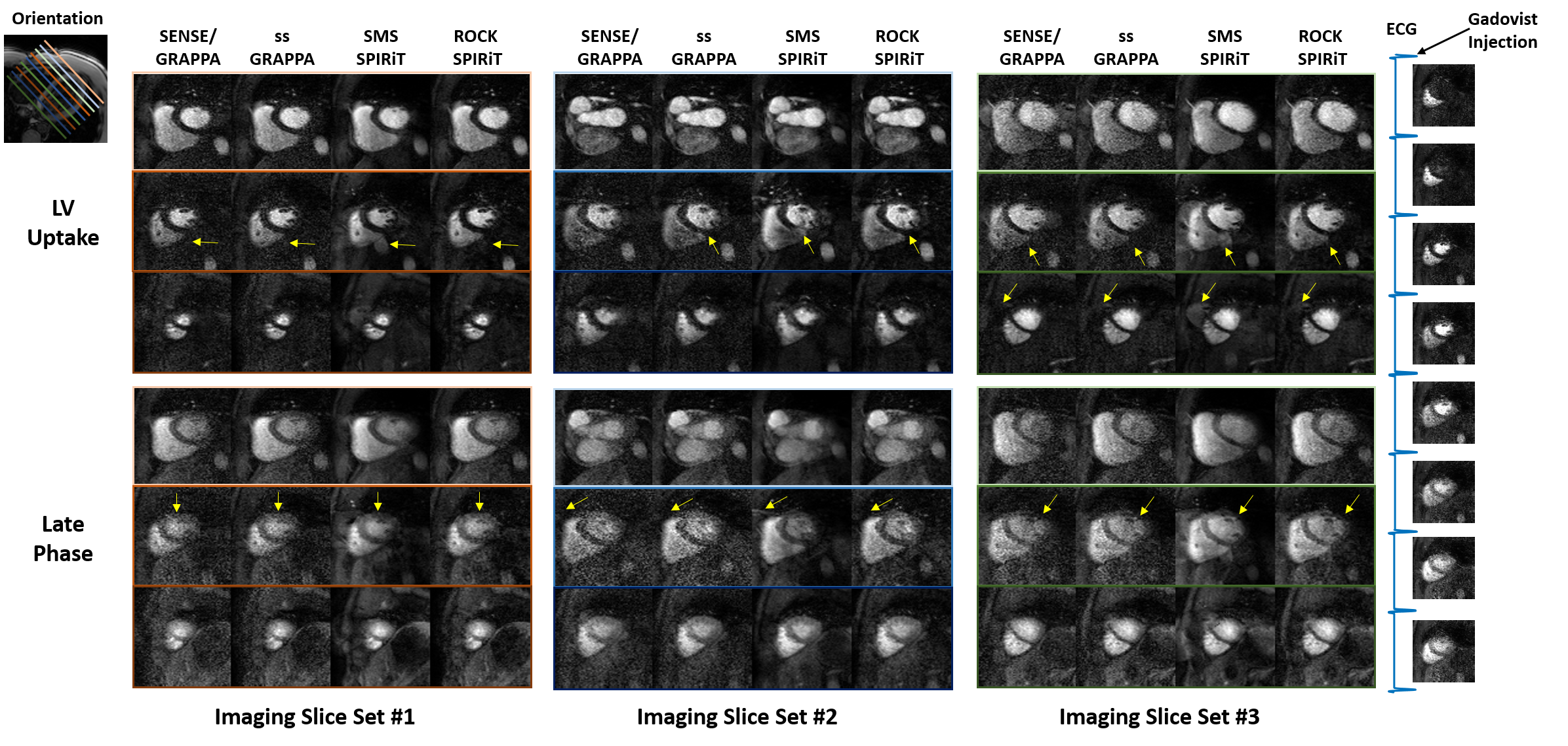
Fig. 4 shows OVS-based perfusion CMR for a highly-accelerated scan (SMS=3×in-plane acceleration=4×partial Fourier=6/8). Noise amplification is visible in SENSE/GRAPPA and ss-GRAPPA reconstructed images. SMS-SPIRiT shows leakage artifacts (yellow arrows). ROCK-SPIRiT reduces noise and improves image quality compared to other methods.
Discussion
In this study, we proposed a new regularized SMS reconstruction technique called ROCK-SPIRiT. This allows further denoising than SENSE/GRAPPA and ss-GRAPPA by enabling regularization. ROCK-SPIRiT also reduces leakage compared to SMS-SPIRiT.Conclusion
ROCK-SPIRiT provides improved image quality for highly accelerated SMS CMR, reducing leakage and noise amplification compared to existing methods.Acknowledgements
Funding: Grant support: NIH R00HL111410, NIH P41EB027061, NIH 1S10OD017974-01 and NSF CAREER CCF-1651825.References
- Weingartner, S., et al., Simultaneous multislice imaging for native myocardial T-1 mapping: Improved spatial coverage in a single breath-hold. Magnetic Resonance in Medicine, 2017. 78(2): p. 462-471.
- Weingärtner, S., S. Moeller, and M. Akcakaya. Feasibility of Ultra-high Simultaneous Multi-slice and In-plane Accelerations for Cardiac MRI Using Outer Volume Suppression and Leakage-Blocking Reconstruction. 2018. Annual Meeting of the International Society of Magnetic Resonance in Medicine.
- Yang, Y., et al., Reduced field of view single-shot spiral perfusion imaging. Magnetic Resonance in Medicine, 2018. 79(1): p. 208-216.
- Blaimer, M., et al., Accelerated volumetric MRI with a SENSE/GRAPPA combination. Journal of Magnetic Resonance Imaging, 2006. 24(2): p. 444-450.
- Cauley, S.F., et al., Interslice Leakage Artifact Reduction Technique for Simultaneous Multislice Acquisitions. Magnetic Resonance in Medicine, 2014. 72(1): p. 93-102.
- Yang, Y., et al., Whole-heart spiral simultaneous multi-slice first-pass myocardial perfusion imaging. Magnetic Resonance in Medicine, 2019. 81(2): p. 852-862.
- Breuer, F.A., et al., Controlled aliasing in parallel imaging results in higher acceleration (CAIPIRINHA) for multi-slice imaging. Magnetic Resonance in Medicine, 2005. 53(3): p. 684-691.
- Lustig, M. and J.M. Pauly, SPIRiT: Iterative Self-consistent Parallel Imaging Reconstruction From Arbitrary k-Space. Magnetic Resonance in Medicine, 2010. 64(2): p. 457-471.
- Zhang, T., J.M. Pauly, and I.R. Levesque, Accelerating Parameter Mapping with a Locally Low Rank Constraint. Magnetic Resonance in Medicine, 2015. 73(2): p. 655-661.
- Akcakaya, M., et al., Low-dimensional-Structure Self-Learning and Thresholding: Regularization Beyond Compressed Sensing for MRI Reconstruction. Magnetic Resonance in Medicine, 2011. 66(3): p. 756-767.
Figures
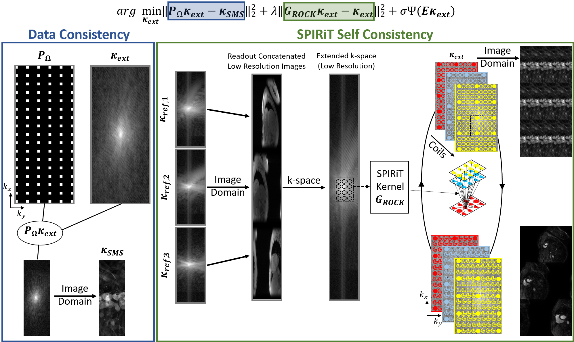
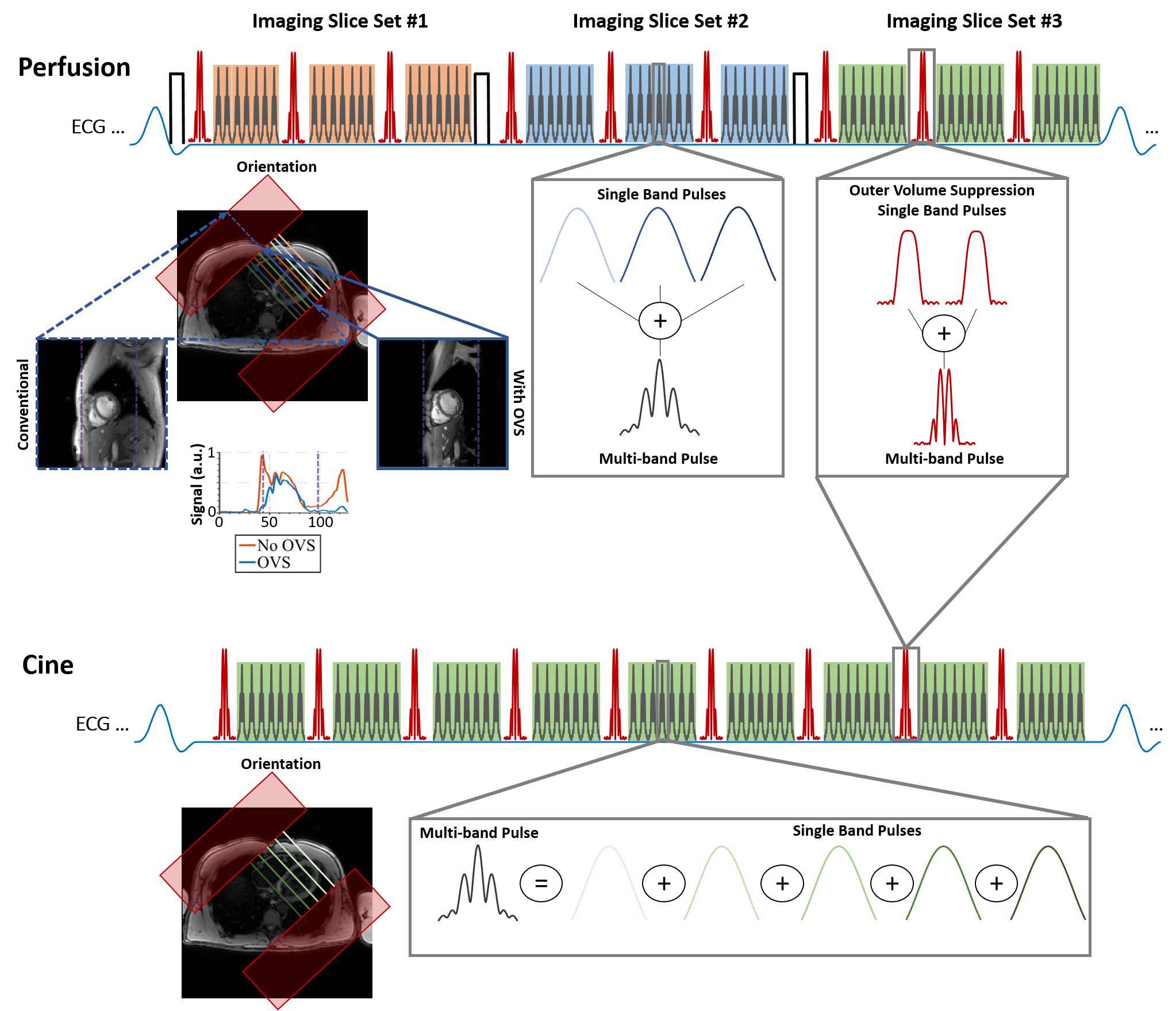
Figure 2: OVS-based sequences used for perfusion and cine imaging. Sum of sinc pulses at different center frequencies are utilized to enable CAIPIRINHA shifts in SMS acceleration. Two slab-selective single band OVS pulses are combined into a multiband OVS pulse to saturate signals from chest and back simultaneously. These are interleaved within the imaging window. Maroon rectangles in short-axis view represent the rest slabs.