0769
Rapid Whole-Heart CMR with Single Volume Super-Resolution1University College London, London, United Kingdom, 2Great Ormond Street Hospital, London, United Kingdom, 3University and ETH Zurich, Institute for Biomedical Engineering, Zurich, Switzerland, 4University of Oulu, Oulu, Finland
Synopsis
Three-dimensional (3D), whole heart, balanced steady state free precession (WH-bSSFP) sequences provides excellent delineation of both intra-cardiac and vascular anatomy. However, they are usually cardiac triggered and respiratory navigated, resulting in long acquisition times (10-15minutes). Here, we propose a machine-learning single-volume super-resolution reconstruction (SRR), to recover high-resolution features from rapidly acquired low-resolution WH-bSSFP data. We show that it is possible to train a network using synthetically down-sampled WH-bSSFP data. We tested the network on synthetic test data and 40 prospective data sets, showing ~3x speed-up in acquisition time, with excellent agreement with reference standard high resolution WH-bSSFP images.
Background
Three-dimensional (3D), whole heart, balanced steady state free precession (WH-bSSFP) sequences provide delineation of both intra-cardiac and vascular anatomy. However, they are usually cardiac triggered and respiratory navigated, resulting in long acquisition times (10-15 minutes).Here, we propose significant speed-ups using a deep-learning ‘single volume’ super-resolution reconstruction (SRR), to reconstruct high-resolution data from rapidly acquired low-resolution WH-bSSFP images. The benefits of SRR is that no additional imaging information is required and it can be performed as a simple post-processing step.
The aims of this study were to:
- Assess the accuracy of deep learning single volume SRR for recovering high-resolution data from synthetically down-sampled WH-bSSFP data.
- Assess the feasibility of using deep learning single volume SRR for reconstruction of prospectively acquired low-resolution WH-bSSFP data.
- Compare acquisition time, image quality and accuracy of vessel diameter measurements from single volume SRR, compared to low-resolution and reference standard high-resolution WH-bSSFP images.
Methods
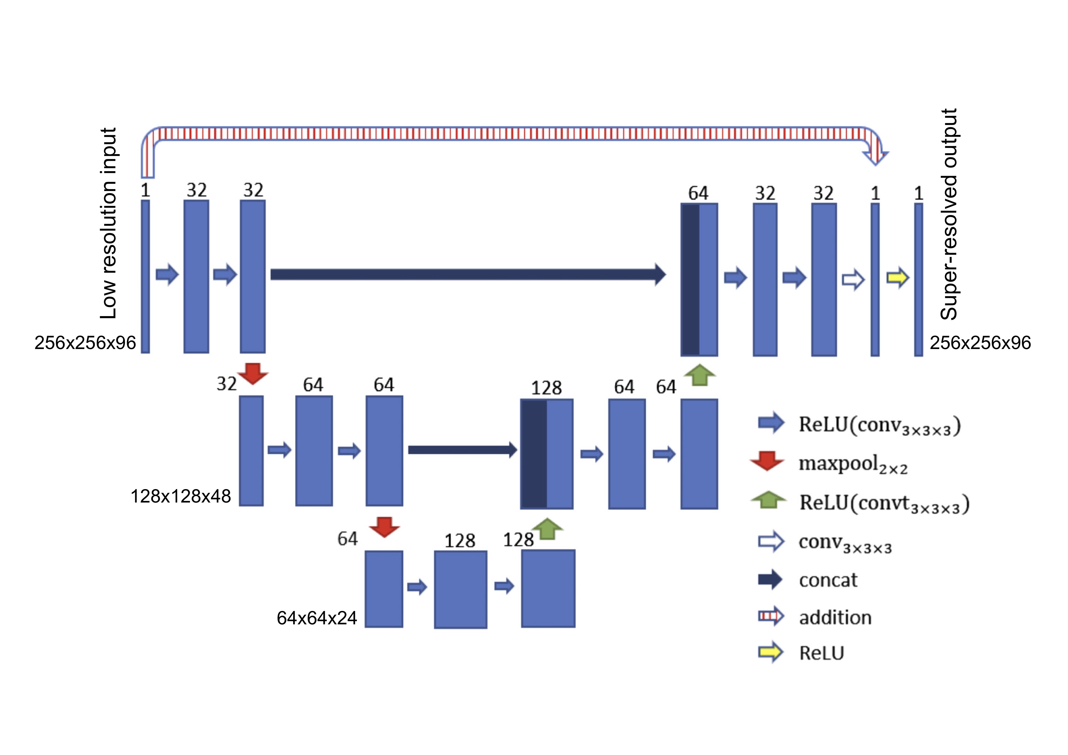
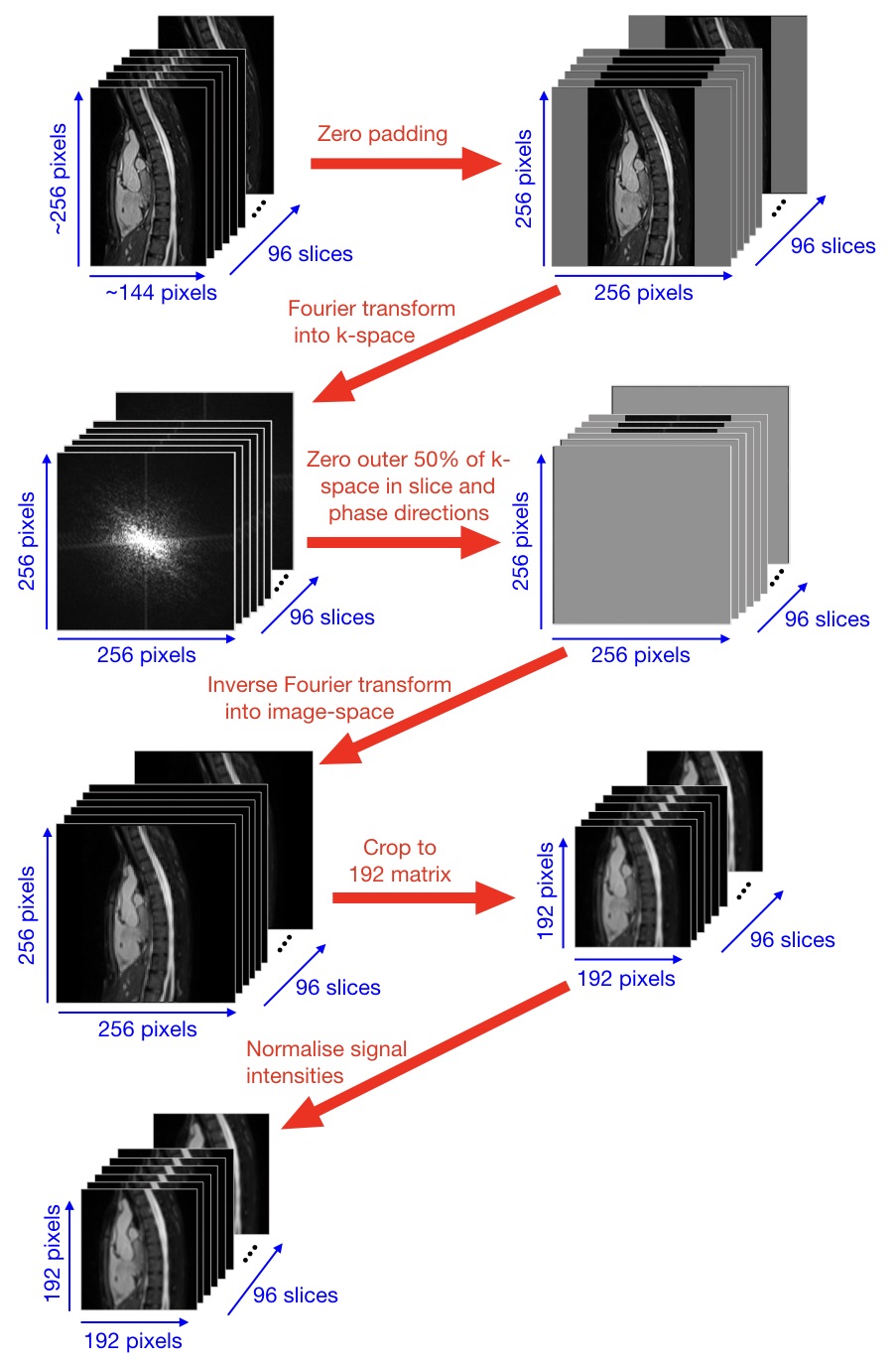
Training of NetworkA 3D residual U-Net [1] was trained with synthetic training data consisting of paired high-resolution and corresponding low-resolution WH-bSSFP images (see Figure 1 for chosen network architecture). 'Synthetic’ low-resolution training data was created from reference standard high-resolution WH-bSSFP images, collected from 500 previously scanned children and adults with paediatric heart disease or congenital heart disease, CHD (age: 26.3±13.4years). Low-resolution data was created by simulating 50% slice resolution and 50% phase resolution (performed in MATLAB 2016b - see Figure 2 for more details). The total training time took ~38 hours on a Titan XP GPU with 12Gb memory.
Testing Using Synthetic Test Data
The trained network was validated using 'synthetic' test data (created in the same way as the training data) from 25 previously unseen children and adults with CHD (age: 27±12years). The low-resolution WH-bSSFP images and resulting super-resolved WH-bSSFP images were compared to the ground truth, high-resolution data using root mean square error (RMSE) and Structural Similarity Index (SSIM).
Testing Using Prospective Data
The technique was also tested in 40 prospective children and adults with CHD (age: 27±14 years). Low-resolution (LR) WH-bSSFP and high-resolution (HR) WH-bSSFP data were both acquired on all subjects. The trained network was then used to perform SRR on the LR data.
Multi-planar reformats (MPR’s) of the Ascending Aorta (AAo), Descending Aorta (DAo), Main Pulmonary Artery (MPA), Left Pulmonary Artery (LPA) and Right Pulmonary Artery (RPA), were made separately from all WH-bSSFP data sets.
Qualitative image scores were made in all MPR images (n=600 - 40patients x 5vessels x 3techniques) for sharpness of vessel borders, image distortion and residual artefacts (Likert scale 1-5; 1=worst, 5-best). Quantitative image scores were also made for edge sharpness (ES), signal-to-noise ratio (SNR) and relative contrast (RC). Vessel diameter measurements were also made in all MPR images.
Results
Results from Synthetic Test DataComparing the reference HR WH-bSSFP data to the SR WH-bSSFP gave an SSIM of 0.93±0.11 and RMSE of 30.8±30.4 x10⁻³.
Results from Prospective Data
HR WH-bSSFP and LR WH-bSSFP were successfully acquired in all 40 patients. Total acquisition time for HR data was 488±138s, and for LR data was 173±54s, representing a speed-up of 2.9±0.8. The SRR took ~60s per volume.
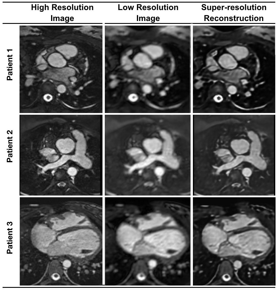
Image quality after SRR was excellent with significant improvements compared to the LR WH-bSSFP images and good agreement with the HR WH-bSSFP. See Figure 3 for examples of image quality.
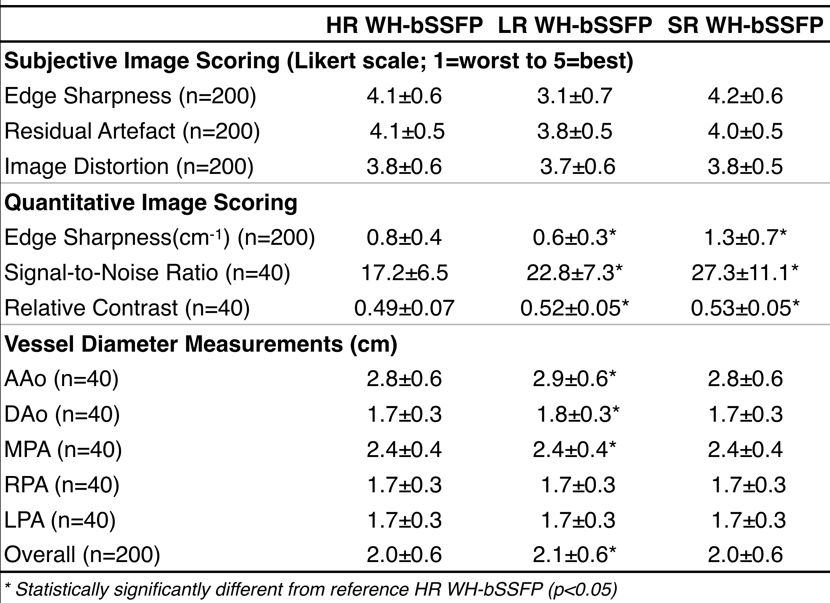
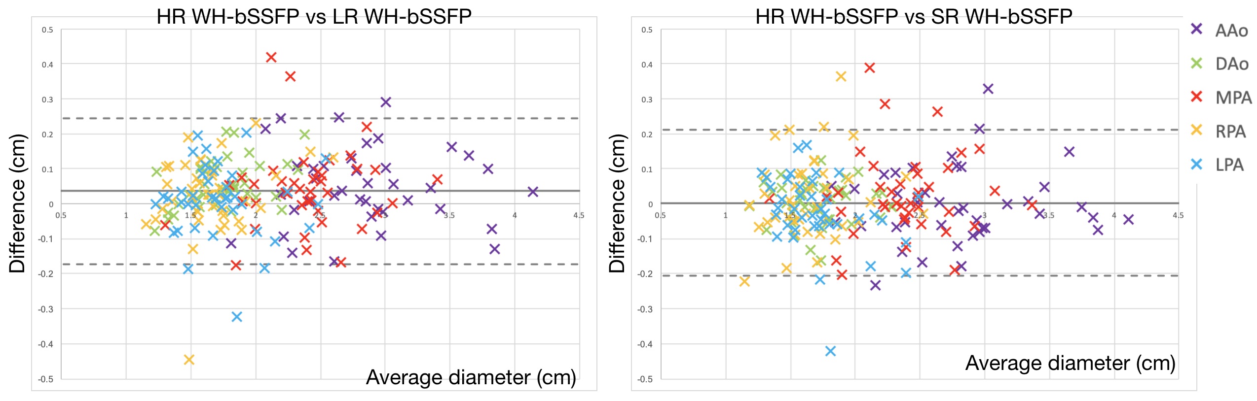
Figure4 shows a table with the qualitative image scores, quantitative image scores, as well as vessel diameter measurements. Qualitative image scores showed SR images had better edge sharpness, fewer residual artefacts and less image distortion than LR images, with similar scores to HR data. Quantitative image scores showed SR images had significantly better edge sharpness than LR or HR images, with significantly better SNR and RC compared to HR data, and similar SNR and RC to LR data. In terms of vessel diameters, the LR images showed significantly higher diameter measurements than the HR images in the AAo, Dao, MPA, as well as over all vessels. Over all vessels the bias was 0.5mm with limits of agreement from -1.7mm to 2.4mm. The SR diameter measurements showed no statistically significant differences in any vessel compared to the measurements from the HR images, with no bias and limits of agreement from -2.0mm to 2.0mm. See Figure 5 for Bland Altman analysis of the vessel diameter measurements comparing the LR and SR techniques to the HR data.
Conclusion
In this study we were able to successfully train a machine learning network to perform single volume SRR to recover high-resolution data from low-resolution Whole Heart data. We were able to train a network using synthetic training data from retrospective high-resolution WH data, in 500 patients. The resultant network provided very high SSIM values (0.93), and low RMSE values (0.03) in synthetic SR data, compared to HR WH images. Additionally, in prospectively acquired low resolution data, SRR resulted in excellent qualitative and quantitative image quality, with no differences in vessel diameters compared to reference standard HR WH images.This method allowed reduction in acquisition time for 3D Whole Heart bSSFP data set from ~8.2minutes to ~2.9minutes. This can be simply applied as a post-processing step of reconstruction, with no complex changes to the acquisition protocol.
Acknowledgements
We would like to thank our funders: The Royal Society-EPSRC, BHF and UKRI.References
1. Hauptmann A, Arridge S, Lucka F, Muthurangu V, Steeden JA: Real-time cardiovascular MR with spatio-temporal artifact suppression using deep learning–proof of concept in congenital heart disease. Magnetic Resonance in Medicine 2019, 81:1143-1156.Figures