0767
Permittivity-Tunable Ceramic Technology for Largely Improving B1 fields and SNR for Broad MR Imaging Applications1CMRR, Radiology, University of Minnesota, Minneapolis, MN, United States, 2Department of Engineering Science and Mechanics, Pennsylvania State University, University Park, PA, United States, 3Center for NMR Research, Neurosurgery, Pennsylvania State College of Medicine, Hershey, PA, United States
Synopsis
We present an innovative technique of tunable ultrahigh dielectric constant (tuHDC) ceramics incorporating RF coil(s) for MR imaging applications. The ceramic has a very high permittivity tunability of 2000-15000 by varying the ceramic temperature between few to 40 °C to achieve optimal performance at the nuclide Larmor frequency of interest, resulting in larger B1 field and SNR improvements for 1H MRI at 1.5T clinic scanner and 17O MRSI at 10.5T human scanner. We found a large denoising effect in 17O MRSI, which further boosts the SNR gain. The technology should benefit for biomedical research and clinical diagnosis.
Introduction
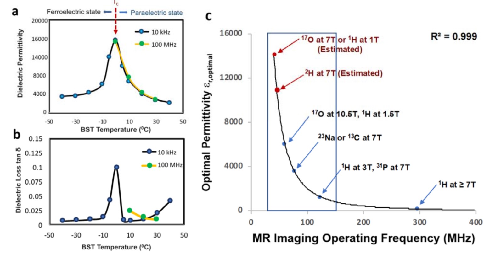
Achieving higher spatiotemporal and spectral resolutions for MRI and MRSI is of importance for assessing neural microstructural properties, brain function and energy metabolism in vivo. A new engineering approach of high dielectric constant (HDC) materials has shown new utility to effectively improve the RF coil transmission efficiency and reception field 1-4. However, the optimal performance of HDC materials for 1H MRI application on clinical scanners (≤ 3T) or low-gyromagnetic ratio X-nuclear MRSI even at UHF requires an ultrahigh permittivity with low loss 5-7. In this work, we introduce an innovative technique of highly tunable-permittivity ultrahigh HDC (tuHDC) ceramic incorporating a RF coil. The permittivity can be tuned from approximately 2000 to >10000 by varying the block temperature (Tb) with a safe range: ±10 °C of the room temperature, to ensure an optimal permittivity and performance for a variety of MR imaging applications across different field strengths.Methods
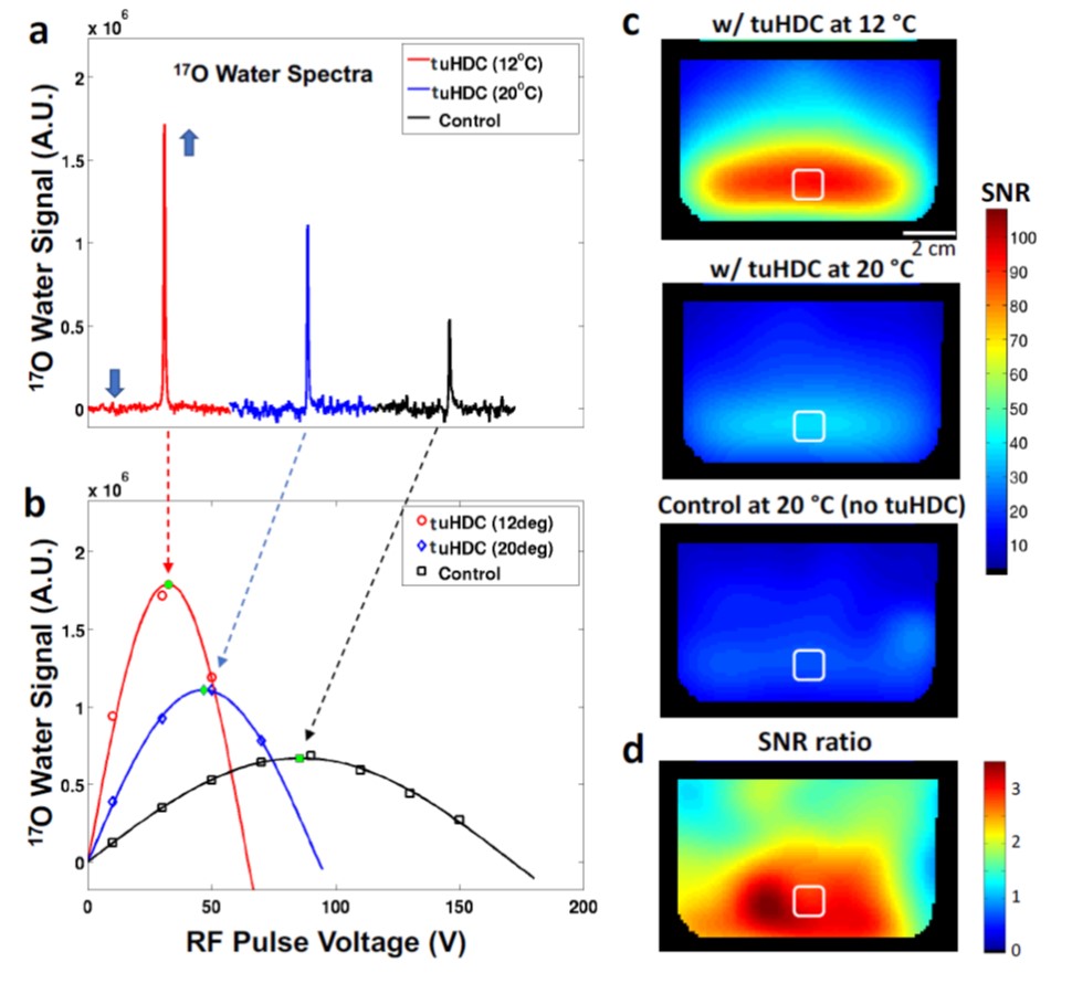
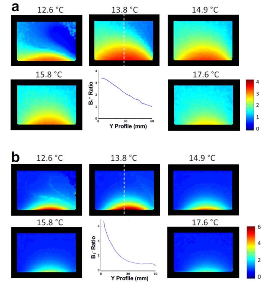
A circular tuHDC ceramic (8-cm diameter, 2.1-cm thickness) was made of composite barium strontium titanate (BST) material (Ba0.6Sr0.4TiO3) having an extremely large relative permittivity (εr) tuning range from ~2000 at 40 °C to >10000 at ~5 °C, εr » 4700 at room temperature (20°C, Fig. 1a), and a very low dielectric loss (tanδ< 0.03 at ~100 MHz, Fig. 1b). 17O MRSI experiments were carried out on a Siemens 10.5T whole-body/88-cm human scanner with a single-loop 17O RF coil (15 cm diameter). The 3D 17O-CSI data were acquired (TR = 0.2 s, RF (hard) pulse = 2 ms, FA = 90°, 3D phase encoding matrix: 9×9×7, spectral bandwidth = 30 kHz, FOV = 10×10×10 cm3, number of complex points = 1024). 1H MRI study was conducted on Siemens 1.5T whole-body/70-cm human scanner with 16-channel receiver array, and MR images were acquired using 2D FLASH sequence (TR/TE = 200 / 2.5 ms, FA = 20°, voxel size = 2×2×7 mm3). To quantify B1 fields at varied BST temperatures, we collected multiple datasets with varied RF pulse voltages using the water phantom (9-cm diameter, 7.2-cm height, 77 mM NaCl). The BST block temperature was controlled from 10 to 20 °C using the temperature controller. The block was placed between the phantom and temperature controller and was surrounded by Styrofoam for heating insulation. All results were compared to the control condition without the use of the tuHDC block at room temperature.Results
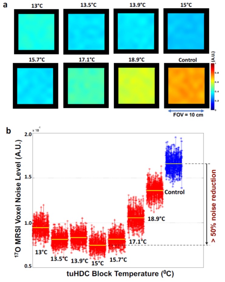
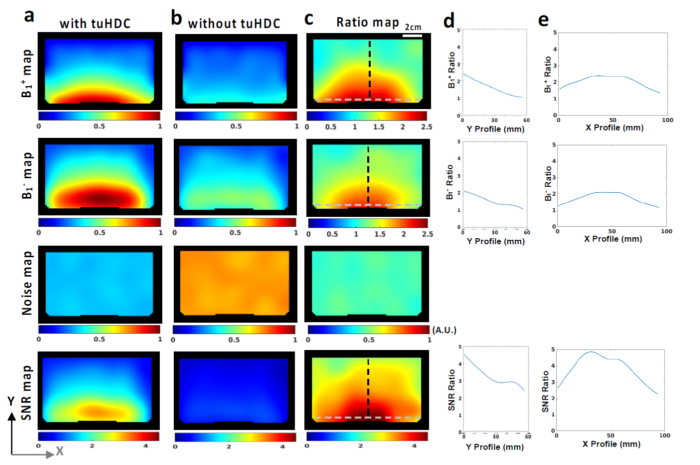
Figure 2 shows the representative 3D 17O spectra of the central voxel acquired under three conditions at 10.5T (Fig. 2a), and the signal intensity profiles as the function of RF pulse voltage (Fig. 2b) that were used to determine RF transmission efficiency (B1+: inversely proportional to the regressed voltage for reaching 90° FA) and detection sensitivity (B1-: proportional to the maximum signal intensity with 90° FA). Compared to control condition, the tuHDC block led to a substantial improvement in B1+, B1-, and a strikingly large noise reduction (denoising effect) at Tb=12 °C as well as room temperature (Fig. 2b), resulting in a large increase in SNR (Figs. 2c and 2d). Figure 3 further depicts temperature-dependent noise patterns of the 17O MRSI, showing a global noise reduction with the tuHDC block that decreased 55% noise level between 14-16 °C compared to control. Taken together, Fig. 4 reveals that the maximum improvement of B1+, B1-, and denoising observed at Tb =15°C leads to > 400% SNR improvement in the region near the block. Based on this result, the estimated optimal permittivity for 10.5T 17O MRSI application at Tb =15°C was εr » 6030 (Fig. 1a), which was the highest permittivity ceramic employed for MR imaging application. Figure 5 further demonstrates temperature-dependent B1 patterns of 1H MR images at 1.5T clinical scanner, showing a significant contribution of tuHDC block to MR signal enhancement below room temperature compared to control. As analogous with the results of 17O MRSI, the maximum B1 improvements of 1H MRI was observed around ~14 °C, which corresponds to the optimal permittivity of ~ 6400 (Fig. 1a). The 1D profile indicates a greater B1+ (> 200%) (Fig. 5a) and B1- (α SNR) improvement (400-500%) (Fig. 5b) in the sample region near the block.Discussion
This work demonstrates an innovative technology of BST-based tuHDC ceramics for achieving large improvements in B1 fields, noise reduction, thus, unprecedented SNR gains for various human MRI scanners with field strength from 1.5T to 10.5T. Owing to similar RF operating frequencies between 1H MRI at 1.5T (64 MHz) and 17O MRSI at 10.5T (61 MHz), the tuHDC offers similar improvements under optimal permittivity condition. Therefore, the key guidance for optimal design is to follow the regressed relationship between the operating (or Larmor) frequency and er,optimal as summarized in Fig. 1c. In this regard, the large permittivity tunability of the BST-based tuHDC ceramic could be employed for broad MRI and MRSI applications across the field strength from 1.5T to 10.5T (Fig. 1c) covering most nuclides of interest.Conclusion
As a robust and cost-effective engineering solution, the tuHDC technique can significantly benefit broad MR imaging applications of various spin nuclides with biological interest at different field strengths. This technology advancement should be critical for basic and clinical research and more importantly for diagnostic imaging.Acknowledgements
This work was supported in part by NIH grants of U01 EB026978, R01 MH111413, R01 CA240953, R24 MH106049, S10 RR026783, P41 EB027061, P30 NS076408.References
1. Yang QX, Mao W, Wang J, Smith MB, Lei H, Zhang X, Ugurbil K, Chen W. Manipulation of image intensity distribution at 7.0 T: passive RF shimming and focusing with dielectric materials. J Magn Reson Imaging 2006;24(1):197-202.
2. Haines K, Smith NB, Webb AG. New high dielectric constant materials for tailoring the B1+ distribution at high magnetic fields. J Magn Reson 2010;203(2):323-327.
3. Webb AG. Dielectric materials in magnetic resonance. Concepts in Magnetic Resonance Part A 2011;38A(4):148-184.
4. Yang QX, Wang J, Wang J, Collins CM, Wang C, Smith MB. Reducing SAR and enhancing cerebral signal-to-noise ratio with high permittivity padding at 3 T. Magn Reson Med 2011;65(2):358-362.
5. Lee BY, Zhu XH, Rupprecht S, Lanagan MT, Yang QX, Chen W. Large improvement of RF transmission efficiency and reception sensitivity for human in vivo(31)P MRS imaging using ultrahigh dielectric constant materials at 7T. Magn Reson Imaging 2017;42:158-163.
6. Rupprecht S, Sica CT, Chen W, Lanagan MT, Yang QX. Improvements of transmit efficiency and receive sensitivity with ultrahigh dielectric constant (uHDC) ceramics at 1.5 T and 3 T. Magn Reson Med 2018;79(5):2842-2851.
7. Koolstra K, Bornert P, Brink W, Webb A. Improved image quality and reduced power deposition in the spine at 3 T using extremely high permittivity materials. Magn Reson Med 2018;79(2):1192-1199.
Figures