0728
Improved contextual fibre growth for generating white matter numerical phantoms with realistic microstructure
Ross Callaghan1, Daniel C Alexander1, Marco Palombo1, and Hui Zhang1
1Department of Computer Science & Centre for Medical Image Computing, University College London, London, United Kingdom
1Department of Computer Science & Centre for Medical Image Computing, University College London, London, United Kingdom
Synopsis
We present an improved version of the ConFiG white matter numerical phantom generator to create realistic white matter microstructure. Building on ConFiG’s novel fibre growth algorithm, the enhancement incorporates a dynamic growth network and global optimisation of fibre positions. Resulting phantoms represent a significant improvement over those from the original ConFiG algorithm, with realistic morphology and an increase in packing density of up to 30%. These improved phantoms result in much more realistic simulated diffusion MRI signals, reducing RMSE to real data by ten times. This improvement demonstrates the potential of ConFiG as a computational model of white matter microstructure.
Introduction
The aim of this work is to improve an existing white matter (WM) numerical phantom generator, ConFiG1, and show that this improvement enables ConFiG to produce realistic microstructure leading to realistic simulated diffusion MRI (dMRI) signals. WM numerical phantoms are often used to validate dMRI microstructure models or as the basis of computational models of WM microstructure. However, these phantoms typically oversimplify the true microstructure of WM and often cannot represent complex fibre configurations such as dispersion and crossing bundles. The original ConFiG algorithm attempted to address this by growing fibres on a static network of connected nodes, avoiding overlap between fibres while respecting morphological priors. This approach produced realistic appearing white matter phantoms, however the low maximum density achievable limited its applicability. Here we present two key changes to ConFiG which greatly improve the density achieved. We demonstrate that this improvement leads to realistic phantoms in terms of both microstructural morphology and simulated diffusion MRI signals.Proposed Improvements to ConFiG
The first improvement to ConFiG is incorporating a dynamic growth network in which extra nodes are added to the network around fibres which have grown. This removes the limitation of the static network which is dependent on initialisation and could lead to fibres getting stuck in regions in which the initial network was too sparse. The addition of points enables the network to be adaptively dense in regions where fibres exist and future fibres would therefore be likelier to become stuck.Secondly, because fibres grow on a discrete network of nodes, the final fibre positions may be suboptimal. The random placement of nodes in the network means that certain positions on some of the fibres may be too close to other fibres, causing fibres to shrink their diameter to prevent overlap. To mitigate against this, we include a global optimisation step at the end of the growth in a procedure similar to MEDUSA2. In essence, fibre nodes that are closer to another fibre than their target diameter will be repelled to allow their diameter to move closer to their target diameter.
Experiments
Impact of improvementsTo quantify these improvements, four scenarios of interest were generated using several variants of the original ConFiG algorithm that include these improvements either one at a time or all at once:
- one bundle of parallel fibres, target density 75%
- one bundle with Watson distributed fibres (κ=8), target density 75%
- two perpendicular crossing bundles, intrabundle target density 40%
- three mutually perpendicular crossing bundles, intrabundle target density 30%
To investigate the impact of the improvements on dMRI simulation, a comparison was made between real dMRI signals and simulations from ConFiG phantoms. The NODDI model3 was fitted to a WM ROI in the corpus callosum of a Human Connectome Project (HCP)4 subject to give initialisation parameters for ConFiG to generate phantoms using both the original and improved ConFiG algorithms. The dMRI signal was simulated in the phantoms using Camino5 with identical simulation conditions in both cases and the measurement scheme corresponding to the HCP dMRI sequence6.
Comparison to real data
To assess the morphology of ConFiG phantoms, microstructure measurements were performed on each phantom to mimic processing of real ex-vivo EM data as described by Lee et al.7. Diameter distributions were calculated by cutting each fibre with a plane perpendicular to the centreline of the fibre and finding the circle equivalent diameter for the resulting cross-section (Figure 4c).
To assess the quality of its dMRI signal prediction, simulations in improved ConFiG phantoms were compared to dMRI data from three WM ROIs, as described in the previous section.
Results and Discussion
Impact of improvementsThe dynamic network improves the density by 4-11%. Global optimisation improves density by 17-22%. Both enhancements together improve the density by 19-29%. These results are summarised in Figure 1 along with virtual histology demonstrating the increased density in Figure 2. This improved performance is comparable to the state of the art, MEDUSA, with particularly good performance in the crossing fibre configurations.
Improved ConFiG produces much more realistic dMRI signals than original ConFiG, reducing the root mean squared error to the real data by nearly ten times. This is shown in the direction averaged signals in Figure 3.
Comparison to real data
The microstructural morphology produced by ConFiG is comparable to real examples from ex vivo tissue as shown in Figure 4. Whilst this is only a comparison to a single real tissue sample, it demonstrates the ability of ConFiG to produce fibres with realistic morphology.
ConFiG also produces realistic simulated dMRI signals when compared to the three WM ROIs in Figure 5. We show that ConFiG generates realistic dMRI signals, particularly in regions of high fibre density where the original ConFiG would have failed.
Conclusion
The ConFiG improvements presented demonstrate the potential of ConFiG phantoms to be used for validating existing dMRI models or as the basis of a computational model WM microstructure by generating highly realistic WM numerical phantoms and associated dMRI signals.Acknowledgements
This work is supported by the EPSRC-funded UCL Centre for Doctoral Training in Medical Imaging (EP/L016478/1) and the Department of Health’s NIHR-funded Biomedical Research Centre at University College London Hospitals. This work was supported by EPSRC grants EP/M020533/1 and EP/N018702/1.References
- Callaghan, R., Alexander, D. C., Zhang, H. & Palombo, M. Contextual Fibre Growth to Generate Realistic Axonal Packing for Diffusion MRI Simulation. in Information Processing in Medical Imaging. IPMI2019. Lecture Notes in Computer Science, vol 11492 (eds. Chung, A., Gee, J., Yushkevich, P. & Bao, S.) (Springer, Cham, 2019)
- Ginsburger, K. et al. MEDUSA : A GPU-based tool to create realistic phantoms of the brain microstructure using tiny spheres. Neuroimage 193, 10–24 (2019).
- Zhang, H., Schneider, T., Wheeler-Kingshott, C. A. & Alexander, D. C. NODDI: Practical in vivo neurite orientation dispersion and density imaging of the human brain. Neuroimage 61, 1000–1016 (2012).
- Van Essen, D. C. et al. The Human Connectome Project: A data acquisition perspective. NeuroImage(2012).
- Hall, M. G. & Alexander, D. C. Convergence and Parameter Choice for Monte-Carlo Simulations of Diffusion MRI. IEEE Trans. Med. Imaging 28, 1354–1364 (2009).
- Sotiropoulos, S. N. et al. Advances in diffusion MRI acquisition and processing in the Human Connectome Project. Neuroimage 80, 125–143 (2013).
- Lee, H. H. et al. Along-axon diameter variation and axonal orientation dispersion revealed with 3D electron microscopy : implications for quantifying brain white matter microstructure with histology and diffusion MRI. Brain Struct. Funct. (2019).
Figures
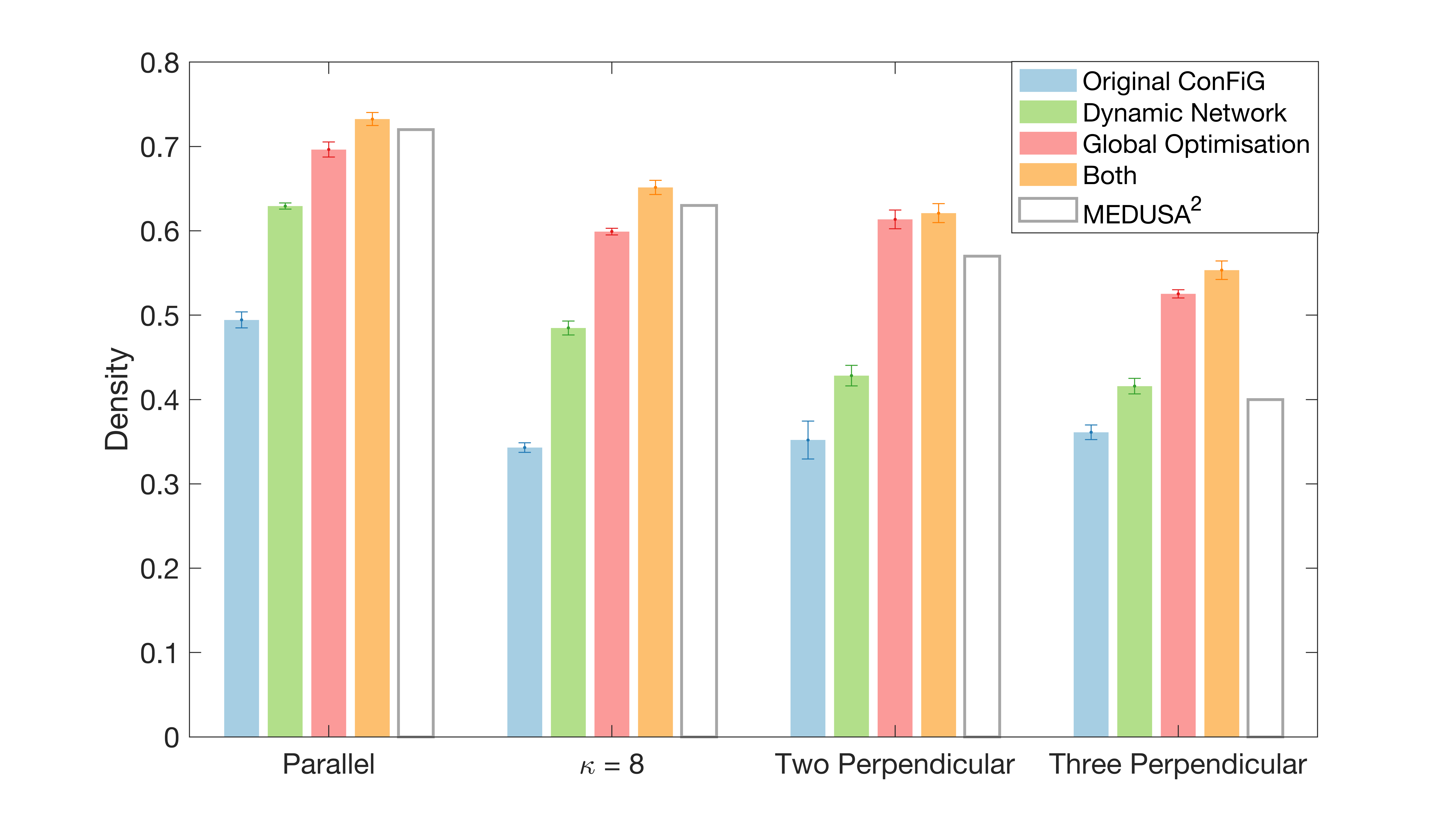
Figure 1. Demonstration of the impact of the improvements to ConFiG. Top: Measured mean density of three different phantoms for original ConFiG and each of the proposed improvements. Error bar shows ± standard error on the mean. MEDUSA values are taken from Fig. 14 in Ginsberger et al.2
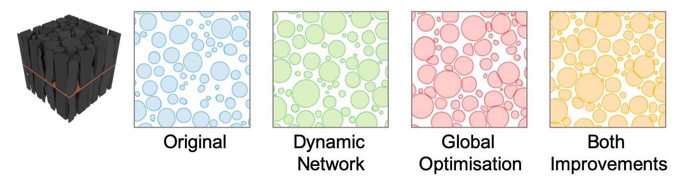
Figure 2. Virtual histology performed on one of the parallel phantoms in each of the experiments demonstrating visually the increased density. Leftmost image shows the phantom and the cutting plane used to produce the virtual histology on the right.
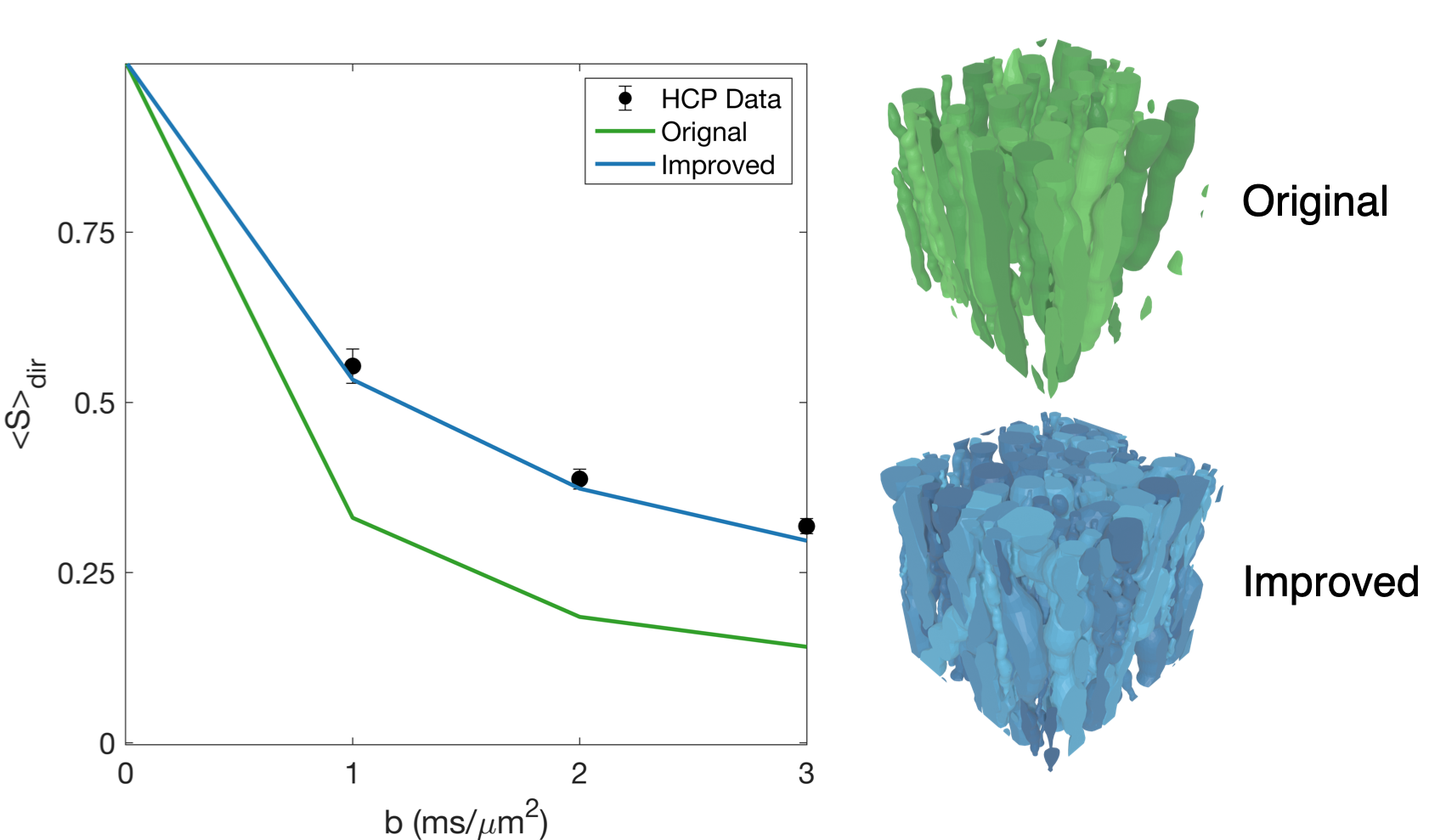
Figure 3. Left: Direction averaged signal for real HCP data (± standard deviation over ROI) and simulated data from original and improved ConFiG showing that improved ConFiG can produce realistic dMRI signals. Right: The original and improved ConFiG phantoms used to generate the signal on the left. Simulations performed with 105 spins, 2000 timesteps, diffusivity 2.0 μm2/ms and HCP measurement scheme6.
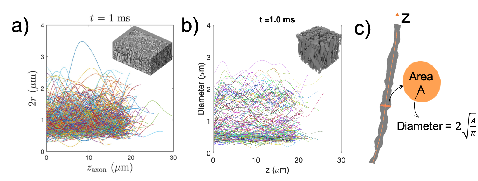
Figure 4. a) Along fibre diameter variation in ex vivo mouse corpus callosum, reproduced from Lee et al.7. b) Along axis diameter variation in the phantom inset demonstrating the ability of ConFiG to generate realistic microstructure. c) the process used to produce b), the cutting plane is moved along the fibre axis, z, and the area of each slice gives a circle-equivalent diameter.
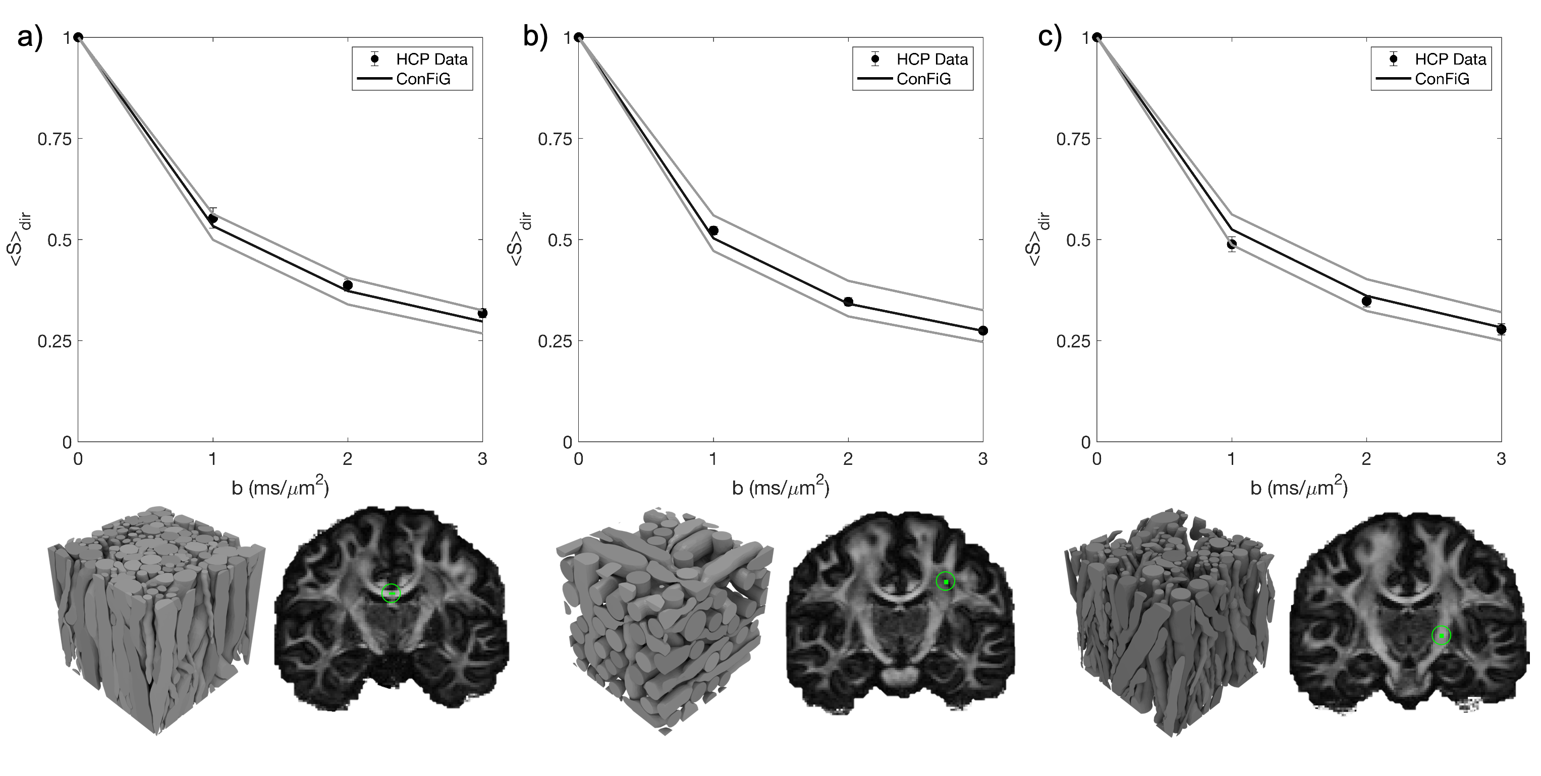
Figure 5. Examples of simulated signals from improved ConFiG phantoms and real dMRI signal from three ROIs in a HCP subject: a) an area in midbody of corpus callosum, b) an area in which three crossing bundles are expected and c) an area in the internal capsule. Black line represents default ConFiG phantoms while grey lines represent ConFiG phantoms generated with radii scaled by 5%.