0680
Construction of water-fat separation deep learning model combined with multi-echo nature of gradient-recalled echo sequence1School of Information Engineering, Wuhan University of Technology, Wuhan, China, 2United Imaging of Scientific Instruments, Shanghai, China, 3State Key Laboratory of Magnetic Resonance and Atomic and Molecular Physics, Wuhan Institute of Physics and Mathmatics, Innovation Academy for Precision Measurement Science and Technology, Wuhan, China, 4School of Civil Engineering & Architecture, Wuhan University of Technology, Wuhan, China
Synopsis
We proposed a novel deep learning network architecture (MEBC-RCAN) for water-fat separation based on multi-echo GRE sequence. The network architecture contains three main components: the first part is Multi-Echo Bidirectional Convolutional (MEBC) to explore the correlations of successive images in multi-echo GRE; the second part is Residual Channel Attention (RCA) network to mimic the iterative optimization in traditional water-fat separation method; and the third part is Multi-Layer Feature Fusion (MLFF) to combine separation information learned from every RCA network. The results show that the proposed network could effectively obtain the high-quality water and fat images from clinical multi-echo GRE data.
INTRODUCTION
In the clinical application of MRI, strong fat signal is often detrimental to the identification and diagnosis of lesions, such as inflammation, edema, tumors, etc. Therefore, a large number of methods have been developed to suppress, augment, or separate water and fat signals for MR imaging and spectroscopy, including CHESS, water excitation, STIP and Dixon Techniques1. Recently, Dixon methods gain in popularity as it can get both water and fat images. Compare the traditional In-phase/Out-of-phase imaging2, these methods can get more accurate water or fat images through sophisticated mathematical processing. However, long reconstruction time is still problems in these methods.In recent years, deep learning methods have been applied to the MRI water-fat separation applications due to their accuracy and efficiency3,4,5,6. Unlike traditional water-fat separation methods, deep learning based methods can implicitly learn the priori information (TEs, frequency shifts of multi-peak fat spectrum, etc.) from input data without prior specification. However, these adopted network architectures are generally versatile and are not optimized for multi-echo water-fat separation task7. The goal of this work is to design a novel deep learning network architecture, which obtained high-equality water and fat images from the multi-echo GRE sequence by jointly learning the correlations of successive images in multi-echo GRE and the iterative optimization characteristic of the traditional water-fat separation methods.
METHODS
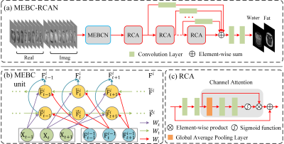
Multi-Echo Bidirectional Convolutional Residual Channel Attention Network (MEBC-RCAN) architecture is shown in Figure 1(a), it includes of 3 components:1) Multi-Echo Bidirectional Convolutional Network evolving over echoes and iterations (MEBCN) is shown in Figure 1(b), the main function of this component is to extract the features for the different echo time images. The MEBCN comprised the convolutions of three kind of different data: the input raw data of the overall architecture (purple arrows); the feature representations learned from the previous iteration of the unit (red arrows); and the feature representations learned from the previous echo or next echo (light green arrows). We simultaneously conduct convolution in the forward and backward direction of the echo sequence. Summing the features extracted from both directions as the final output of the MEBC unit of the-th echo at iteration step.
2) Residual Channel Attention block (RCA, Figure 1(c)), which contains two stacked convolution layers, a skip connection and channel attention mechanism for iterative water-fat separation. The channel attention mechanism by learning channel descriptor to allow the network to adaptively assign different weights to the feature representations.
3) Multi-Layer Feature Fusion(MLFF), which effectively combining the separation feature information learned from every iteration steps of RCA block by element-wise summing (Figure 1(a)). Then, we employ two stacked convolution layers to obtain the final high equality signals from the integrated separation feature information.
Reference water and fat signals used for training network were created using a conventional method8. The quality of the outputs of the network were evaluated by the three quantitative metrics: mean square error (MSE), peak signal-to-noise ratio (PSNR), structural similarity index (SSIM).
RESULTS
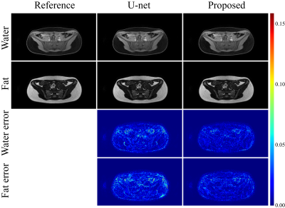
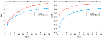
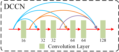
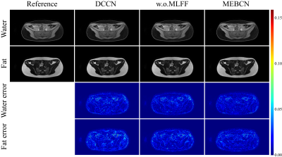
Table 1 and Figure 2 show the comparison of the conventional U-net-based methods3,4 and this new MEBC-RCAN method. We can also find that the quantitative curves of PSNR and SSIM of the proposed method are both higher than U-net-based methods after curves tend to stabilize, and no overfitting is shown (Figure 3).In the ablation experiment of investigating the effects of MECN block and MLFF mechanism, we designed a densely connected convolutional network (DCCN) block with a parameter size (242,128) roughly equivalent to the MECN block (159,936) (Figure 4). By replacing the MECN block with the DCCN block or removing the MLFF mechanism, the performance becomes worse compared with the origin network (which contains MEBCN, RCAs and MLFF, Table 2). Visual assessment find the proposed method has fewer bright spots and red highlights in the water and fat error maps (Figure 5).
DISCUSSION & CONCLUSION
A new deep learning network architecture (MEBC-RCAN) was proposed for water-fat separation task. This new network could learn both the correlations of successive echoes in multi-echo GRE and the iterative water-fat separation process effectively. Even if sufficient multi-echo data and plenty of time are needed during the training process, the proposed method has certain application value to water-fat separation in clinical study benefit from its high efficiency with high quality.Acknowledgements
We gratefully acknowledge financial support by the National Natural Science Foundation of China (11705274), and National Major Scientific Research Equipment Development Project of China (81627901).References
1. Del Grande F, Santini F, Herzka D A, et al. Fat-suppression techniques for 3-T MR imaging of the musculoskeletal system[J]. Radiographics, 2014, 34(1): 217-233.
2. Dixon WT. Simple proton spectroscopic imaging. Radiology. 1984;153:189-194.
3. Andersson J, Ahlström H, Kullberg J. Separation of water and fat signal in whole-body gradient echo scans using convolutional neural networks[J]. Magnetic resonance in medicine, 2019, 82(3):1177-1186.
4. Goldfarb JW, Craft J, Cao JJ. Water-fat separation and parameter mapping in cardiac MRI via deep learning with a convolutional neural network[J]. Journal of Magnetic Resonance Imaging, 2019.
5. Cho JJ, Park HW. Robust water-fat separation for multi-echo gradient-recalled echo sequence using convolutional neural network[J]. Magnetic resonance in medicine, 2019, 82(1):476-484.
6. Zhang T, Chen Y, Vasanawala S, Bayram E. Dual echo water-fat separation using deep learning. In Proceedings of the 26th Annual Meeting of ISMRM, Paris, France, 2018. Abstract 5614.
7. Ronneberger O, Fischer P, Brox T. U-Net: convolutional networks for biomedical image segmentation. In Proceedings Part III of the 18th International Conference of MICCAI, Munich, Germany, 2015. p. 234-241.
8. Hernando D, Kellman P, Haldar JP, Liang ZP. Robust water/fat separation in the presence of large field inhomogeneities using a graph cut algorithm. Magn Reson Med. 2010;63:79-90.
Figures

