0672
Disentangling time series between gray matter and non-gray matter tissue using deep neural network improves resting state fMRI data quality1Cleveland Clinic Lou Ruvo Center for Brain Health, Las Vegas, NV, United States, 2Department of Psychology and Neuroscience, University of Colorado, Boulder, CO, United States
Synopsis
The fluctuation introduced by head motion, cardiac and respiratory fluctuations and other noise sources considerably confounds the interpretation of resting-state fMRI data. These noise fluctuations widely spread the whole brain regardless of the kinds of brain tissues, however, neural activity is more likely limited to gray matter tissue. Considering that the contribution of neural activity varies in different brain tissues, we hypothesized that disentangling gray matter and non-gray matter time series can clean fMRI data and improve the data quality. With such a hypothesis, we proposed a deep neural network method to denoise resting state fMRI data.
Introduction
The fluctuation introduced by head motion, cardiac and respiratory fluctuations and other noise sources considerably confounds the interpretation of resting-state fMRI data [1]. The complex mechanism between the noise source and their contributed fluctuation in fMRI data makes it challenging to clean fMRI data. These noise fluctuations widely spread the whole brain regardless of the kinds of brain tissues, however, neural activity is more likely limited to gray matter tissue. Considering that the contribution of neural activity varies in different brain tissues, we hypothesized that disentangling gray matter (GM) and non-gray matter (non-GM) time series can clean fMRI data and improve the data quality. With such a hypothesis, we proposed a deep neural network method to denoise resting state fMRI data.Methods
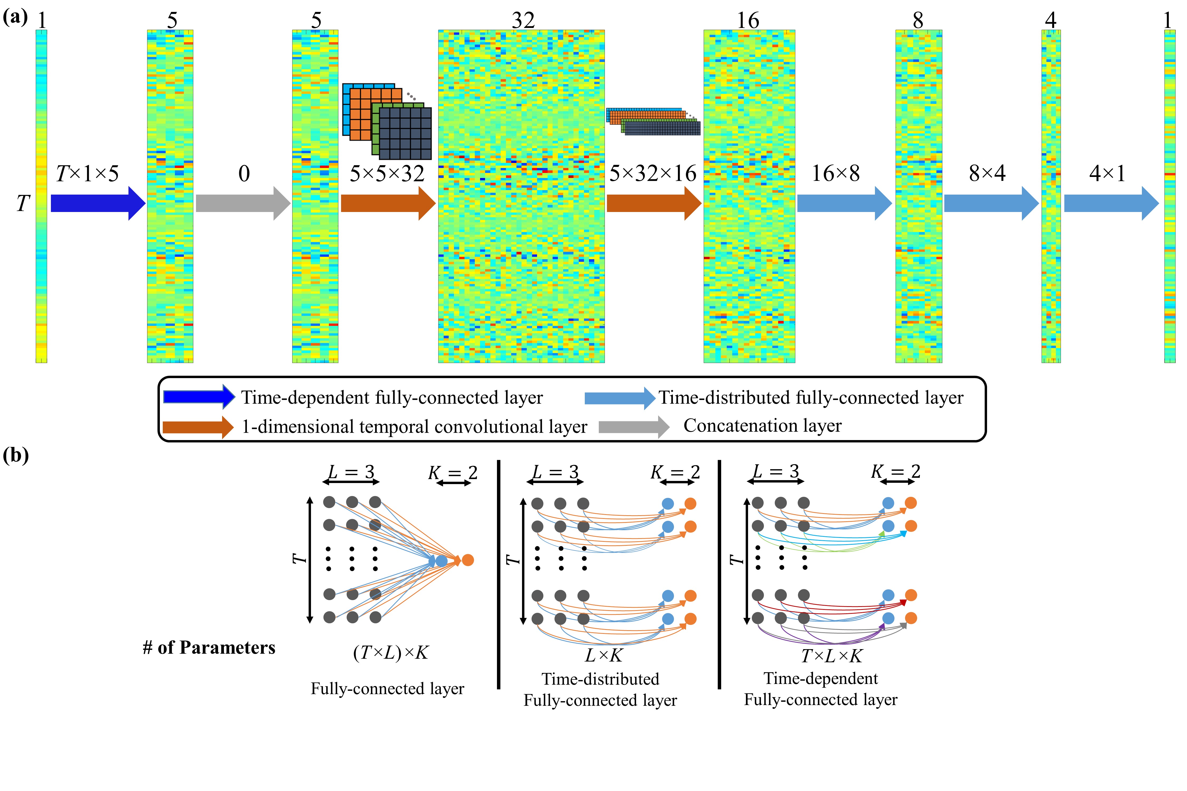
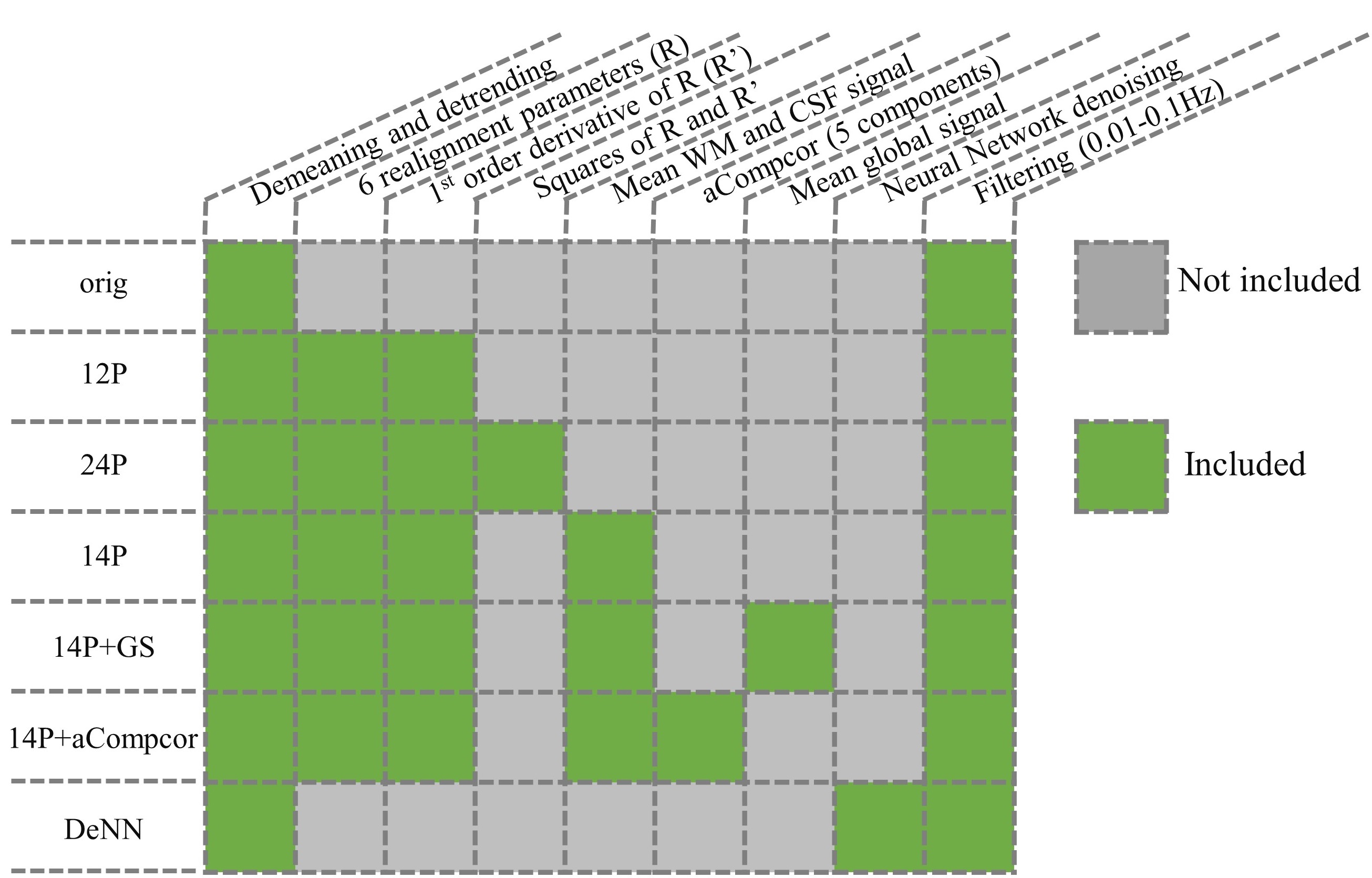
The structural MRI and resting state fMRI (rsfMRI) data used in this study are publicly available in ADNI database (http://adni.loni.usc.edu/). Both T1 and rsfMRI data were normalized to MNI template space. rsfMRI timeseries were divided to two categories, namely GM timeseries and non-GM (white matter and ventricle) time series, based on segmented T1 images. Each GM voxel timeseries is randomly paired with one non-GM voxel timeseries and each pair of time series is treated as one sample to optimize the deep neural network. The neural network is trained on each subject separately and generates a subject-specific model. There are about 50,000 samples for each subject, which is enough to train our neural network. The neural network consists of seven layers as shown in Fig.1a. The first layer is a time-dependent fully-connected layer, which has a fully-connected layer for each time point but with different parameters. This layer can be treated as a deep learning based scrubbing technique to remove spike artifacts. However, unlike scrubbing removes time points and requires arbitrary hard threshold, such a layer keeps all time points since they can be informative and learns from the data to properly separate signals from artifacts. Then a concatenation layer is used to remove the dummy (2nd) dimension, followed by two temporal convolutional layers with 32 and 16 filters, respectively. The temporal convolutional layers play a role as low-pass filtering but without a fixed frequency threshold. Finally three time-distributed fully-connected layers, which have a fully-connected layer with the same parameters for all time points, are used and output the denoised timeseries. A schematic plot of fully-connected layer, time-distributed fully-connected layer and time-dependent fully-connected layer is shown in Fig.1b. The neural network is optimized by minimizing the correlation between GM timeseries and non-GM timeseries in each pair for the purpose of disentangling timeseries between tissues. Along with this denoising neural network (DeNN), nuisance regression was also performed for comparison. The strategies used in each denoised time series were listed in Fig.2.Result
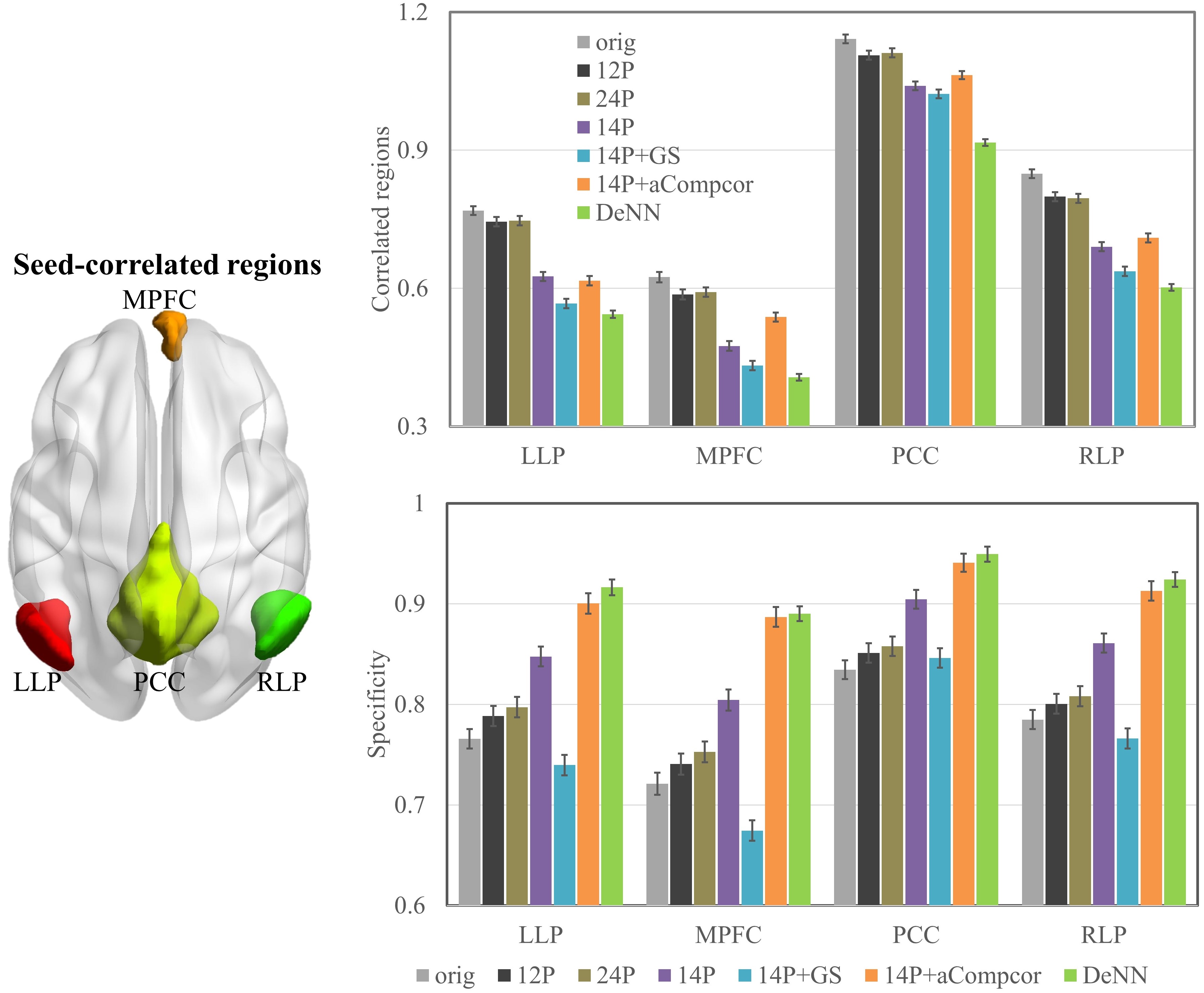
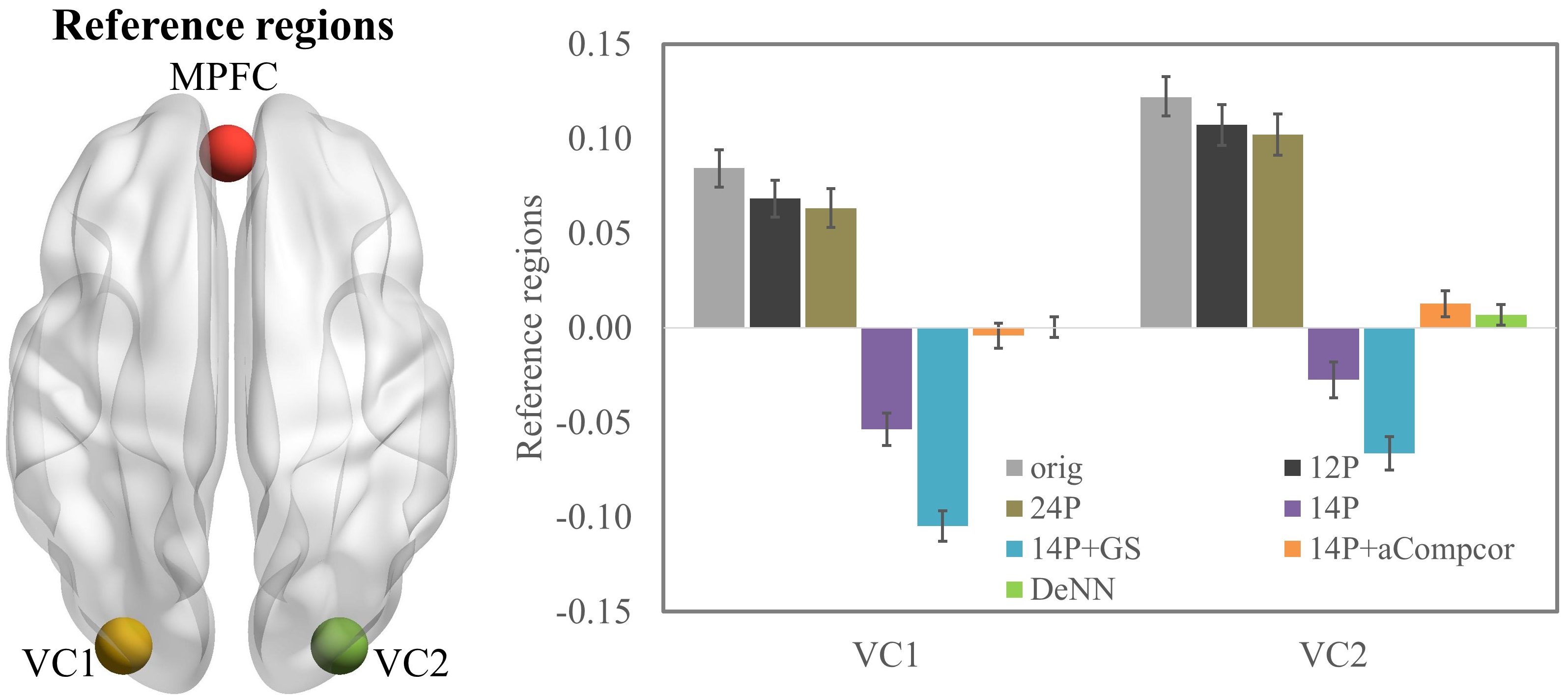
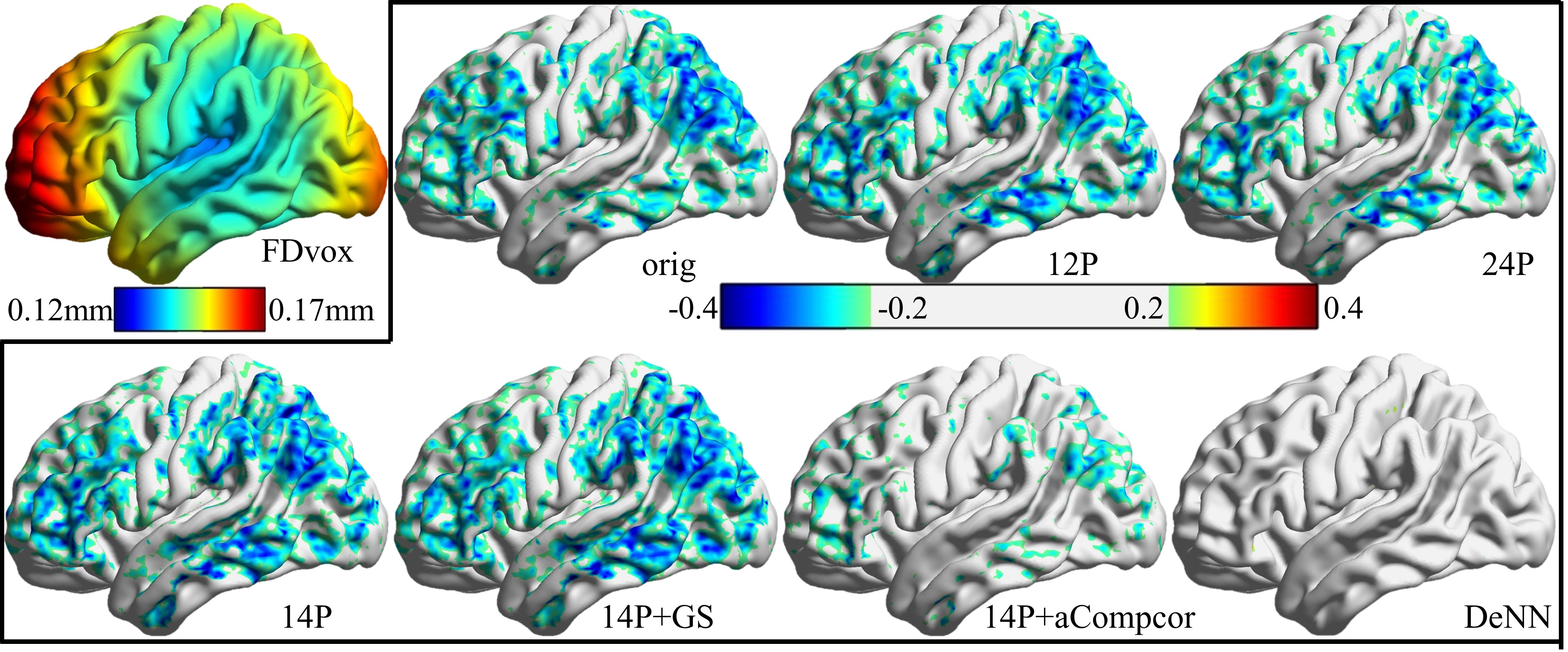
To examine the specificity [2] of each denoising methods, we compared the connectivity values of four regions within default mode network with the posterior cingulate cortex (PCC) seed (-7, -55, 27). The proposed DeNN network has the highest specificity for all these four regions, including left lateral parietal cortex (LLP), medial prefrontal cortex (MPFC), PCC and right lateral parietal cortex (RLP) (see Fig.3). In addition, MPFC (-1, 49, -2) was shown to be functionally unrelated to two visual reference regions (10-mm spheres around (-30, -88, 0) and (30, 88, 0)) [3]. As shown in Fig.4, we observed that orig, 12P, 24P timeseries have significantly positive bias between MPFC and visual reference regions, and both 14P and14P+GS have significantly negative bias. In contrast, DeNN denoised time series has weakest correlation between MPFC and bilateral visual reference regions. Furthermore, voxelwise fractional amplitude of low frequency fluctuation (fALFF) [4] and voxelwise framewise displacement (FDvox) [5] were calculated for all the subjects. The group-level voxelwise correlation between fALFF and FDvox was calculated and shown in Fig.5. The correlation map for original data suggested that FDvox overall is anti-correlated with fALFF. Such a relation remains for 12P and 24P data, and is more severe for 14P and 14P+GS. In contrast, the anti-correlation between FDvox and fALFF is alleviated in 14P+aCompcor, and is even weaker for DeNN. For DeNN denoised data, there is no voxel passing the correlation threshold 0.2.Discussion
In this study, we proposed a deep neural network method to denoise resting state fMRI data by disentangling timeseries between different brain tissues. Instead of generating a set of nuisance regressors as in traditional denoising methods, this method directly outputs the denoised timeseries and remains the degrees of freedom in the original data. Compared with other methods, DeNN method does not have either positive or negative bias with reference regions, has the highest the specificity for the regions within default mode network, and has the best performance in alleviating the correlation between fALFF and quality control measurement.Conclusion
A robust and automated deep neural network framework is proposed to reduce noise fluctuation from multiple noise sources by disentangling timeseries between brain tissues.Acknowledgements
This research project was supported by the NIH (grant 1R01EB014284 and COBRE grant 5P20GM109025), Young Investigator Award from Cleveland Clinic, a private grant from Peter and Angela Dal Pezzo, a private grant from Lynn and William Weidner, and a private grant from Stacie and Chuck Matthewson. Data collection and sharing for this project was funded by the Alzheimer's Disease Neuroimaging Initiative (ADNI) (National Institutes of Health Grant U01 AG024904) and DOD ADNI (Department of Defense award number W81XWH-12-2-0012).References
[1] Caballero-Gaudes, C., Reynolds, R.C., 2017. Methods for cleaning the BOLD fMRI signal. Neuroimage 154, 128-149.
[2] Weissenbacher, A., Kasess, C., Gerstl, F., Lanzenberger, R., Moser, E., Windischberger, C., 2009. Correlations and anticorrelations in resting-state functional connectivity MRI: a quantitative comparison of preprocessing strategies. Neuroimage 47, 1408–1416.
[3] Van Dijk, K.R., Hedden, T., Venkataraman, A., Evans, K.C., Lazar, S.W., Buckner, R.L., 2010. Intrinsic functional connectivity as a tool for human connectomics: theory, properties, and optimization. J. Neurophysiol. 103, 297–321.
[4] Q.H. Zou, C.Z. Zhu, Y. Yang, X.N. Zuo, X.Y. Long, Q.J. Cao, Y.F. Wang, Y.F. Zang. An improved approach to detection of amplitude of low-frequency fluctuation (ALFF) for resting-state fMRI: fractional ALFF J. Neurosci. Methods, 172 (2008), pp. 137-141, 10.1016/j.jneumeth.2008.04.012
[5] Yan, C., et al, 2013. A comprehensive assessment of regional variation in the impact of head micromovements on functional connectomics. NeuroImage 76 183-201.
Figures