0654
Spiral real-time cardiac MR imaging using a GSTF-based pre-emphasis1Department of Diagnostic and Interventional Radiology, University Hospital Würzburg, Würzburg, Germany, 2Comprehensive Heart Failure Center Würzburg, Würzburg, Germany, 3Siemens Healthcare, Erlangen, Germany, 4Department of Internal Medicine I, University Hospital Würzburg, Würzburg, Germany
Synopsis
An automatic pre-emphasis based on the Gradient System Transfer Function (GSTF) was applied to real-time cardiac MR imaging at 3T to compensate for deviations of spiral k-space trajectories. This yielded cine series with coverage of the whole heart in free-breathing, less than 40 s total scan time and a temporal resolution of 50 ms. The developed framework was compared to a gated Cartesian acquisition in multiple breath-holds, in one healthy volunteer and one patient suffering from cardiac arrhythmia.
Introduction
Non-Cartesian data acquisition suffers from imperfections of the dynamic gradient system, which lead to k-space misregistrations and consequently to image artifacts. Measuring the systems impulse response in terms of the Gradient System Transfer Function (GSTF) and using it for trajectory correction can mitigate image artifacts not only in post-processing but also as a pre-emphasis1,2.In this study, the workflow developed in [2] was applied to spiral real-time cardiac functional MR imaging in free-breathing and the results were compared to an ECG-gated Cartesian cardiovascular product sequence with multiple breath-holds, as typically applied in clinical routine.
Methods
The first order GSTF self-terms of the x-, y- and z-axis of a 3T system (MAGNETOM Prismafit, Siemens Healthcare, Erlangen, Germany) were determined by measuring the phase evolutions in two parallel slices of a spherical phantom. 12 triangular input gradients with varying duration were switched as test waveforms (TR = 1 s, read-out BW = 100 Hz/px, dwell-time = 8.7 µs, slice thickness = 3 mm, slice distance from isocenter = ± 16.5 mm, flip-angle = 90°, 100 measurements)1,3,4. The pre-emphasis of the applied spiral read-out gradients was realized by a projection on the physical x-, y- and z-axis and a subsequent multiplication with the respective inverse GSTF in the frequency domain.Slew-rate optimized spiral gradients with variable density were generated using an open source Matlab toolbox (Hargreaves B., https://mrsrl.stanford.edu/~brian/vdspiral/) and implemented in a 2D FLASH sequence. Ten consecutively acquired and equally distributed spiral interleaves form a three-fold undersampled k-space, which represents one time frame during image reconstruction (TR = 4.96 ms, temporal resolution < 50 ms). The inter-frame k-space angle increment was defined as φ = (2·GA)/10, with the golden angle GA = (2·π)/(1+√5) ≈ 111.25°. Further measurement parameters were: TE = 0.84 ms, read-out BW = 320 Hz/px, dwell-time = 2.2 µs, flip-angle = 15°, FOV = 480 mm × 480 mm, in-plane spatial resolution = 1.34 mm × 1.34 mm, slice thickness = 8 mm.
The framework was tested in one healthy volunteer and one patient suffering from heart failure with reduced ejection fraction (HFrEF) and intermittent arrhythmia. An ECG-gated Cartesian cardiovascular sequence with multiple breath-holds served as reference. All investigations were performed in short-axis orientation covering the whole heart by means of 11 slices. Spiral acquisitions were performed in free-breathing and 3.5 s scan-time per slice (i.e. all slices < 40 s measurement time). The Cartesian reference required breath-holds of 10 s per slice, leading to a total measurement time of 3-5 min including additional pauses for breathing and recovery.
For the Cartesian reference technique the measurement parameters were adjusted individually to match the spatial resolution of the spiral acquisition and to avoid aliasing: volunteer / patient: TE = 2.36 ms / 2.29 ms, TR = 4.70 ms / 4.60 ms, read-out BW = 707 Hz/px, dwell-time = 3.4 µs, flip-angle = 15°, FOV = 310 mm × 253 mm / 360 mm × 294 mm, temporal resolution = 42.2 ms / 27.4 ms, in-plane spatial resolution = 1.49 mm × 1.49 mm / 1.73 mm × 1.73 mm, slice thickness = 8 mm, T-GRAPPA = 2.
Spiral acquisitions were reconstructed offline using GRAPPA operator gridding (GROG)5 and a low rank plus sparse compressed sensing model6.
Results
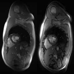
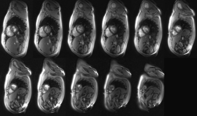
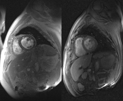
Video 1 shows the results of both methods in a midventricular slice of the healthy volunteer in short-axis orientation: the breath-held gated Cartesian reference technique on the left and two adjacent R-R intervals of the free-breathing spiral real-time sequence on the right. In general, comparable image quality can be observed. No artifacts corresponding to trajectory errors were traceable. Solely, off-resonance in regions with fat tissue led to blurring artifacts and a slight jitter associated with residual aliasing remained. Respiratory motion was resolved and can be monitored in the image series. The ejection fraction (EF) was 63.8 % for the Cartesian reference and 60.4 % for the spiral real-time technique.Video 2 assembles all short-axis slices of the healthy volunteer by means of one synchronized R-R interval using the spiral real-time technique in free-breathing. Video 3 displays the cine series of a midventricular slice of the patient suffering from HFrEF in accordance to video 1. Apparent arrhythmia led to corrupted triggering and thus impaired image quality in the gated Cartesian reference technique, while the arrhythmic cardiac kinetics could be resolved by the spiral real-time sequence. Quantification led to an EF of 21.5 % for the Cartesian reference and 23.0 % for the spiral real-time technique.
Discussion & Conclusion
The GSTF-based pre-emphasis serves as a robust setup to mitigate trajectory errors when using non-Cartesian k-space trajectories. A framework was established to resolve cardiac motion in real-time with 50 ms temporal resolution. Whole heart coverage can be achieved in less than 40 s in free-breathing, which increases time efficiency as well as patient comfort. Furthermore, the proposed real-time imaging allows for the omission of ECG-gating and thereby enables proper cardiac cine imaging in patients with cardiac arrhythmia. Quantitative analysis yielded similar ejection fractions for both methods.Funding
Comprehensive Heart Failure Center Würzburg, Grant BMBF 01EO1504Acknowledgements
No acknowledgement found.References
1. Stich M. et al., Gradient waveform pre-emphasis based on the gradient system transfer function, Magnetic Resonance in Medicine, 80(4):1521-1532 (2018)
2. Eirich P. et al., Spiral imaging using a fully automatic pre-emphasis based on the Gradient System Transfer Function (GSTF), Abstract #3850, ISMRM 2019
3. Liu H. et al., Accurate Measurement of Magnetic Resonance Imaging Gradient Characteristics, Materials, 7:1-15 (2014)
4. Campbell-Washburn A. E. et al., Real-Time Distortion Correction of Spiral and Echo Planar Images Using the Gradient System Impulse Response Function, Magnetic Resonance in Medicine, 75(6):2278-2285 (2016)
5. Seiberlich N. et al., Non-Cartesian data reconstruction using GRAPPA operator gridding (GROG), Magnetic Resonance in Medicine, 58:1257-1265 (2007)
6. Otazo M. et al., Low-rank plus sparse matrix decomposition for accelerated dynamic MRI with separation of background and dynamic components, Magnetic Resonance in Medicine, 73(3):1125-1136 (2015)
Figures


