0652
Analysis of Physiological Brain Shift and Optic Chiasm in the Closed Cranium due to Postural Position
Etsuko Kumamoto1, Shigeto Hayashi1,2, Ari Shinojima3, Koshi Yokota4, and Eiji Kohmura5
1Kobe University, Kobe, Japan, 2Department of Neurosurgery, Hyogo Emergency Medical Center, Kobe, Japan, 3Keio University, Tokyo, Japan, 4Japan Aerospace Exploration Agency, Tsukuba, Japan, 5school of medicine, Kobe University, Kobe, Japan
1Kobe University, Kobe, Japan, 2Department of Neurosurgery, Hyogo Emergency Medical Center, Kobe, Japan, 3Keio University, Tokyo, Japan, 4Japan Aerospace Exploration Agency, Tsukuba, Japan, 5school of medicine, Kobe University, Kobe, Japan
Synopsis
Methodological analysis related to physiological brain shift in the closed cranium is lacking. The spaceflight-associated neuro-ocular syndrome is attributed to the upward shift of the brain. We analyzed the relationship of brain shift and optic nerve shift by using MR volume data acquired in different body positions. The movement and rotation of each voxel, divided into 20 × 20 × 20 pixels3, were calculated using the block matching method. Experimental results show that the optic nerve transforms and deforms with the movement of the brain because of a change in body position.
Introduction
There have been many reports on brain shift during craniotomy, but methodological analysis related to physiological brain shift in the closed cranium has been lacking. We analyzed brain shift and transformation using MR volume data acquired in different body positions[1]. Comparing the MR brain images of astronauts who stayed in outer space for six months and two weeks, we found that the narrowing of the central sulcus and the upward shift of the brain occurred more in long-stay astronauts than in short-stay ones[2]. Shinojima suggests that the spaceflight-associated neuro-ocular syndrome, which has also been detected in astronauts, is attributed to the upward shift of the brain[3]. In the present study, we analyze the relationship between the shift of brain tissue by changing the posture and the optic chiasm.Materials and Methods
Acquisition of MR images: Three-dimensional MR images of the head of six healthy volunteers (22-, 34-, 38-, and 46-year-old males and 29- and 51-year-old females) were acquired using 3.0T MRI with fast spin echo at four different postures. The imaging conditions were as follows: TR/TE: 2,500 ms/270 ms; slice thickness: 1 mm; field of view: 256 × 256 mm2; slice gap: 0.5 mm; acquisition matrix: 256 × 256; and spatial matrix: 512 × 512 × 400. The patient positions were supine, prone, left lateral decubitus, and right lateral decubitus.Volume registration: Two steps of volume registration of MRI volume data acquired in different posture was performed. In first step, registration of the whole head was performed based on the 3D phase-only correlation method[4] with a coarse-to-fine strategy was performed. Next step, semi-automatic registration was performed using normalized cross-correlation with three semicircular canals, including the left and right internal auditory meatus because these did not move by posture changes.
Measuring the shift of the brain and the optic chiasm: The volumes at the supine, prone, left lateral decubitus, and right lateral decubitus positions were divided into voxels of 20 × 20 × 20 pixels3. Each voxel at the prone and right lateral decubitus positions was registered using supine and left division voxels as templates, and the 3D movement and rotation of each voxel were recorded. The coronal and sagittal planes were set so that the optic chiasm could be observed well. The angles of the optic chiasm and the optic chiasm stem were measured as shown in Figure 1.
Results and Discussion
Figure 2 shows that the cerebrum was slightly displaced toward the occipital region or the left side. However, the displacements were different for each place. It was suggested that the cerebrum was deformed. Figure 3 shows the average of the shift transformation in the posture changes from the left lateral decubitus to the right lateral decubitus and from the supine to the prone positions. The superficial parts of the brain, the frontal lobe, the parietal lobe, the temporal lobe, and the occipital lobe, as well as the deep parts of brain, the corona radiate, the base ganglia, and the brain stem, behave differently with posture change. The average amount of shift transformation of the superficial parts tends to be smaller than that of the deep parts, as the tissue in the superficial parts moves with deformation. The deep parts of the brain collectively shift to the downward direction. Figure 4 shows the angular variation of the optic chiasm. The changes in the angle of the optic chiasma when the posture is changed has a large individual difference. It is expected that some stress is applied to the optic nerve because of the change in body position.Conclusion
The brain parenchyma was deformed as a result of the transformation in body position and was displaced in the cerebrospinal fluid. The optic nerve transforms and deforms with the movement of the brain because of the change in body position. A more detailed analysis will be conducted in the future.Acknowledgements
No acknowledgement found.References
- E. Kumamoto , S. Hayashi , K. Matsuda , et.al., Noninvasive Analysis of Brain Shift Transformation in Closed Cranium using MR Images Acquired in Different Body Positions, Proc. of 26th ISMRM, 2088, 2018.
- D. R. Roberts, M. H. Albrecht, H. R. Collins, D. Asemani, et. Al, Effects of Spaceflight on Astronaut Brain Structure as Indicated on MRI, New English Journal, Vol. 377, 1746-1753, 2017. DOI: 10.1056/NEJMoa1705129
- A. Shinojima, I. Kakeya, S. Tada, Association of Space Flight With Problems of the Brain and Eyes, JAMA Ophthalmology, Vol. 136, 1075-1076, 2018. DOI: 10.1001/jamaophthalmol.2018.2635
- Y. Tajima, et al. Fast and Robust 3D Correspondence Matching and Its Application to Volume Registration, IEICE TRANS. INF. & SYS., Vol.96, No.4, 826-835, 2013. DOI: 10.1587/transinf.E96.D.826
Figures

Figure 1 Angles of the coronal right, the coronal left and sagittal are measured for analyze the shift of the optic chiasm.
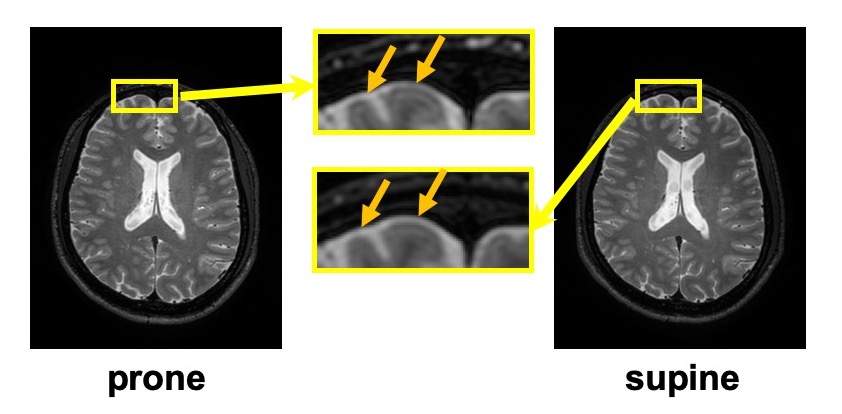
Figure 2 Registered MR image of supine and prone positions of male #3. In the magnified image, the cerebral parenchyma shifts forward in prone position.
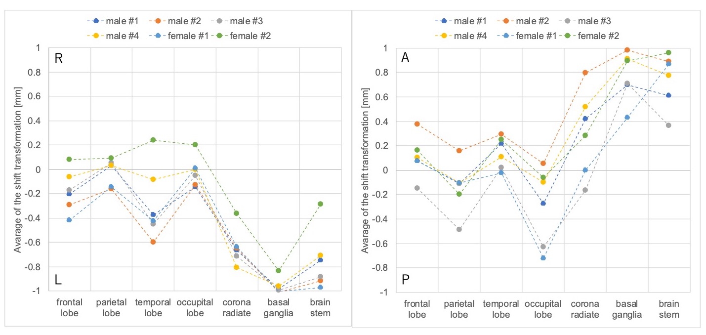
Figure 3 Average of the shift transformation of each tissue in brain. (a) the posture change from left lateral decubitus to right lateral decubitus in LR direction (b) the posture change from supine to prone in AP direction
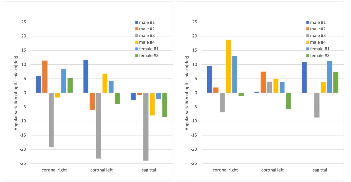
Figure 4 Angular variation of optic chiasm. (a) the posture change from left lateral decubitus to right lateral decubitus (b) the posture change from supine to prone in AP direction