0651
Evaluation of Cerebrospinal Fluid Dynamics in endoscopic third ventriculostomy for treating obstructive hydrocephalus with 4D Flow MRI1Department of Medical Imaging,, Jinling Hospital , Medical School of Nanjing University, Nanjing, China, 2GE Healthcare, MR Research, Beijing, P.R. China, Nanjing, China
Synopsis
We aim to investigate the clinial value of 4D flow MRI in assessing cerebrospinal fluid(CSF)dynamics. The optimal velocity encoding factor(VENC) and high test-retest reproducibility was firstly obtained in CSF measurements for healthy volunteers. Ensured by thses, 4D flow MRI has been further applied to evaluate the CSF dynamics for patients with obstructive hydrocephalus before and after endoscopic three ventriculostomy(ETV). Notable cerebrospinal fluid flow at the stoma has been found, indicating that a new cerebrospinal fluid circulation pathway was established. Our study thus demonstrated that 4D flow MRI is an effective tool to assess CSF dynamics quantitively.
Introdution
Obstructive hydrocephalus is a common disease. Endoscopic third ventriculostomy (ETV) has been widely used in the treatment of obstructive hydrocephalus1. At present, phase contrast MR cine imaging is often used to evaluate cerebrospinal fluid (CSF) dynamics in clinical, but it has some limitation including small imaging range2-3.4D flow MRI ,as a relative novel imaging technique, can show CSF flow in a cardiac cycle completely and quantitatively measure of CSF flow parameters in any reconstruction plane4-5. This technique has been used in cardiovascular research. According to thess intrinsic characteristics of 4D flow, we assume that 4D flow MRI might have the potential to evaluate the changes of CSF before and after ETV. Therefore, the main goal of this study was to investigate the feasibility of 4D flow in the evaluation of CSF dynamics in ETV for treating obstructive hydrocephalus . To achieve this goal, firstly ,the optimal velocity encoding factor(VENC) and the reproducibility of 4D flow measurement were explored. Secondly, this technique was further applied to evaluate the changes of CSF dynamics before and after ETV .
Materials and Methods
SubjectsWith the approval of the local ethics board, ten healthy volunteers(4 females, age from 24 to 35 years) and 5 patients(2 females, age from 22 to 38 years) with obstructive hydrocephalus were included in this study.
To optimize the VENC, 5 healthy volunteers were randomly selected for 4D flow MRI examination with three different VENC values.With this optimal VENC potentially obtained, each of ten healthy subjects underwent 4D flow MRI scans twice in three months to assess the test-retest reproducibility of this technique. For each patient, 4D flow MRI were performed twice before and after ETV to measure the CSF flow , mean velocity and peak velocity.
MRI experiment
MRI experiments were performed using a 3.0-T scanner (GE, DISCOVERY750, USA) with a 32-channel head coil. All 4D flow scans were acquired with the scan parameters including: TE/TR = 2.6 ms/5.1 ms, flip angle = 8°, FOV = 220 x 220 mm2, matrix size = 192 x 192, velocity encoding factor (VENC) = 3 cm/s,5 cm/s and 10cm/s,slice thickness = 2.4mm. The total scan time was about 10~17 mins depending on the heart rate.
Data analysis
4D flow data analysis was performed using the dedicated software (cvi42, Circle Cardiovascular Imaging, Inc, Calgary, Canada). For each scan, region of interest (ROI) based measurements of cerebrospinal fluid flow (ml/s), mean velocity (cm/s) and peak velocity (cm/s) were performed in basal cistern or stoma. The CSF images with three different VENC values were independently reviewed in a double blind method by two experienced radiologists.
The reproducibility was assessed by two-way mixed intra-class correlation coefficient (ICC) analysis. Paried-t test was also applied to estimate the difference of CSF dynamics parameters before and after ETV . Significance threshold was set as p<0.05.
Result
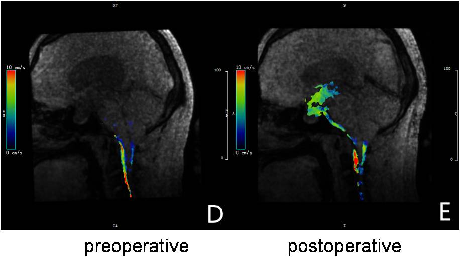
For healthy volunteers, when the VENC value was 5cm/s, the flow of CSF in the basal cistern, pontine cistern and medullary cistern is the clearest (Fig 1), and the flow curve in each cardiac cycle is a biphasic waveform. With the optimal VENC of 5cm/s, high reproducibility of 4D flow mearuerment was obtained over two sacns with ICC of 0.8.Ensured with high reproducibility of 4D flow measurement, for patients with obstructive hydrocephalus after ETV , we found that the corresponding CSF flow was obvious at the stoma by 4D flow MRI(Fig 2). The peak velocity is 4.62cm/s, mean velocity is 2.75cm/s , and the cerebrospinal fluid flow is 0.51ml/s.Moreover, compared to these measuerd preoperatively, statistical differences were found in postoperative measurement (all p<0.05).
Discussion and Conclusion
In this study, we mainly investigated the CSF dynamics for patients in ETV for treating obstructive hydrocephalus with 4D flow MRI. By comparing multiple VENC values, the VENC value of 5cm has shown the optimal performance in the CSF. With this value, test-retest of 4D flow MRI was performed in healthy controls over two scans. High reproducibility of CSF dynamic parameter was achieved. Ensured by these, this technique showed a new cerebrospinal fluid circulation pathway in the evaluation of the patency of stoma CSF dynamics for patients before and after ETV. It thus indicates that 4D flow MRI provided a basis for preoperative diagnosis and efficacy evaluation of diseases such as obstructive hydrocephalus.In conclusion, 4D flow MRI can be considered an effective tool in the evaluation of cerebrospinal fluid dynamics in endoscopic third ventriculostomy for treating obstructive hydrocephalus.
Acknowledgements
References
1.Bilginer B,Oguz KK,Akalan N. Endoscopic third ventriculostomy for malfunction in previously shunted infants[J]. Childs NervSyst, 2009, 25: 683-688.
2.Bargallo N, Olondo L, Garcia AI, et al.Functional analysis of third ventriculostomy patency by quantification of csf stroke volume by using cine phase-contrast MR imaging.AJNR Am J Neturoradiology, 2005, 26(10):2514-2521.
3.BATTAL B, KOCAOGLU M, ULAKBASI N, et al. Cerebrospinal fluid flow imaging by using phase contrast MR technique[J]. BrJ Radiol, 2011, 84( 1004): 758-765.
4.Andreas Stadlbauer, Erich Salomonowitz, Wilma van der Riet, et al. Insight into the patterns of cerebrospinal fluid flow in the human ventricular system using MR velocity mapping [J]. Neuroimage, 2010,51:42-52.
5.Andreas Stadlbauer, Erich Salomonowitz, Christian Brenneis, et al. Magnetic resonance velocity mapping of 3D cerebrospinal fluid flow dynamics in hydrocephalus: preliminary results[J]. Eur Radiol, 2012, 22:232–242.
Figures
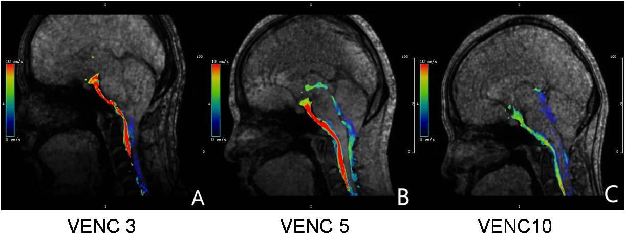
Fig.1 CSF flow of one healthy subject underwent 4D flow MRI scans with three different VENC values