0645
Cyclic Intracerebral Coherent Motion on Peripheral-Pulse-Gated Ultra-Low VENC MRI: Noninvasive Depiction of Glymphatic Flow?1Uniformed Services University of the Health Sciences, Bethesda, MD, United States, 2Walter Reed National Military Medical Center, Bethesda, MD, United States, 3GE Global Research, Niskayuna, NY, United States
Synopsis
A combination of pressure gradients from arterial pulsatility, respiratory cycles, and resistance changes is thought to drive convective influx of CSF into paraarterial spaces for rapid exchange with ISF, followed by efflux into paravenous spaces toward arachnoid granulations, meningeal lymphatics, or cranial nerves. Visualization of this phenomenon was attempted with peripheral-pulse-gated phase contrast sequences at VENC = 5 mm/s (gradient echo) and 0.24 mm/s (spin echo) in four healthy adults using an ultra-high-performance MAGNUS gradient coil. Very slow intracerebral coherent motion was depicted, cerebropetal during systole, cerebrofugal during diastole, possibly reflecting bulk flow in paravascular spaces of the glymphatic system.
Introduction
The glymphatic system is a clearance pathway for extracellular proteins and waste products in the mammalian brain; it depends on astrocytic AQP4 channel-mediated exchange between the extracellular interstitial fluid (ISF) and paravascular cerebrospinal fluid (CSF) compartments.(1) A combination of pressure gradients from arterial pulsatility, respiratory cycles, and resistance changes is thought to drive convective influx of CSF into paraarterial spaces for rapid exchange with ISF, followed by efflux into paravenous spaces toward arachnoid granulations, meningeal lymphatics, or cranial nerves.(2-3)In vivo studies have tracked the movement of intrathecally administered tracers from CSF into ISF over the course of minutes to hours, using fluorescent tracers in mice or gadolinium-based contrast agents in mice and humans.(4-8) It would be beneficial to be able to assess changes in glymphatic flow without invasive procedures or extended imaging. While one human study has purported to use ultra-fast <100 ms whole-brain 3D BOLD fMRI to visualize cerebral glymphatic pulsations,(9) we propose ultra-low VENC MRI as an alternative noninvasive method.
Methods
Under an IRB-approved protocol, four awake healthy adult volunteers were imaged in 3T MRI with a MAGNUS (Microstructure Anatomy Gradient for Neuroimaging with Ultrafast Scanning) insert, which is a head-only ultra-high-performance gradient coil that can deliver simultaneous 200 mT/m and 500 T/m/s on each axis.(10) While designed primarily for benefits in high b-value low TE diffusion weighted imaging, it can also be applied to phase contrast imaging with shorter TE at ultra-low VENC, beyond what is normally feasible on a commercial 3T scanner.Two peripheral-pulse-gated motion-sensitive sequences of the brain were acquired: 1) a fast gradient echo phase contrast sequence with VENC = 5 mm/s (TE=7.2 ms; TR = 13.1 ms) and 2 views per segment (ΔT = 52 ms) at 27-29 time points over the cardiac cycle and 2) a modified DTI sequence using a phase sensitive reconstruction with VENC = 0.24 mm/s (b = 2000 s/mm2) at 8 time points over the cardiac cycle. Because velocity-encoding gradients serve as diffusion-encoding gradients for the magnitude reconstruction, the latter sequence will be called SCIMI (Simultaneous Coherent-Incoherent Motion Imaging).
Results
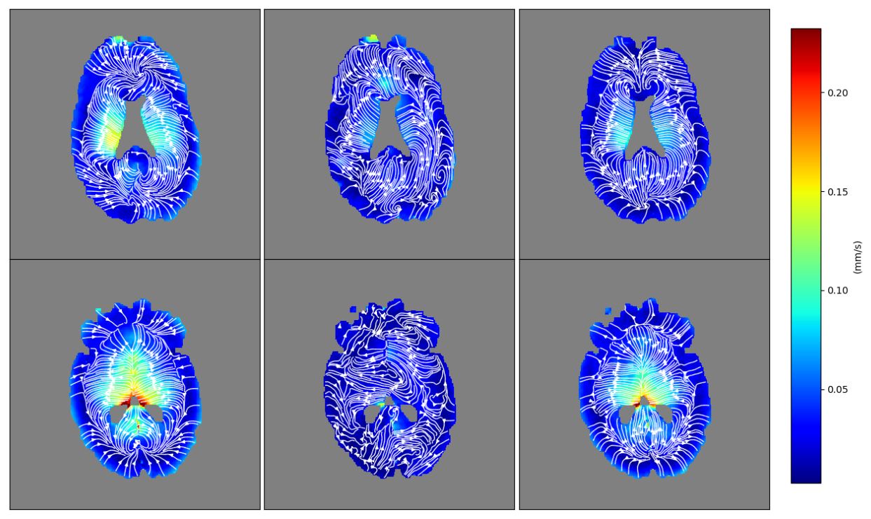
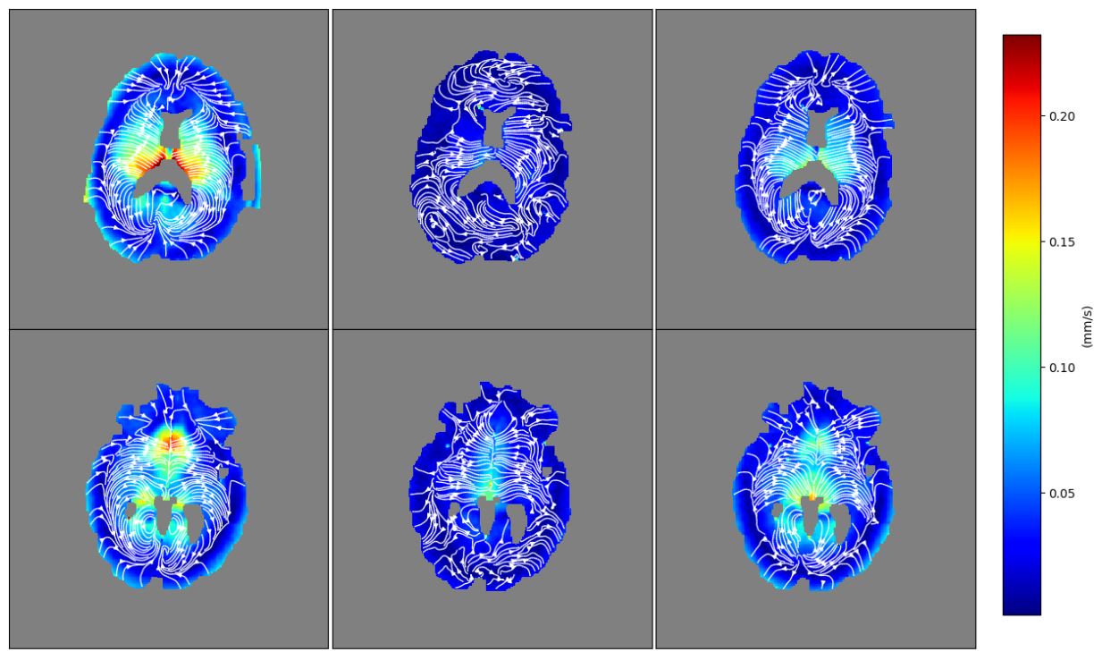
On the peripheral-pulse-gated low VENC (5 mm/s) phase contrast sequence, coherent motion is seen in the lateral ventricles which is directed toward the brain parenchyma in the early cardiac cycle and away from the brain parenchyma in the late cardiac cycle (see Figure 1). Fainter signal or phase shifts in the brain parenchyma is consistent with very slow sub-mm/s flow.On the peripheral-pulse-gated ultra-low VENC (0.24 mm/s) SCIMI sequence, coherent motion is depicted in the cerebral parenchyma, which is directed away from the ventricles and CSF spaces in the early cardiac cycle then toward the ventricles and CSF spaces in the late cardiac cycle (see Figures 2-5). This bulk flow is on the order of 0.1 mm/s and highest in the central/ventral brain.
Discussion
To the best of our knowledge, this is the first depiction of cardiac cyclic intracerebral coherent motion using ultra-low VENC phase contrast MRI. This may represent visualization of pulsatile bulk flow in the paravascular spaces, cerebropetal in systole and then cerebrofugal in diastole. Unilateral internal carotid arterial ligation studies in mice as well as mathematical modeling of paravascular-interstitial flow and resistance have shown the importance of arterial pulsation in driving glymphatic peristalsis as steady pressure-driven flow is not sufficient.(11-12) Regarding glymphatic flow in the interstitial space, which has been postulated to represent a combination of coherent (convection) and incoherent (diffusion) motion, an analysis of data from real-time iontophoresis experiments in mice/rats suggested an upper limit for superficial convective bulk flow velocity of 50 μm/min (0.0008 mm/s), which may remain undetectable, even with VENC = 0.24 mm/s.(13)We do not believe that SCIMI is visualizing cardiac cyclic cerebral parenchymal deformation or displacement, which has been shown to be oriented in the opposite direction (i.e. expansion or cerebrofugal in systole and contraction or cerebropetal in diastole) by previous studies utilizing cardiac-gated phase contrast, pencil-excitation M-mode , and 3D DENSE MRI techniques.(14-16) We also do not believe that SCIMI is visualizing cardiac cyclic intravascular movement, because our velocity range is well below expected values for cerebral arteriolar (2.4 mm/s) and capillary (0.8 mm/s) blood flow,(17) yet we do not see evidence of aliasing. In the end, measurements of differences between wakefulness and slow wave sleep are needed to prove the utility of SCIMI in the noninvasive assessment of glymphatic flow. If successful, animal scanners may be used to replicate mouse experiments based on injected tracers: awake versus asleep, young versus old, adrenergic blockade, anesthetic use, loss of perivascular AQP4 localization.(18-20)
Conclusion
Phase sensitive reconstruction of a modified DTI sequence (SCIMI) with ultra-low VENC = 0.24 mm/s and peripheral pulse gating in four healthy adult volunteers depicted very slow cardiac cyclic intracerebral coherent motion on the order of 0.1 mm/s. The streamlines were oriented cerebropetal during systole and cerebrofugal during diastole, which may reflect pulsatile bulk flow in the paravascular spaces of the glymphatic system. Our plans for further study include quantification and comparison of this finding during wakefulness versus slow wave sleep, as well as animal models of impaired glymphatic clearance (e.g. AQP4 knockout mice).Acknowledgements
The views expressed in this abstract are those of the authors and do not reflect the official policy or position of the Uniformed Services University of the Health Sciences, Walter Reed National Military Medical Center, the Department of Defense, or the U.S. Government.References
1. Iliff JJ, Wang M, Liao Y, et al. A paravascular pathway facilitates CSF flow through the brain parenchyma and the clearance of interstitial solutes, including amyloid β. Sci Transl Med. 2012 Aug 15;4(147):147ra111.
2. Jessen NA, Munk AS, Lundgaard I, et al. The Glymphatic System: A Beginner's Guide. Neurochem Res. 2015 Dec;40(12):2583-99.
3. Plog BA, Nedergaard M. The Glymphatic System in Central Nervous System Health and Disease: Past, Present, and Future. Annu Rev Pathol. 2018 Jan 24;13:379-394.
4. Iliff JJ, Lee H, Yu M, et al. Brain-wide pathway for waste clearance captured by contrast-enhanced MRI. J Clin Invest. 2013 Mar;123(3):1299-309.
5. Eide PK, Ringstad G. MRI with intrathecal MRI gadolinium contrast medium administration: a possible method to assess glymphatic function in human brain. Acta Radiol Open. 2015 Nov 17;4(11):2058460115609635.
6. Ringstad G, Vatnehol SAS, Eide PK. Glymphatic MRI in idiopathic normal pressure hydrocephalus. Brain. 2017 Oct 1;140(10):2691-2705.
7. Ringstad G, Valnes LM, Dale AM, et al. Brain-wide glymphatic enhancement and clearance in humans assessed with MRI. JCI Insight. 2018 Jul 12;3(13).
8. Eide PK, Ringstad G. Delayed clearance of cerebrospinal fluid tracer from entorhinal cortex in idiopathic normal pressure hydrocephalus: A glymphatic magnetic resonance imaging study. J Cereb Blood Flow Metab. 2019 Jul;39(7):1355-1368.
9. Kiviniemi V, Wang X, Korhonen V, et al. Ultra-fast magnetic resonance encephalography of physiological brain activity - Glymphatic pulsation mechanisms? J Cereb Blood Flow Metab. 2016 Jun;36(6):1033-45.
10. Foo TK, Tan ET, Vermilyea ME, et al. Highly efficient head-only magnetic field insert gradient coil for achieving simultaneous high gradient amplitude and slew rate at 3.0T (MAGNUS) for brain microscopic imaging. Magn Reson Med. (2019) DOI:10.1002/mrm.28087 (in press).
11. Iliff JJ, Wang M, Zeppenfeld DM, et al. Cerebral arterial pulsation drives paravascular CSF-interstitial fluid exchange in the murine brain. J Neurosci. 2013 Nov 13;33(46):18190-9.
12. Faghih MM, Sharp MK. Is bulk flow plausible in perivascular, paravascular and paravenous channels? Fluids Barriers CNS. 2018 Jun 15;15(1):17.
13. Ray L, Iliff JJ, Heys JJ. Analysis of convective and diffusive transport in the brain interstitium. Fluids Barriers CNS. 2019 Mar 6;16(1):6.
14. Poncelet BP, Wedeen VJ, Weisskoff RM, et al. Brain parenchyma motion: measurement with cine echo-planar MR imaging. Radiology. 1992 Dec;185(3):645-51.
15. Maier SE, Hardy CJ, Jolesz FA. Brain and cerebrospinal fluid motion: real-time quantification with M-mode MR imaging. Radiology. 1994 Nov;193(2):477-83.
16. Adams AL, Kuijf HJ, Viergever MA, et al. Quantifying cardiac-induced brain tissue expansion using DENSE. NMR Biomed. 2019 Feb;32(2):e4050.
17. Vieni C, Ades-Aron B, Conti B, et al. Effect of intravoxel incoherent motion on diffusion parameters in normal brain. Neuroimage. 2019 Sep 30;204:116228.
18. Kress BT, Iliff JJ, Xia M, et al. Impairment of paravascular clearance pathways in the aging brain. Ann Neurol. 2014 Dec;76(6):845-61.
19. Xie L, Kang H, Xu Q, et al. Sleep drives metabolite clearance from the adult brain. Science. 2013 Oct 18;342(6156):373-7.
20. Hablitz LM, Vinitsky HS, Sun Q, et al. Increased glymphatic influx is correlated with high EEG delta power and low heart rate in mice under anesthesia. Sci Adv. 2019 Feb 27;5(2):eaav5447.
Figures