0644
Compressed sensing accelerated 4D flow magnetic resonance imaging of the cerebrospinal fluid.1University of Cologne, Faculty of Medicine and University Hospital Cologne, Institute for Diagnostic and Interventional Radiology, Cologne, Germany, 2Philips GmbH, Hamburg, Germany
Synopsis
Cerebrospinal fluid (CSF) flow dynamics are relevant parameters in the diagnosis of neurological diseases and can be accessed by three-dimensional time-resolved phase-contrast MRI (4D flow MRI). However, these measurements are accompanied by long scan times making acquisition acceleration necessary to accomplish clinical feasibility. The aim of this study was to evaluate the feasibility of compressed sensing (CS) acceleration in 4D flow MRI of the CSF. CS factors 4 to 10 were compared against the conventional SENSE in 16 healthy subjects. Preliminary results show feasibility of CS factor 6 with comparable image and velocity data quality.
Introduction
Cerebrospinal fluid (CSF) dynamics constitute a complex flow system, which is yet unsatisfactory understood. The physiologic function of the CSF and the diagnosis of pathologies in neurological diseases, e.g. normal pressure hydrocephalus (NPH)1 or Chiari Malformation (CM)2 are subject of recent investigations3. So far, the clinical diagnostic standard to evaluate CSF hydrodynamics are time-resolved 2D phase-contrast MRI acquisitions with velocity encoding in feet-head direction. However, due to the complex flow patterns of CSF, 2D velocity measurements may not be sufficient to fully capture CSF hydrodynamics and to investigate correlations to clinically relevant pathologies. Patients may show vortices and spatial-dependent peak velocities, which can only be adequately represented by time-resolved three-dimensional velocity-encoded phase-contrast MRI (4D flow MRI)4-6. However, 4D flow MRI acquisitions are accompanied by long acquisition times and are therefore rarely used in clinical routine. To overcome these limitations several advanced acceleration techniques were proposed, e.g. exploiting spatial-temporal correlations7-9 or compressed sensing (CS)10,11, which were mainly evaluated in the cardiovascular area. To improve clinical feasibility this study evaluates the applicability of CS acceleration for 4D flow MRI of the CSF. Therefore, acquisitions with different CS acceleration factors were compared with the conventional acquisition method using SENSE parallel imaging.Methods
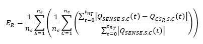
4D flow of the CSF was acquired in 16 healthy subjects on a clinical 3T system (Ingenia, Philips Healthcare, Best, The Netherlands). A sagittal three-dimensional, time-resolved phase-contrast MRI image volume covering the cervical CSF was acquired applying retrospective triggering using a wireless pulse oximeter. All imaging parameters are shown in Table 1. The acquisition was repeated for different acceleration methods (SENSE and CS) and factors (RSENSE=3.75, RCS=4, 6, 8, 10). The order of the acquisition was randomized to avoid systematic errors due to progressing session time and therefore increased subject motion or physiologic variations (e.g. heart rate) of the subjects. In 10 of the 16 subjects the SENSE acquisition was additionally repeated at the end of the session to analyze the scan-rescan reproducibility. Images were reconstructed on the scanner by the vendor supplied software package (Compressed SENSE, Philips Healthcare). All image post-processing was performed off-line using GTFlow (Gyrotools LLC, Zurich, Switzerland). Velocity data were extracted by manually drawn regions of interest (ROI) in transversal 2D planes at the level of vertebrae C1 – C7, respectively. ROIs were drawn at the time point with the most apparent systolic flow in the SENSE acquisition for each subject and copied to all time points and CS acquisitions. The position of the ROIs was adjusted, if necessary. Peak velocity, forward volume flow, backward volume flow and the absolute net flow were extracted. To compare the temporal deviations between flow curves the time-accumulated flow error ER, averaged over all contours and subjects, similar as proposed by Giese et al.12, was calculated as shown in Equation 1 with the corresponding number of subjects nS, contour levels nC and time points nT. QSENSE,S,C(t) and QCSR,S,C(t) correspond to the flow rates through the contour C of subject S at time-point t in the SENSE and CS factor R measurements (R = 4, 6, 8, 10), respectively.Results
The acquisition times averaged over all subjects were 10.35 min (SENSE), 9.52 min (CS4), 6.41 min (CS6), 4.88 min (CS8), 3.86 min (CS10), 10.62 min (repetition of SENSE). The reconstruction of a CS6 acquisition took approximately 10 min. Figure 1 shows resulting magnitude and velocity images exemplary in one subject. Decreasing image quality can be observed with increasing CS factor. Figure 2 shows the time-accumulated flow error for all CS accelerations factors. An increasing error with increasing acceleration can be observed. Table 2 shows results from the linear correlation for peak velocity, forward volume flow, backward volume flow and the absolute net flow between SENSE and CS accelerated images. The tendency of an increasing deviation from the SENSE acquisition with increasing CS acceleration factor can be depicted for all flow parameters.Discussion
The image quality of the magnitude and velocity images for CS acceleration factors 4 and 6 are comparable to SENSE accelerated images. The small R² for the maximum velocity in both acceleration techniques and factors can be explained by a higher noise level and increased artifacts as the maximum velocity is defined by one single voxel. All other three parameters show distinctly higher R² and therefore better scan-rescan reproducibility. Regarding the flow error, the image quality and the flow parameters, CS4 and CS6 show best accordance with SENSE accelerated acquisitions. The limitations of this study are a small study cohort, especially for repeated SENSE acquisitions and the relative high SENSE acceleration, which will be addressed in future.Conclusion
CS factors 4-10 and SENSE acceleration in CSF 4D flow MRI were compared and evaluated regarding image quality and flow parameters in 16 healthy volunteers. Preliminary results show, that a CS6 acceleration of CSF 4D flow is feasible and enables a scan time reduction of about 40% compared to conventional SENSE acquisition. Studies in larger cohorts and patients are necessary to investigate if CS acceleration enables the replacement of conventional 2D PCMRI acquisitions by 4D flow MRI in the diagnosis of neurological diseases and facilitates the detailed analysis of CSF flow patterns.Acknowledgements
No acknowledgement found.References
1. Bradley WG, Jr., Scalzo D, Queralt J, et al. Normal-pressure hydrocephalus: evaluation with cerebrospinal fluid flow measurements at MR imaging. Radiology 1996;198(2):523-529.
2. Greitz D. Unraveling the riddle of syringomyelia. Neurosurg Rev 2006;29(4):251-263; discussion 264.
3. Alves T, Ibrahim ES, Martin BA, et al. Principles, Techniques, and Clinical Applications of Phase-Contrast Magnetic Resonance Cerebrospinal Fluid Imaging. Neurographics 2017;7(3):199-210.
4. Stadlbauer A, Salomonowitz E, Brenneis C, et al. Magnetic resonance velocity mapping of 3D cerebrospinal fluid flow dynamics in hydrocephalus: preliminary results. Eur Radiol 2012;22(1):232-242. 5. Brandner S, Buchfelder M, Eyuepoglu IY, et al. Visualization of CSF Flow with Time-resolved 3D MR Velocity Mapping in Aqueductal Stenosis Before and After Endoscopic Third Ventriculostomy : A Feasibility Study. Clin Neuroradiol 2018;28(1):69-74.
6. Bunck AC, Kroeger JR, Juettner A, et al. Magnetic resonance 4D flow analysis of cerebrospinal fluid dynamics in Chiari I malformation with and without syringomyelia. Eur Radiol 2012;22(9):1860-1870.
7. Carlsson M, Toger J, Kanski M, et al. Quantification and visualization of cardiovascular 4D velocity mapping accelerated with parallel imaging or k-t BLAST: head to head comparison and validation at 1.5 T and 3 T. J Cardiovasc Magn Reson 2011;13:55.
8. Jung B, Stalder AF, Bauer S, Markl M. On the undersampling strategies to accelerate time-resolved 3D imaging using k-t-GRAPPA. Magn Reson Med 2011;66(4):966-975.
9. Knobloch V, Boesiger P, Kozerke S. Sparsity transform k-t principal component analysis for accelerating cine three-dimensional flow measurements. Magn Reson Med 2013;70(1):53-63.
10. Hsiao A, Lustig M, Alley MT, et al. Rapid pediatric cardiac assessment of flow and ventricular volume with compressed sensing parallel imaging volumetric cine phase-contrast MRI. AJR Am J Roentgenol 2012;198(3):W250-259.
11. Neuhaus E, Weiss K, Bastkowski R, et al. Accelerated aortic 4D flow cardiovascular magnetic resonance using compressed sensing: applicability, validation and clinical integration. J Cardiovasc Magn Reson 2019;21(1):65.
12. Giese D, Wong J, Greil GF, et al. Towards highly accelerated Cartesian time-resolved 3D flow cardiovascular magnetic resonance in the clinical setting. J Cardiovasc Magn Reson 2014;16:42.
Figures