0610
In-plane simultaneous multi-segment imaging: example employing diffusion-weighted imaging using a 2D RF pulse1Center for MR Research,University of Illinois at Chicago, Chicago, IL, United States, 2Department of Bioengineering, University of Illinois at Chicago, Chicago, IL, United States, 3Department of Radiotherapy,Cancer Center,Guangdong Provincial People's Hospital & Guangdong Academy of Medical Science, Guangzhou, China, 4Departments of Radiology and Neurosurgery, University of Illinois at Chicago, Chicago, IL, United States
Synopsis
Reduced field of view (rFOV) imaging offers several advantages, including high spatial resolution and reduced image distortion. We propose an in-plane simultaneous multi-segment (IP-SMS) imaging method to extend the benefits of rFOV to full FOV imaging. Unlike the conventional simultaneous multi-slice imaging, IP-SMS performs in-plane simultaneous multi-segment excitation by utilizing the periodic replicas of excitation profile of a 2D RF pulse, followed by parallel segment reconstruction using a set of “virtual” coil sensitivity profiles. We have demonstrated the IP-SMS imaging technique on phantoms and human brains where high-resolution diffusion images were obtained with minimal distortion.
Introduction:
Simultaneous multi-slice (SMS) imaging has been increasingly used in fMRI and diffusion imaging1. These applications typically rely on single-shot echo planar imaging (ssEPI) due to its fast imaging speed. However, ssEPI suffers from in-plane geometric distortion caused by off-resonance effects. To mitigate image distortion while achieving a high spatial resolution, sequences with a reduced field of view (FOV) have been employed2, 3. The limited FOV, however, is not suitable for applications requiring whole-brain coverage. In a technique known as image-domain segmented EPI (iSeg-EPI), Sui et al. proposed to acquire a series of reduced-FOV (rFOV) images, or segments within the same plane and combine them to produce a slice4. An obvious disadvantage is the considerably lengthened scan times due to the need to acquire multiple in-plane segments sequentially. Recently, a high-speed technique was developed by integrating a multiband excitation technique with a two-dimensional (2D) RF pulse5. Although this technique considerably shortens the scan times, it limits the number of slices per TR due to signal saturation and increased specific absorption rate (SAR). Herein, we report a novel approach to accelerating iSeg-EPI using in-plane simultaneous multi-segment (IP-SMS) imaging and apply this technique to high-resolution diffusion imaging with reduced image distortion.Methods:
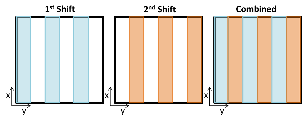
IP-SMS: Unlike conventional SMS imaging where multiple slices within a volume are simultaneously excited, IP-SMS excites multiple segments (or bands) within a slice using a 2D RF excitation pulse. These segments are separately reconstructed and combined to form a final image of a slice. In doing so, the final image can have the full benefits (e.g., high resolution and reduced image distortion) of rFOV imaging without the lengthy scan times of acquiring the individual segments sequentially as in iSeg-EPI.Sequence design: A 2D RF pulse was designed by employing a fly-back EPI-like excitation k-space trajectory. Discrete sampling of excitation k-space along the blipped direction resulted in periodic replicas of the excitation profile (Figure 1). By blipping along the phase-encoding direction, multiple in-plane segments in the image domain were simultaneously excited and used to accelerate acquisition of iSeg-EPI. Eleven sub-pulses with a time-bandwidth product (TBP) of 3.0 were modulated by an envelope pulse with a TBP of 6.3 and a pulse width of 21ms (Figure 1a). The spatial response of the 2D RF pulse (Figure 1b) was used as a weighting function in image reconstruction. Complete coverage of full FOV was obtained by shifting the 2D excitation pattern. The shift was accomplished by frequency selectivity based on a small GY gradient during the sub-pulse. The slight tilt in the resultant side bands was negligible in comparison with slice thickness (~5%). The 2D RF pulse was implemented in a diffusion-weighted (DW) ssEPI pulse sequence for experimental demonstration. Fat suppression was achieved by a frequency-selective saturation pulse preceding the 2D RF pulse.
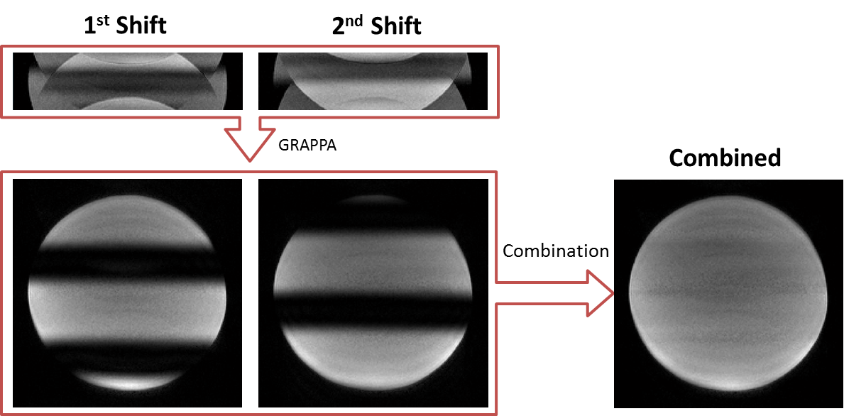
Image reconstruction: As illustrated in Figure 2, the spatially localized patterns in different shifts can complement the sensitivity maps produced by physical receiving coils. Thus, the total number of “virtual” coils is Nc x Ns (where Nc is the number of physical receiving coils and Ns is the number of shifts necessary to sweep the prescribed full FOV)5. The multiplied sensitivity profiles were employed to reconstruct the individual segments using GRAPPA. After all segments were reconstructed, the magnitudes of all segments were combined to form the full-FOV image using the simulated 2D RF excitation profile as a weighting function.
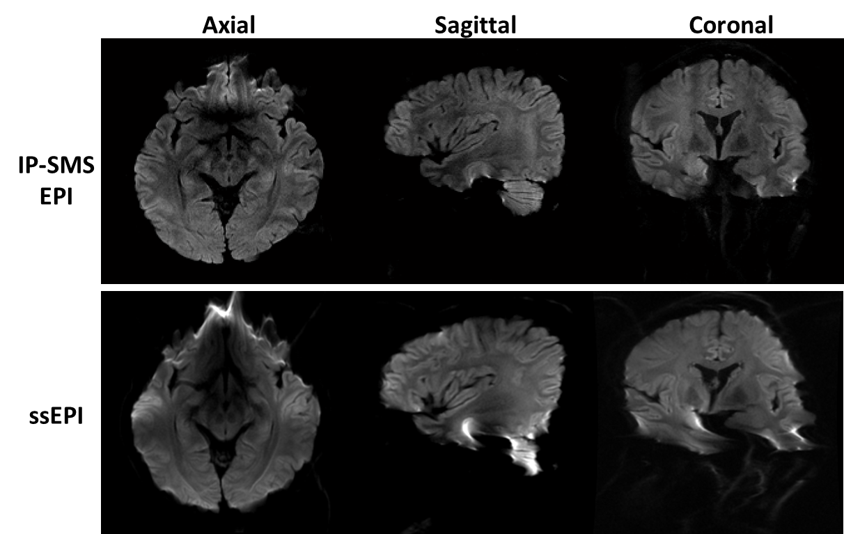
Experiments: Two experiments were conducted on a 3T scanner (GE MR750) with an 8-channel head coil. In the first experiment, a water phantom was used to validate the IP-SMS technique. In the second experiment, DW brain images were acquired from a healthy human volunteer in axial, sagittal, and coronal planes, respectively, to demonstrate the application of IP-SMS sequence for diffusion imaging. The key parameters were: in-plane multiband factor = 3, Ns = 2, acquisition FOV = 200x50 mm2 (i.e., 4-fold under-sampling), spatial resolution = 1x1x5 mm3, TR/TE = 4000/73.8 ms, and b = 1000 s/mm2. For comparison, images in the same imaging planes were also acquired using conventional DW-ssEPI with a full FOV (200x200 mm2).
Results:
Figure 3 shows the phantom results in which the steps involved in IP-SMS are illustrated. Sixteen [8 (Nc) x 2 (Ns)] “virtual” coils successfully resolved the aliasing caused by the 4-fold under-sampling. In the human brain experiment, high-resolution IP-SMS DW images in axial, sagittal, and coronal planes (3 segments; 2 shifts) exhibited good image quality with considerably reduced image distortion as compared to the conventional DW ssEPI images, especially at the frontal and temporal lobes (Figure 4).Discussion and conclusion:
In this study, the conventional SMS concept was extended to in-plane acceleration imaging. This allows the use of rFOV imaging to cover a full FOV with the benefits of high spatial resolution and reduced image distortion. The proposed IP-SMS technique takes advantage of periodic replicas of the excitation profile of 2D RF pulse, “virtual” coil sensitivity maps, and image combination with known weighting functions from the Bloch simulations. The IP-SMS sequence, which does not escalate SAR and is not subject to the slice limitations as in other competing sequences5, can be further extended to other applications beyond diffusion imaging.Acknowledgements
This work was supported in part by the National Institutes of Health (5R01EB026716-01 and 1S10RR028898-01; ZX is not supported or associated with these grants). The authors are grateful to Dr. Y. Sui for providing the original iSeg-EPI pulse sequence and technical assistance.References
1. Barth M, Breuer F, Koopmans PJ, et al. Simultaneous multislice (SMS) imaging techniques. Magn Reson Med. 2016; 75: 63-81.
2. Feinberg DA, Hoenninger JC, Crooks LE, et al. Inner volume MR imaging: Technical concepts and their application. Radiology. 1985; 156 (3): 743-747.
3. Mansfield P, Ordidge RJ, Coxon R. Zonally magnified EPI in real time by NMR. J Phys E. 1988; 21: 275-280.
4. Sui Y, Damen FC, Xie K, et al. Image Domain Segmented Diffusion Imaging Using A 2D Excitation RF Pulse for Distortion Reduction. ISMRM. 2014.
5. Taviani V, Alley MT, Banerjee S, et al. High-resolution diffusion-weighted imaging of the breast with multiband 2D radiofrequency pulses and a generalized parallel imaging reconstruction. Magn Reson Med. 2017; 77: 209-220.
Figures