0600
The total ellipse of the heart: Cardiac CINE imaging using frequency-modulated bSSFP and the elliptical signal model1Departement of Diagnostic and Interventional Radiology, University Hospital Würzburg, Würzburg, Germany
Synopsis
Balanced steady state free precession sequences are well suited for cardiac imaging as they are fast, yield high signal and provide excellent contrast between blood and myocardium. To avoid the typical banding artifacts in such sequences, conventionally, multiple phase-cycled acquisitions are performed and combined to one image. Using a frequency-modulated bSSFP sequence the acquisition of many off-resonances can be performed in one single scan and a model-based reconstruction using the elliptical signal model performs well to reconstruct such measurements. Therefore, it allows the reconstruction of multiple off-resonance states for multiple heart phases from a single frequency-modulated bSSFP measurement.
Purpose/Introduction
Cardiac imaging is a main application of fast MRI imaging techniques. To depict the heart in the different phases of the cardiac cycle the data acquisition needs to be fast, yield high signal and provide excellent contrast between blood and myocardium. Balanced steady state free precession (bSSFP) sequences offer all these advantages. Unfortunately, they are sensitive to field inhomogeneities, which lead to banding artifacts, that can considerably reduce the image quality1. Conventionally, this is resolved by acquiring multiple phase-cycled datasets. The banding artifacts are hereby shifted across the FOV and a combination of the phase-cycled acquisitions can provide banding free images. To describe the signal behavior of bSSFP with off-resonance (or in different phase-cycled images) an elliptical signal model was proposed2. The model can be fitted to data from phase-cycled bSSFP acquisitions and was so far utilized for the reconstruction of banding free images3, estimating underlying tissue parameters4 and to create artificial contrasts based on these parameters5. All procedures are based on the acquisition of multiple phase-cycled measurements, which significantly increases scan time. As shown before a frequency-modulated acquisition can acquire the same information in a single measurement6,7. For very slow modulations the elliptical signal model can serve as prior knowledge to reconstruct different phase-cycled images from this one frequency-modulated measurement8. Here, the elliptical signal model was applied in a model- based reconstruction of a frequency modulated cardiac bSSFP measurement with a radial trajectory to obtain images of different heart phases and different phase-cycles from one single measurement.Subjects and Methods
Radial bSSFP measurements were performed in a healthy volunteer on a 3T MRI system (Siemens MAGNETOM Prisma) using a 30-channel body array coil. One scan featured a shift in the offset frequency for each projection, covering a total offresonance range of 360°. A second measurement without modulation was conducted for comparison. Other imaging parameters were: TE 1.4ms, TR 2.8ms, flip angle 36°, resolution 2.2x2.2x10mm3, total acquisition time 19s. Readouts of all measurements were retrospectively assigned to 20 cardiac phases using the ECG information recorded during the measurement. The frequency-modulated data was additionally binned into 23 different off-resonance states, the equivalent to 23 phase-cycled images. Undersampled k-spaces for each cardiac phase and off-resonance state were received using GROG9. Iteratively, the elliptical signal model was enforced in a MAP10-like algorithm in the image space, while data consistency was preserved in k-space to reconstruct fully sampled images of all 20 x 23 combinations. Resulting images of each heart phase were combined using sum of squares to obtain banding free images.Results
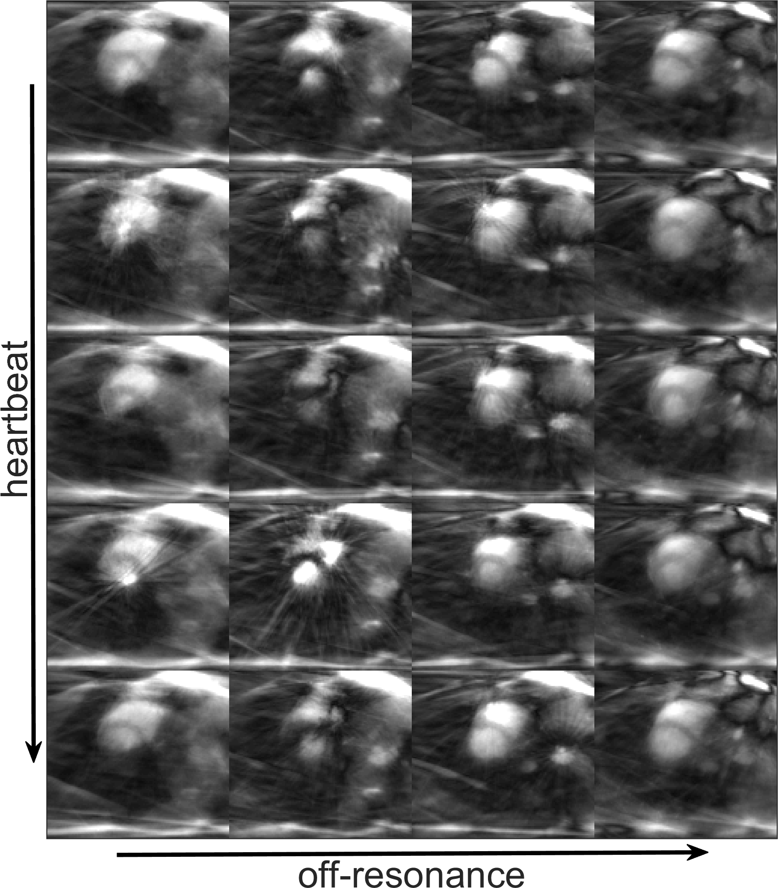
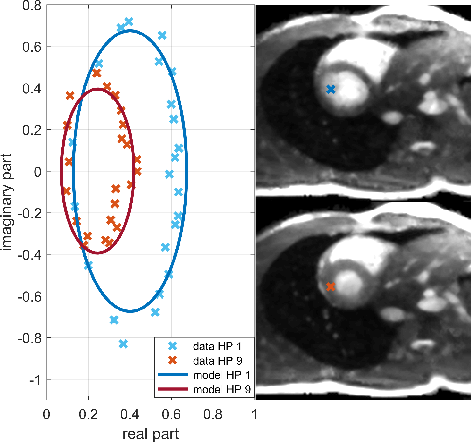
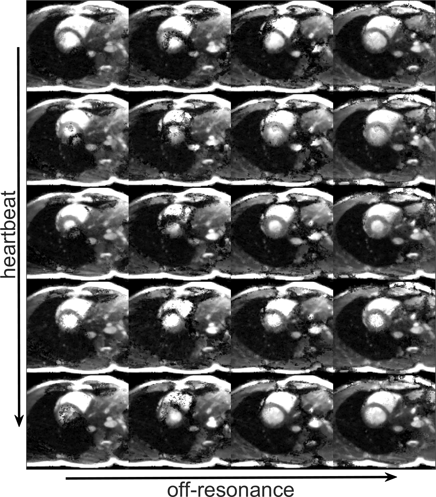
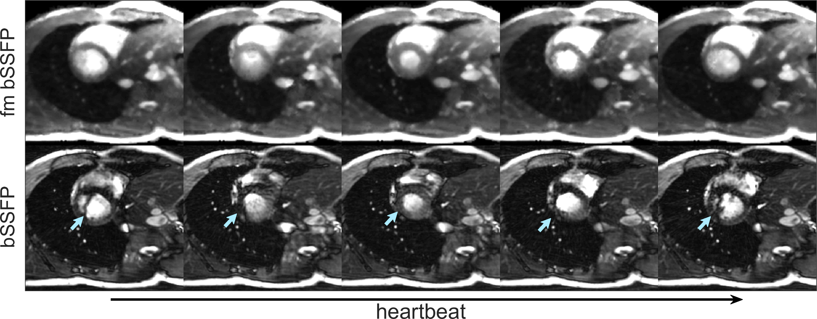
Initially reconstructed images show strong undersampling artifacts and no anatomical details are discernable. Examples for four different off-resonances and five heart phases are shown in Figure 1. An exemplary fit of the elliptical signal model to the off resonance data in one pixel at two different heart phases is shown in Figure 2. The results after 12 iterations of the reconstruction algorithm are shown in Figure 2, no undersampling artifacts remain and a good anatomical representation is given. The banding artifact shifts across the field of view with changing off-resonance, as in phase-cycled imaging. Minor noise amplification occurs in areas where high flow velocities and banding artifact coincide. The combination of the 23 phase-cycled images of one heart phase can reliably remove all banding artifacts and provide quality images, as shown in Figure 3a. The final images are comparable to the standard bSSFP images in Figure 3b and retain all the advantages, like high signal and favorable contrast for cardiac imaging.Discussion/Conclusion
The elliptical signal model is well suited for the reconstruction of frequency-modulated cardiac bSSFP acquisitions. It allows the reconstruction of different phase-cycled images for all heart phases from a single radial measurement. The advantages of bSSFP, acquisition speed, high signal level and good contrast, are available even in cases of high field inhomogeneity. Main limitation of the frequency-modulated acquisition is the necessity for a slow modulation to retain high signal and the validity of the signal model. As the acquisition in cardiac CINE imaging is spread over multiple heart beats this condition can be met. Other suitable application include 3D and high-resolution imaging. An extension to a free-breathing measurement is also feasible.Acknowledgements
No acknowledgement found.References
1. Schär M, Kozerke S, Fischer SE, Boesiger P. Cardiac SSFP imaging at 3 Tesla. Magn. Reson. Med. 2004;51:799–806
2. Xiang Q-S, Hoff MN. Banding artifact removal for bSSFP imaging with an elliptical signal model. Magn. Reson. Med. 2014;71:927–933
3. Hoff MN, Andre JB, Xiang Q-S. Combined geometric and algebraic solutions for removal of bSSFP banding artifacts with performance comparisons. Magn. Reson. Med. 2017;77:644–654
4. Shcherbakova Y, van den Berg CAT, Moonen CTW, Bartels LW. PLANET: An ellipse fitting approach for simultaneous T1 and T2 mapping using phase-cycled balanced steady-state free precession. Magn. Reson. Med. 2018;79:711–722
5. Hilbert T, Nguyen D, Thiran J-P, Krueger G, Kober T, Bieri O. True constructive interference in the steady state (trueCISS). Magn. Reson. Med. 2018;79:1901–1910
6. Foxall D l. Frequency-modulated steady-state free precession imaging. Magn. Reson. Med. 2002;48:502–508
7. Benkert T, Ehses P, Blaimer M, Jakob PM, Breuer FA. Dynamically phase-cycled radial balanced SSFP imaging for efficient banding removal. Magn. Reson. Med. 2015;73:182–194
8. Slawig A, Wech T, Köstler H. Reconstruction of all offresonance states in DYPR bSSFP using an elliptical signal model. In: Book of Abstracts ESMRMB 2019. Rotterdam, Niederlande.
9. Seiberlich N, Breuer F, Blaimer M, Jakob P, Griswold M. Self-calibrating GRAPPA operator gridding for radial and spiral trajectories. Magn. Reson. Med. 2008;59:930–935
10. Tran‐Gia J, Stäb D, Wech T, Hahn D, Köstler H. Model‐based Acceleration of Parameter mapping (MAP) for saturation prepared radially acquired data. Magn. Reson. Med. 2013;70:1524–1534
Figures