0597
High Spatiotemporal Resolution Motion-Resolved MRI using XD-GRASP-Pro1Biomedical Engineering and Imaging Institute and Department of Radiology, Icahn School of Medicine at Mount Sinai, New York, NY, United States, 2Gordon Center for Medical Imaging, Massachusetts General Hospital, Harvard Medical School, Boston, MA, United States, 3Department of Radiology, University of Wisconsin Madison, Madison, WI, United States
Synopsis
This work presents a free-breathing motion-resolved golden-angle image reconstruction method called XD-GRASP-Pro, which extends the original XD-GRASP (eXtra-Dimensional Golden-angle RAdial Sparse Parallel MRI) method with imProved reconstruction performance through an additional self-estimated/calibrated low-rank subspace-constraint. The temporal basis used to construct the subspace is estimated from an intermediate reconstruction step on the low-resolution portion of radial k-space, which eliminates the need of using auxiliary data or a physical signal model that is not always available. XD-GRASP-Pro were tested for high spatiotemporal resolution motion-resolved liver MRI.
INTRODUCTION:
The original GRASP (Golden-angle RAdial Sparse Parallel) imaging technique (1) relies entirely on a 1D total-variation (TV) constraint enforced along the dynamic dimension to remove streaking artifacts caused by undersampling. Despite the convincing performance of temporal TV regularization as demonstrated in prior studies (1-4), it can cause noticeable blurring with visible residual artifacts at high acceleration rates. The variant of GRASP for motion-resolved MRI, called XD-GRASP (eXtra-Dimensional GRASP) (5), also employs TV constraints along the motion dimensions generated by resorting the golden-angle radial data. Building upon the recently proposed GRASP-Pro (imProved Sparse Golden-Angle Radial imaging) technique (6), we aimed to extend XD-GRASP to XD-GRASP-Pro in this work for high spatiotemporal resolution motion-resolved MRI. This is achieved through a combination of self-estimated subspace-constraint and spatiotemporal TV constraint based on the recent innovation of low-rank subspace modeling (7). In particular, we propose that the temporal basis used to construct the subspace can be estimated from the low-resolution portion of radial k-space without the use of auxiliary data or a physical signal model (which is not always available) as in prior works (8-11). GRASP-Pro/XD-GRASP-Pro represents a different and new way of reconstructing dynamic radial datasets, in which the low-resolution information (with a much lower undersampling factor around the k-space center) is used to guide the reconstruction of full-resolution images. In the following sections, the overall GRASP-Pro/XD-GRASP-Pro framework is presented, followed by the demonstration of the technique in high-resolution motion-resolved liver MRI.METHODS:
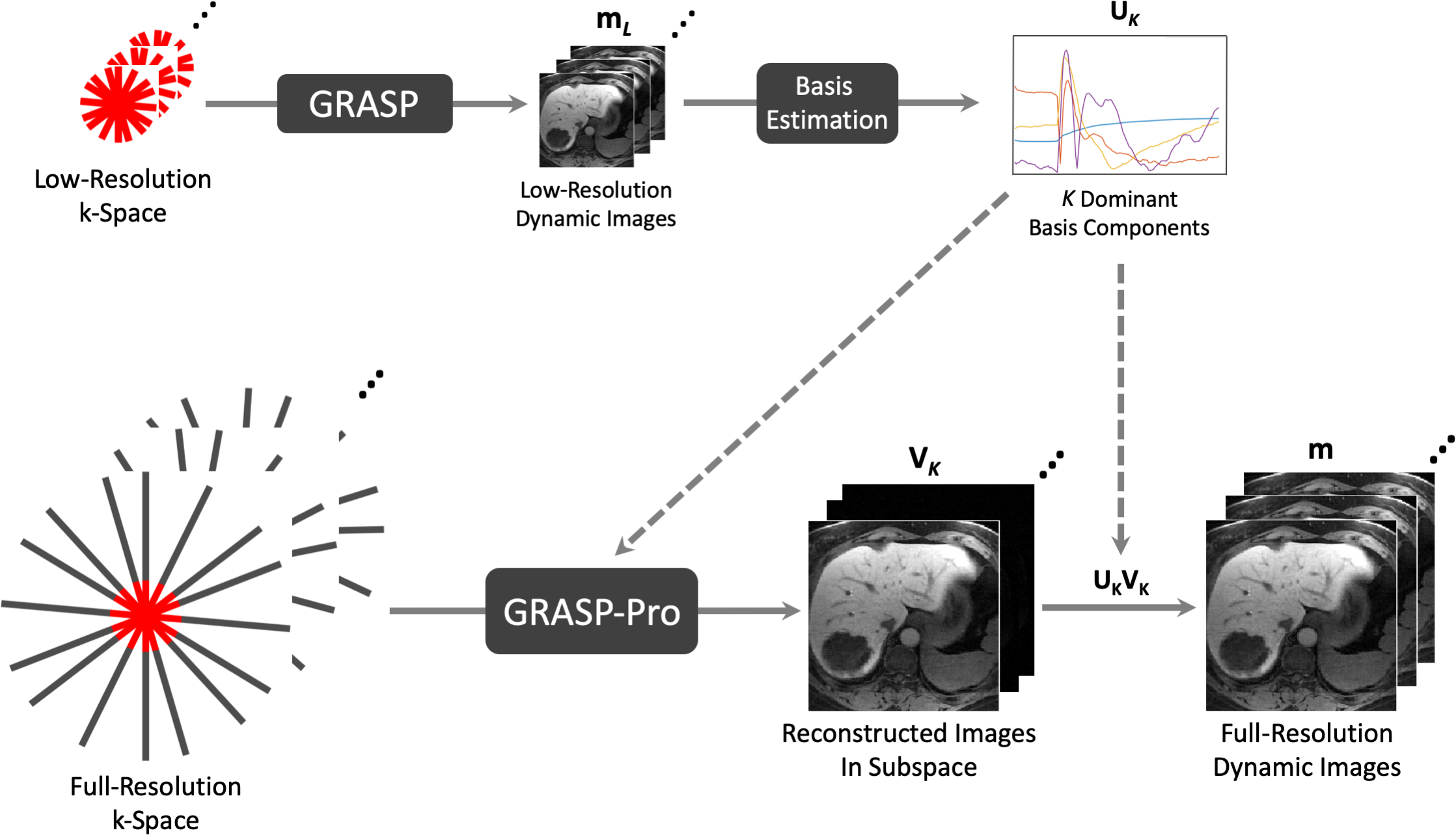
GRASP-Pro & XD-GRASP-Pro reconstruction: Figure 1 shows the overall workflow of GRASP-Pro reconstruction. First, the low-resolution k-space (yL) is extracted for a standard GRASP reconstruction with a spatiotemporal TV constraint (TVt and TVs, Equation 1). Since radial sampling has a much lower undersampling factor around the k-space center compared to the periphery, a low-resolution image-series (mL) can be reliably reconstructed even at high acceleration rates. This low-resolution image-series can be used to estimate a temporal basis (U) for the dynamic images, under the assumption that mL shares similar anatomic structure with the full-resolution images (m). Given the extensive temporal correlation in m, the full-resolution dynamic images can be compressed to a much lower dimensional subspace, represented by only the first K dominant basis components (UK) and associated coefficients (VK). This leads to highly-reduced degrees of freedom and thus improved reconstruction performance (6). Subspace-constrained reconstruction can then be performed to reconstruct VK, the coefficients to represent the fully-resolution image-series under UK (Equation 2). A spatiotemporal TV constraint is added to further improve the reconstruction performance, in which the temporal TV constraint (TVt) is applied along the dynamic image dimension and spatial TV constraint (TVs) is enforced directly on the subspace. With the above notations, it is then straightforward to extend GRASP-Pro to XD-GRASP-Pro with self-navigation to generate one or more motion-resolved dynamic dimensions.$$\tilde{m}_{L}=arg\min_{m_{L}}\frac{1}{2}\parallel{Em_{L}-y_{L}}\parallel_{2}^{2}+\lambda_{1}TV_{t}(m_{L})+\lambda_{2}TV_{s}(m_{L})\quad\quad\quad[1]$$ $$\tilde{V}_{K}=arg\min_{V_{K}}\frac{1}{2}\parallel{E(U_{K}V_{K})-y}\parallel_{2}^{2}+\lambda_{1}TV_{t}(U_{K}V_{K})+\lambda_{2}TV_{s}(V_{K})\quad\quad\quad[2]$$
Evaluation: The proposed technique was evaluated for high-resolution motion-resolved liver MRI. Two free-breathing liver MR datasets were acquired on a 3T MR scanner (MAGNETOM TimTrio, Siemens) with the following imaging parameters: FOV=320x320mm2, voxel size=1x1x5mm3, TR/TE=3.40/1.68ms, flip angle=10o, number of slices=44 with 80% slice partial Fourier. A total of 1000 spokes were acquired in each partition during free-breathing and the total acquisition time was 138 seconds. For each data, both XD-GRASP and XD-GRASP-Pro were performed to reconstruct 25 respiratory phases spinning from end-expiration to end-inspiration (40 resorted spokes in each frame) with a respiratory motion signal extracted from the acquired radial data (5). For XD-GRASP-Pro, a 64x64 central k-space region was first reconstructed to generate the temporal basis for subspace construction, and the first 6 (K=6) dominant basis components were kept for the subsequent subspace-constrained reconstruction.
RESULTS:
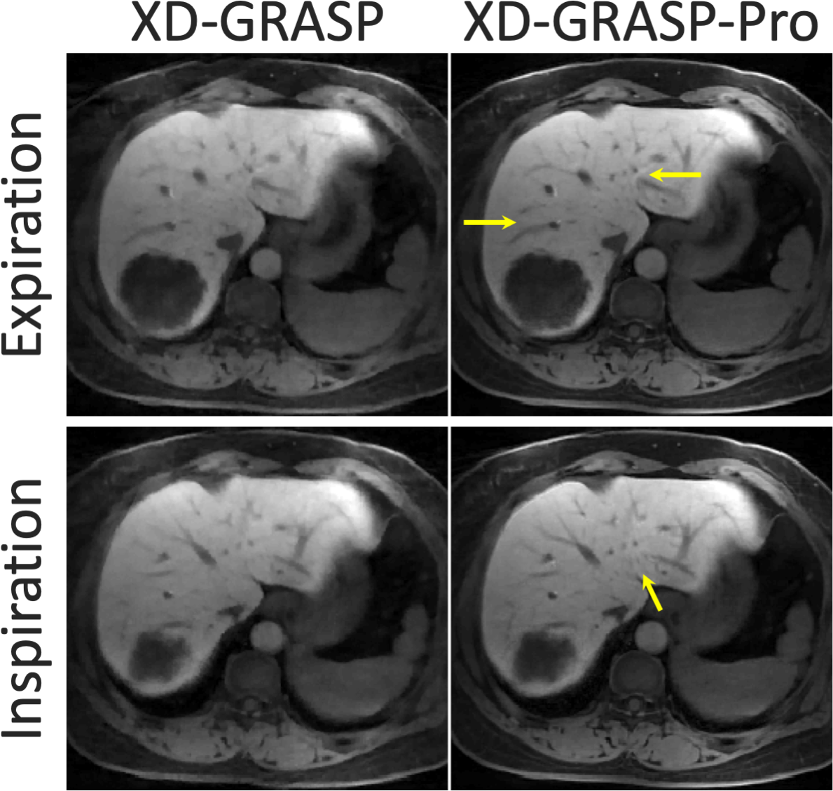
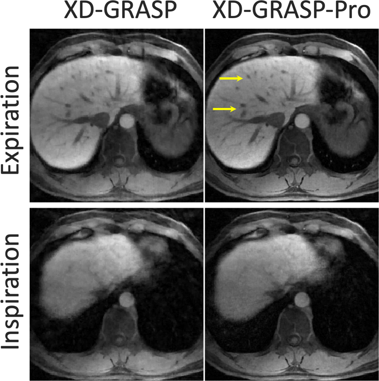
Figure 2 and Figure 3 show the comparison of XD-GRASP and XD-GRASP-Pro in reconstructing the two free-breathing liver MR datasets. Due to the sorting of data into a large number of respiratory phases (25 phases), XD-GRASP resulted in certain image blurring and residual artifacts. This is caused by (1) the high undersampling factor and (2) the reduced uniformity of k-space coverage in each motion phase after data sorting with an increased number of temporal frames. XD-GRASP-Pro, on the other hand, enabled better delineation of the hepatic vessels (yellow arrows) and improved overall image sharpness. Full-respiratory-cycle cine movies (in GIF format) comparing XD-GRASP and XD-GRASP-Pro in the two subjects are shown as Figure 4, in which XD-GRASP-Pro (right column) produced better overall image quality with less residual streaking artifacts compared to XD-GRASP (left column).DISCUSSION:
This work demonstrated the performance of a new reconstruction technique called GRASP-Pro and its variant XD-GRASP-Pro for highly-accelerated sparse golden-angle radial MRI. GRASP-Pro/XD-GRASP-Pro employs a self-calibrating subspace construction, eliminating the need of using auxiliary data or a physical signal model for generating temporal basis. With the incorporation of an explicit subspace constraint, XD-GRASP-Pro is able to reconstruct dynamic radial image series with improved sharpness, less residual artifacts and overall image quality, as shown in our initial results demonstrated for high-resolution motion-resolved liver imaging at high acceleration rates.Acknowledgements
The authors would like to thank Dr. Lihua Chen from the Southwest Hospital in Chongqing, China for sharing the post-contrast golden-angle stack-of-stars liver datasets, which were retrospectively used to test the performance of XD-GRASP-Pro in this work.References
1. Feng L, Grimm R, Block KT, Chandarana H, Kim S, Xu J, Axel L, Sodickson DK, Otazo R. Golden-angle radial sparse parallel MRI: combination of compressed sensing, parallel imaging, and golden-angle radial sampling for fast and flexible dynamic volumetric MRI. Magn Reson Med 2014;72(3):707-717.
2. Adluru G, McGann C, Speier P, Kholmovski EG, Shaaban A, Dibella EV. Acquisition and reconstruction of undersampled radial data for myocardial perfusion magnetic resonance imaging. Journal of magnetic resonance imaging : JMRI 2009;29(2):466-473.
3. Feng L, Srichai MB, Lim RP, Harrison A, King W, Adluru G, Dibella EV, Sodickson DK, Otazo R, Kim D. Highly accelerated real-time cardiac cine MRI using k-t SPARSE-SENSE. Magn Reson Med 2013;70(1):64-74.
4. Adluru G, Awate SP, Tasdizen T, Whitaker RT, Dibella EV. Temporally constrained reconstruction of dynamic cardiac perfusion MRI. Magn Reson Med 2007;57(6):1027-1036.
5. Feng L, Axel L, Chandarana H, Block KT, Sodickson DK, Otazo R. XD-GRASP: Golden-angle radial MRI with reconstruction of extra motion-state dimensions using compressed sensing. Magn Reson Med 2016;75(2):775-788.
6. Feng L, Wen Q, Huang C, Tong A, Liu F, Chandarana H. GRASP-Pro: imProving GRASP DCE-MRI through self-calibrating subspace-modeling and contrast phase automation. Magn Reson Med 2020;83(1):94-108.
7. Liang ZP. Spatiotemporal imaging with partially separable functions. 2007 4th Ieee International Symposium on Biomedical Imaging : Macro to Nano, Vols 1-3 2007:988-991.
8. Zhao B, Haldar JP, Christodoulou AG, Liang ZP. Image Reconstruction From Highly Undersampled (k, t)-Space Data With Joint Partial Separability and Sparsity Constraints. Ieee T Med Imaging 2012;31(9):1809-1820.
9. Zhao B, Lu W, Hitchens TK, Lam F, Ho C, Liang ZP. Accelerated MR parameter mapping with low-rank and sparsity constraints. Magn Reson Med 2015;74(2):489-498.
10. Tamir JI, Uecker M, Chen W, Lai P, Alley MT, Vasanawala SS, Lustig M. T2 shuffling: Sharp, multicontrast, volumetric fast spin-echo imaging. Magn Reson Med 2017;77(1):180-195.
11. Christodoulou AG, Shaw JL, Nguyen C, Yang Q, Xie YB, Wang N, Li DB. Magnetic resonance multitasking for motion-resolved quantitative cardiovascular imaging. Nat Biomed Eng 2018;2(4):215-226.
Figures