0594
Complex-Valued Spatial-Temporal Super-Resolution Combined with Multi-Band Technique on T2*-Weighted Dynamic MRI1Biomedical Engineering, The University of Arizona, Tucson, AZ, United States, 2Medical Imaging, The University of Arizona, Tucson, AZ, United States, 3Siemens healthineers, Tucson, AZ, United States, 4GE Healthcare, Waukesha, WI, United States
Synopsis
We present an approach for improving spatial and temporal resolution of complex-valued T2*-weighted dynamic MRI. Compared with the conventional magnitude-valued super-resolution approaches, our technique utilizes phase information to better recover signal loss caused by susceptibility gradients and generate finer representations of temporal dynamic signal variation. Results from numerical and hybrid simulation show that promising improvements in image resolution, susceptibility artifact reduction and temporal signal variation representation can be achieved using our complex-valued super-resolution MRI scheme when compared to magnitude-valued super-resolution.
Introduction:
Improvement in spatial and temporal resolution directly benefits the sensitivity and specificity of T2*-weighted dynamic MRI, for various applications ranging from dynamic susceptibility contrast to fMRI. However, it is challenging to simultaneously achieve high signal quality and high spatial-temporal-resolution due to trade-offs that exist between resolution, acquisition time and signal-to-noise ratio. One approach to resolve this challenge is to use super-resolution1 to reconstruct high resolution images using spatially-sub-voxel-shifted (along the slice-selection direction) low resolution images2, 3. As shown in recent publications, high spatial-temporal-resolution fMRI could be achieved when combining super-resolution and multi-band MRI (e.g., SLIDER-SMS4, 5). However, susceptibility signal loss in T2*-weighted dynamic MRI data obtained with thick slices has not been well addressed with existing magnitude-only super-resolution reconstruction scheme. In this project, we compared the degree of signal-loss recovery for T2*-weighted dynamic MRI with both complex-valued and magnitude-valued multi-band super-resolution reconstruction schemes.Methods:
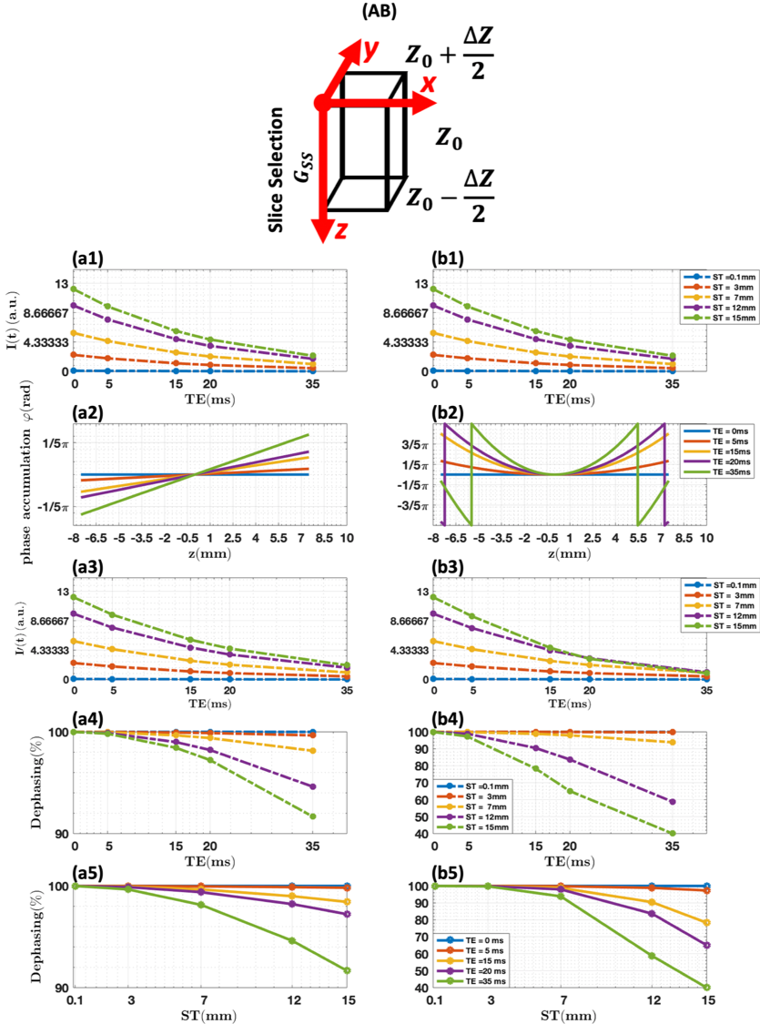
I). Simulation of through-plane susceptibility effect.A simplified simulation of the effects of local through-plane susceptibility gradients on signal intensity was done in a single voxel to evaluate the slice thicknesses in which super-resolution reconstruction would be most effective. Image intensity loss due to spin dephasing caused by susceptibility gradients in the slice-selection direction ($$$G_{ss}$$$) can be represented by6, 7:
$$I = I_{0}\cdot e^{\frac{-t}{T_2^*}}\cdot \sum_{z=z_{0}-\frac{\triangle z}{2}}^{z_{0}+\frac{\triangle z}{2}}p(z)\cdot e^{i\phi(z)}\cdot dz$$
In which, $$$I_{0}$$$ is the initial image intensity without T2* decay and susceptibility gradients; $$$z_{0}$$$ is the center of the voxel, $$$\triangle z$$$ is the slice thickness, $$$p(z)$$$ is a $$$pseudo-rect$$$ slice profile, $$$\phi(z)$$$ is the phase accumulation due to susceptibility gradient and given by $$$\phi(z) = \gamma\cdot G_{ss}\cdot TE\cdot z$$$.
II). Simulation of spatial-temporal super-resolution with multi-band (R=2) technique.
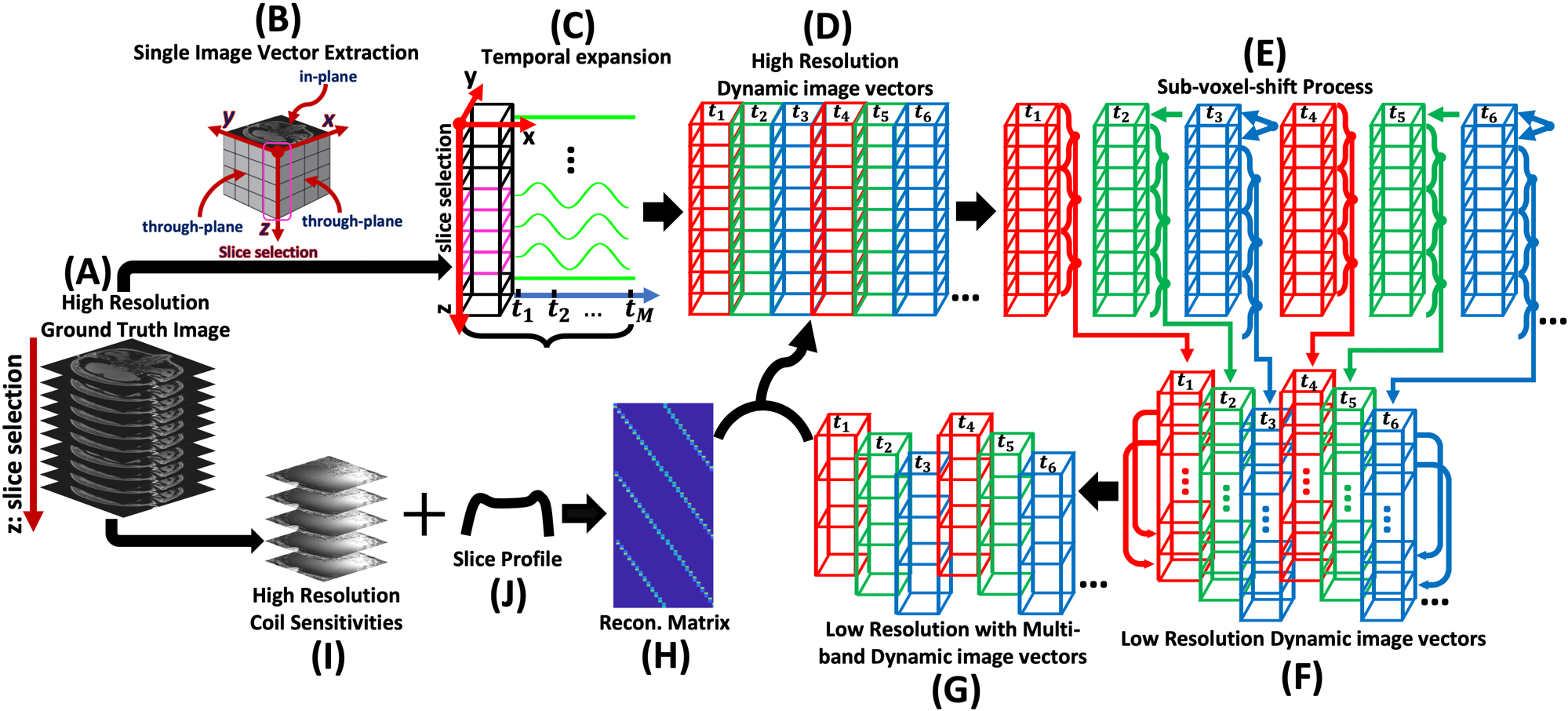
High-resolution 3D static human brain k-space raw data was acquired on the Siemens® 3T scanner with 32-channel head coil. A High-resolution complex-valued image volume was then generated from the raw data using the fast Fourier transform (FFT) as ground truth. An artificially generated sinusoidal signal with relative peak value of 1.3 and valley value of 0.7 (Total 101 time points with arbitrary unit in our simulation) was applied to each of the randomly selected voxels within a chosen target column along the slice-selection direction; the rest of the voxels within the target column were repeated 101 times without any variation in signal intensity. After the temporal expansion, every 3 slices were then weighted by their corresponding slice profile portions and summed to form the low-resolution image volume at each time point with the corresponding number of sub-voxel-shifts. Multi-band combined low-resolution dynamic image volumes were finally created by combining the first and the second half of corresponding low-resolution image volumes along the slice-selection direction for all time points. High-resolution coil sensitivities were combined with the slice profile to create the reconstruction matrix. The final reconstruction was done through direct matrix inversion using complex-valued images and magnitude-valued images, respectively. The general workflow is shown in Fig. 1.
Results:
I). Simulation of through-plane susceptibility effect.As shown in Fig. 2, significant dephasing effects can be observed when the acquisition slice thickness increases in both linear (Fig. 2. (a5)) and nonlinear susceptibility gradient (Fig. 2. (b5)) cases, which indicates the feasibility that susceptibility artifact can be effectively reduced by decreasing the slice thickness using super-resolution reconstruction with thick slice acquisition.
II). Simulation of spatial-temporal super-resolution with multi-band (R=2) technique.
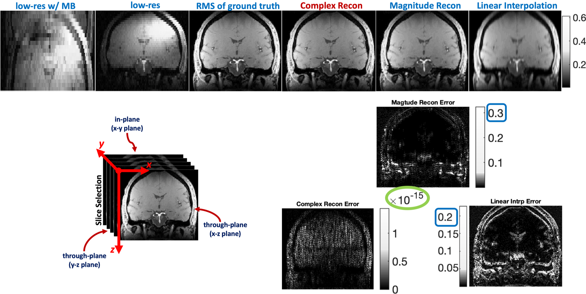
As can be seen from the reconstruction errors in Fig. 3, the complex-valued spatial-temporal super-resolution reconstructed image best matches the ground truth image, with the maximum difference in the order of 10-15, whereas the reconstruction errors of the magnitude-valued super-resolution reconstructed image and the linear interpolated image are both in the order of 10-1. The reconstruction error from the complex-valued spatial-temporal super-resolution is mostly noise and the susceptibility artifact is minimized; however, the reconstruction errors from the magnitude-valued super resolution contain high-frequency structure with significant remaining residual susceptibility artifacts.
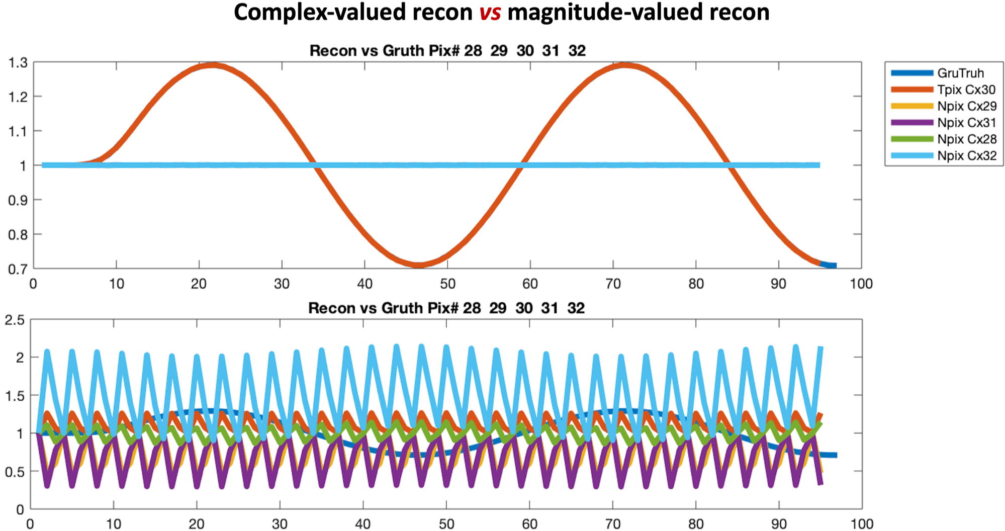
It is indicated in Fig. 4 that the input temporal sine signal variation can be accurately recovered at the targeted voxels and that the signal fluctuations are small in the neighboring voxels for the complex-valued spatial-temporal super-resolution. However, the temporal variations reconstructed at target voxels via magnitude-valued super-resolution are generally unusable, and significant signal fluctuations are also present in the neighboring voxels.
Discussion:
Image reconstruction results have shown that the proposed complex-valued spatial-temporal super-resolution reconstruction can effectively recover signal loss caused by susceptibility dephasing artifacts and can generate finer representations of temporal dynamic signal variation compared to traditional magnitude-valued super-resolution reconstruction. It has been demonstrated that phase information can be utilized in spatial-temporal super-resolution reconstruction to significantly reduce the susceptibility artifact, benefiting applications relying on T2* contrast. Our proposed method can be integrated with multi-band schemes to further improve scan efficiency.Acknowledgements
No acknowledgement found.References
1. Tsai, R.Y.; Huang, T.S. Multiframe image restoration and registration. In Advances in Computer Vision and Image Processing; JAI Press: Greenwich, CT, USA, 1984; Volume 1, pp. 317–339.
2. Li L, Wang W, Luo H, Ying S. Super-Resolution Reconstruction of High-Resolution Satellite ZY-3 TLC Images. Sensors 2017; 17(5).
3. Van Reeth E, Tham IWK, Tan CH, Poh CL. Super-resolution in magnetic resonance imaging: A review. Concepts Magn. Reson. Part A 2012; 40A(6): 306–325.
4. Setsompop K, Fan Q, Stockmann J, et al. High-resolution in vivo diffusion imaging of the human brain with generalized slice dithered enhanced resolution: Simultaneous multislice (gSlider-SMS). Magn. Reson. Med. 2018; 79(1): 141–151.
5. Vu AT, Beckett A, Setsompop K, Feinberg DA. Evaluation of SLIce Dithered Enhanced Resolution Simultaneous MultiSlice (SLIDER-SMS) for human fMRI. NeuroImage 2018; 164: 164–171.
6. Chen N k, Wyrwicz AM. Removal of intravoxel dephasing artifact in gradient-echo images using a field-map based RF refocusing technique. Magn. Reson. Med. 1999; 42(4): 807–812.
7. Deichmann R, Josephs O, Hutton C, Corfield DR, Turner R. Compensation of susceptibility-induced BOLD sensitivity losses in echo-planar fMRI imaging. NeuroImage 2002; 15(1): 120–135.
Figures