0592
MRI Texture Analysis in the Characterization of Cervical Carcinoma1Department of Diagnostic Radiology, The University of Hong Kong, Hong Kong, Hong Kong, 2Philips Healthcare, Hong Kong, China
Synopsis
MRI texture analysis was performed in 100 patients with cervical carcinoma. TexRAD software was used for texture extraction and analysis on ADC maps and T1c images. Texture features were compared between histological subtypes, tumour grades, FIGO stages and nodal status. Feature selection was achieved with AUC ≥ 0.70. ADC-derived MPP5 was significantly lower in SCC than ACA, Entropy6 derived from both ADC and T1c increased from FIGO I~II to FIGO III~IV, and ADC-derived Entropy3 was higher in positive nodal status than negative. No texture features could differentiate tumour grades with acceptable diagnostic efficiency.
Introduction
Texture analysis has been widely applied in the measurement of the spatial distribution derived from grey-level intensities on oncological images. Texture features extracted from texture analysis are emerging as promising imaging biomarkers for quantifying intra-tumour heterogeneity, which are beyond human visual perception [1]. Tumours with diverse histological subtypes and characteristics could present similar signal and morphology on conventional MRI, such as T2-weighted imaging (T2WI) [2]. In contrast, diffusion-weighted imaging (DWI) and contrast-enhanced T1-weighted (T1c) imaging provide deeper perception in tumour microstructure and vasculature. The purpose of this study was to evaluate MRI texture analysis based on DWI and T1c imaging in the characterization of cervical carcinoma (CC), specifically in differentiating histological subtypes, tumour grades, FIGO stages and nodal status.Methods
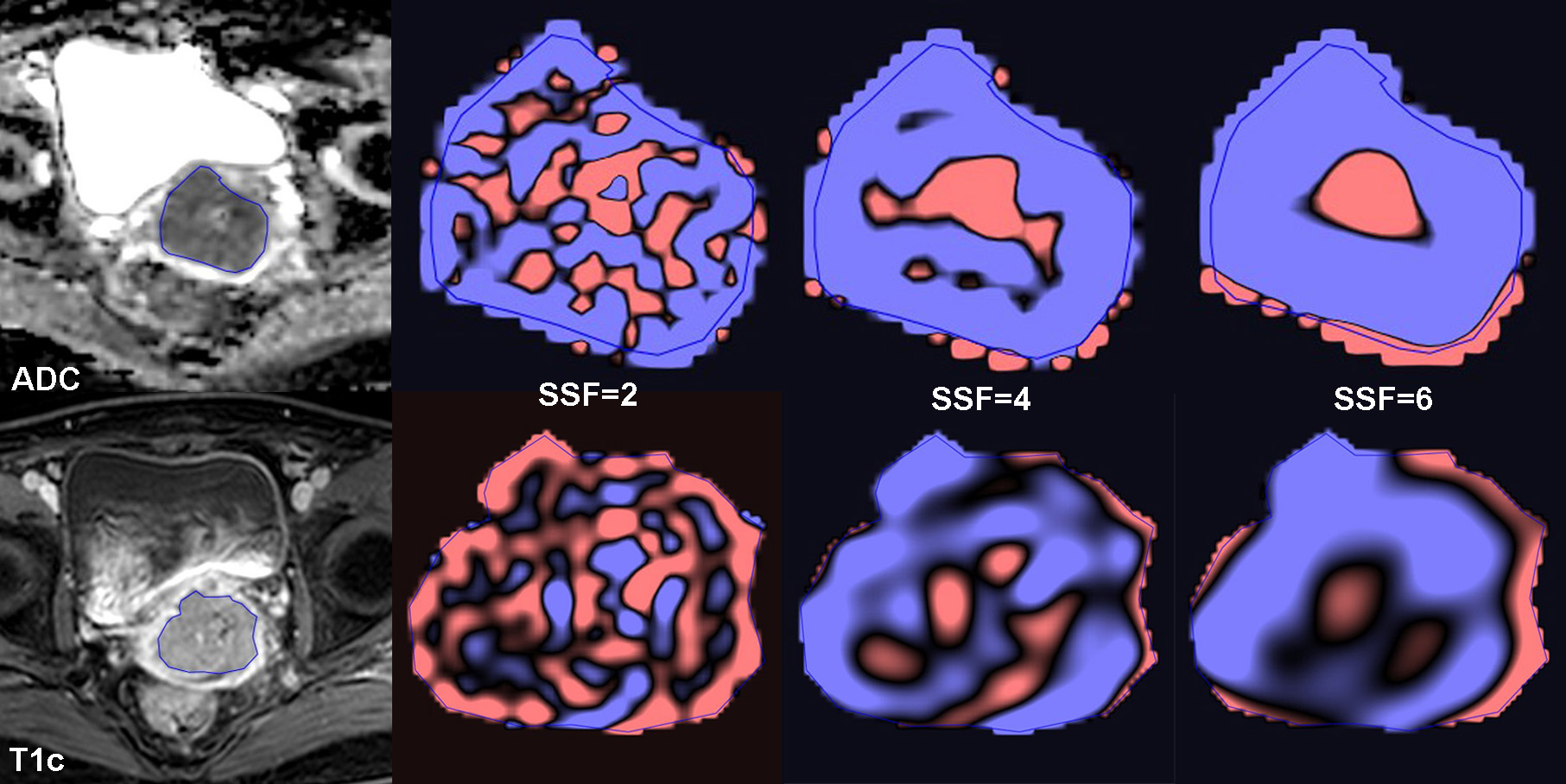
One-hundred patients (55.9 ± 13.5 years) with histological confirmed CC (FIGO stage IB~IVB) were retrospectively enrolled in this study. Pre-treatment MRI include DWI and T1c imaging were acquired on a 3.0T platform (Achieva 3T TX, Philips Healthcare, Best, the Netherlands). The apparent diffusion coefficient (ADC) was derived by using 2 b-values (0 and 1000 s/mm2). MRI texture analysis was performed using propriety software (TexRAD; Cambridge Computed Imaging Ltd, UK). Largest single-slice region of interest (ROI) was manually delineate around the tumour on ADC map and T1c image (Figure 1). Six first-order texture features including mean, standard deviation (SD), entropy, mean of positive pixels (MPP), skewness and kurtosis with 6 spatial scale filters (SSF) were calculated for each ROI. Histological subtypes, tumour grades, FIGO stages and nodal status were dichotomised, and texture features compared. Mann-Whitey U test, receiver operating characteristic (ROC) curve and the area under the curve (AUC) were used for statistical analyses. Feature selection was achieved by AUC ≥ 0.70 as an acceptable diagnostic accuracy. The texture feature with the highest AUC in the same sequence was selected as the best performing texture parameter.Results
ADC MPP5 was significantly lower in squamous cell carcinomas (SCC) than adenocarcinomas (ACA) (p = 0.005, AUC = 0.726). In the differentiation of FIGO stages, ADC and T1c Entropy6 increased significantly from FIGO I~II to FIGO III~IV (p < 0.001, AUC = 0.747; p < 0.001, AUC = 0.730, respectively). As for the nodal status, ADC Entropy3 showed higher value in patients with nodal metastases than those without (p < 0.001, AUC = 0.702). No textures feature was able to differentiate tumour grades with acceptable AUC.Discussion
ADC MPP5 could separate histological subtypes, SCC had lower MPP than ACA. It was reported that ADC-derived mean and MPP were positively correlated with ADC value, and this was in keeping with our previous study that ADC was lower in SCC than ACA [3, 4]. Similar finding was observed in endometrial carcinoma, high T1c MPP4 was able to predict high-risk histological subtype independently [5].ADC and T1c-drived entropy were higher in high FIGO stages (III~IV) and positive nodal status in CC, which corroborated by previous studies in CC and endometrial carcinoma [5, 6]. Both ADC Entropy6 and T1c Entropy6 showed the best performance in the respective group in differentiating FIGO stages. It was argued that features at fine texture scale (SSF = 2) might not represent the biologically significant features because the heterogeneity assessment at that texture scale was more susceptible to the imaging parameters [3]. We hypothesize that at a coarse scale (SSF = 6), more underlying vasculature and microstructure variability could be highlighted to contribute to tumour heterogeneity. Entropy describes the irregularity of grey-level distribution, which reflects the intra-tumoural heterogeneity; hence, in agreement with the findings in this study, CC with advanced stages exhibited higher entropy [7].
Conclusion
Entropy and MPP were helpful in the characterization of CC with acceptable diagnostic efficiency. ADC-derived MPP had potential ability in separating histological subtypes; both ADC and T1c-derived entropy could differentiate FIGO stages; ADC-derived entropy could also distinguish nodal status.Acknowledgements
N.A.References
1. Chitalia RD, Kontos D: Role of texture analysis in breast MRI as a cancer biomarker: A review. J Magn Reson Imaging 2019, 49(4):927-938.
2. Davnall F, Yip CS, Ljungqvist G, Selmi M, Ng F, Sanghera B, Ganeshan B, Miles KA, Cook GJ, Goh V: Assessment of tumor heterogeneity: an emerging imaging tool for clinical practice? Insights Imaging 2012, 3(6):573-589.
3. Hameed M, Ganeshan B, Shur J, Mukherjee S, Afaq A, Batura D: The clinical utility of prostate cancer heterogeneity using texture analysis of multiparametric MRI. International urology and nephrology 2019, 51(5):817-824.
4. Wang M, Perucho JAU, Chan Q, Sun J, Ip P, Tse KY, Lee EYP: Diffusion Kurtosis Imaging in the Assessment of Cervical Carcinoma. Academic radiology 2019 Jul 16. pii: S1076-6332(19)30328-9. doi: 10.1016/j.acra.2019.06.022.
5. Ytre-Hauge S, Dybvik JA, Lundervold A, Salvesen OO, Krakstad C, Fasmer KE, Werner HM, Ganeshan B, Hoivik E, Bjorge L et al: Preoperative tumor texture analysis on MRI predicts high-risk disease and reduced survival in endometrial cancer. J Magn Reson Imaging 2018, 48(6):1637-1647.
6. Guan Y, Li W, Jiang Z, Zhang B, Chen Y, Huang X, Zhang J, Liu S, He J, Zhou Z et al: Value of whole-lesion apparent diffusion coefficient (ADC) first-order statistics and texture features in clinical staging of cervical cancers. Clin Radiol 2017, 72(11):951-958.
7. Yang F, Young L, Grigsby P: Predictive Value of Standardized Intratumoral Metabolic Heterogeneity in Locally Advanced Cervical Cancer Treated With Chemoradiation. International journal of gynecological cancer: official journal of the International Gynecological Cancer Society 2016, 26(4):777-784.
Figures