0591
Application of amide proton transfer in differential diagnosis of mass cervical carcinoma and typical uterine leiomyoma1Department of Radiology, the First Affiliated Hospital of Dalian Medical University, Dalian, China, 2Philips Healthcare, Beijing, China
Synopsis
Amide proton transfer (APT) imaging technology has been applied in the diagnosis of central nervous system tumors to some extent. However, it is only used in the diagnosis of cervical carcinoma in uterine tumors, and there is no study on the differentiation of cervical carcinoma and related tumors with APT. We investigated the value of APT in the differential diagnosis of mass cervical carcinoma and typical uterine leiomyoma.
INTRODUCTION
Cervical cancer is a common gynecologic malignant tumor, originating from cervical squamous epithelium or glandular epithelial cells. Uterine leiomyoma is a common benign tumor, composed of smooth muscle cells and connective tissue, corpus uteri and cervix uterus can occur. Because uterine leiomyoma is prone to a variety of degeneration, it is sometimes difficult to distinguish it from mass cervical carcinoma on MR images. APT imaging technology can provide the information of high resolution free protein and amino compound proton of peptide in vivo to reflect the distribution of protein in the tumor and aid in the differential diagnosis and treatment[1]. Previous studies have shown that APT technology can be used to identify cervical squamous carcinoma and adenocarcinoma[2], as well as neoplastic and infectious mass lesions[3]. It can also be used to explore changes in APT signal intensity among different phases of the menstrual cycle in healthy young women[4]. In this study, we investigated the value of APT in the differential diagnosis of mass cervical carcinoma and typical uterine leiomyoma.METHODS
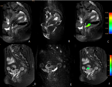
Data of 14 cases of mass cervical cancer and 16 cases of typical uterine leiomyoma confirmed by surgery and pathology were retrospectively analyzed. All patients received 3.0T MRI examination before surgery (including the APT-weighted free-breathing 3D turbo-spin-echo sequence in the Axial orientation, where the TR/TE = 6500/8 ms, FOV = 130 mm, voxel size = 2.0×2.0 mm2, Slice Thick=7.0, B1=2 µT, tsat = 2s, Scan Duration=5min59s,TSE factor=100). Without knowing the pathological results, the two observers identified the lesion area of endometrium through T2WI and DWI. After the fusion of APT image and T2WI, the ROI was delineated in the corresponding lesion area, and APT value was obtained and recorded. Intra-group correlation coefficient (ICC) was used to test the consistency of the two observers' measurements. The difference in APT values between the two groups was analyzed by independent sample t-test, and the diagnostic efficacy was evaluated by ROC analysis.RESULTS
The two observers had good consistency in measuring APT values of the two groups of lesions(ICC>0.75). APT values of the cervical carcinoma group and the typical uterine leiomyoma group were (3.17±0.84)% and (1.57±0.65)%, respectively. The APT images of two patients are showed in Figure 1. APT value of the cervical carcinoma group was higher than that of the typical uterine leiomyoma group, with a statistically significant difference (t=5.784, P < 0.05). The AUC of the APT value was 0.967. The sensitivity was 0.929, the specificity was 0.812, with a cut-off value of 2.125%, and the Youden index was 0.741.DISCUSSION AND CONCLUSIONS
The APT value of the mass cervical carcinoma group was higher than that of the typical uterine leiomyoma group. The reason may be that cervical carcinoma consists of proliferative cells which produce a large amount of free protein, leading to increased APT weighting, or it may be the tumor angiogenesis that resulted in higher APT value[5]. APT value can effectively distinguish mass cervical carcinoma from typical uterine leiomyoma and provide basis for clinical treatment.Acknowledgements
No acknowledgement.References
[1]He YL,LiY,Lin CY,et al. Three-dimensional turbo-spin-echo amide proton transfer-weighted mri for cervical cancer: A preliminary study.J MagnReson Imaging 2019;50:1318-1325.
[2] Meng N, Wang J, Sun J, et al. Using amide proton transfer to identify cervical squamous carcinoma/adenocarcinoma and evaluate its differentiation grade. Magn Reson Imaging 2019;61:9-15.
[3]DebnathA,GuptaRK. Evaluating the Role of Amide Proton Transfer (APT)-Weighted Contrast, Optimized for Normalization and Region of Interest Selection, in Differentiation of Neoplastic and Infective Mass Lesions on 3T MRI.Mol Imaging Biol 2019.
[4] Zhang S, Sun H, Li B, et al. Variation of amide proton transfer signal intensity and apparent diffusion coefficient values among phases of the menstrual cycle in the normal uterus: A preliminary study. Magn Reson Imaging 2019;63:21-28.
[5] Li B,SunH, ZhangS, et al. The utility of APT and IVIM in the diagnosis and differentiation of squamous cell carcinoma of the cervix: A pilot study. MagnReson Imaging 2019;63:105-113.
Figures