0587
3D TSE Amide Proton Transfer and IVIM Imaging for Type I Endometrial Carcinoma: Correlation with Ki-67 Proliferation Status1Peking Union Medical College Hospital, Beijing, China, 2Philips Healthcare China, Beijing, China
Synopsis
This study demonstrates the first attempt of 3D TSE APTw MR imaging for endometrial carcinoma with excellent inter-observer measurement agreement. APT values on 22 type I endometrial carcinoma lesions were moderately positively correlated with Ki-67 labelling index (r = 0.583, p=0.004). APT values of Ki-67 low-proliferation group were significantly lower than high-proliferation group (p=0.016) with AUC 0.768. However, no correlation was found between IVIM-derived parameters and Ki-67 labeling index (Dt, p=0.717; D* p=0.151; f, p=0.153).
INTRODUCTION
Endometrial carcinoma (EC) is the most prevalent gynecologic malignancy in developed countries, with increasing incidence and mortality.1 Identifying reliable imaging and histopathological markers to improve diagnostic accuracy and prognosis is important in therapy strategy. 2 The Ki-67 proliferation index reflects the status of the proliferative activity in tumor tissue and is used to predict the clinical recurrence and outcomes of endometrial carcinoma. 3 Intravoxel incoherent motion (IVIM) imaging could reflect EC with distinctive perfusion and diffusion characteristics, depending on tumor vascularity and cellularity.4 Amide Proton Transfer (APT) MR imaging is a new contrast-agent-free MRI technique that addresses the need for a more confident diagnosis in oncology.5 In 2018, Yukihisa et al reported the first use of 2-Dimensional APTw imaging on endometrial carcinoma, APT signal intensity was positively correlated with the histologic grades of EEA.6 The purpose of our study was to evaluate the feasibility of 3-Dimensional (3D) APTw and IVIM imaging for type I endometrial carcinoma diagnosis, and the correlation with Ki-67 labelling index.METHODS
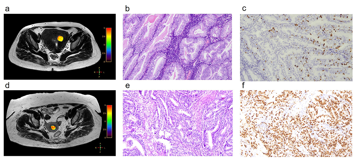
This prospective study was approved by the institutional review board and informed consent were obtained. Between January 2018 and December 2018, 54 consecutive patients suspected of endometrial lesions underwent APTw and IVIM (b values 0, 10, 20, 50, 100, 200, 400 and 800) imaging on a 3T MR scanner (Ingenia 3.0T CX; Philips Healthcare, Best, the Netherlands) equipped with dual radiofrequency (RF) transmission subsystem. 3D turbo spin echo (TSE) sequence was used to acquire APT images, and the APT values were calculated based on asymmetry of z-spectrum, in which saturation RF’s with duration of 2s were applied at ±3.5 ppm with water frequency set at 0 ppm. B0 field maps, generated by the build-in mDIXION TSE sequence, were used for APT calculation correction. The diffusion images for IVIM were acquired with EPI sequence and with fat saturation pulses applied. Least square fitting algorithm was used to calculate the bi-exponential parameters (Dt, D*, f) for IVIM. Images were independently measured by two radiologists on 22 postoperative pathological confirmed of type I endometrial carcinoma lesions with Ki-67 labelling index as well as IVIM-derived parameters. Inter-observer inter-class correlation coefficient (ICC) was computed. Student's t-test and Mann-Whitney U test were used to compare the differences of APT values and IVIM-derived parameters between different histological grades and Ki-67 proliferation groups. Receiver operating characteristic analysis was performed to computationally determine the feasible threshold value, sensitivity and specificity. Pearson’s correlation analysis was performed between the APT values, IVIM-derived parameters and Ki-67 labeling index.RESULTS
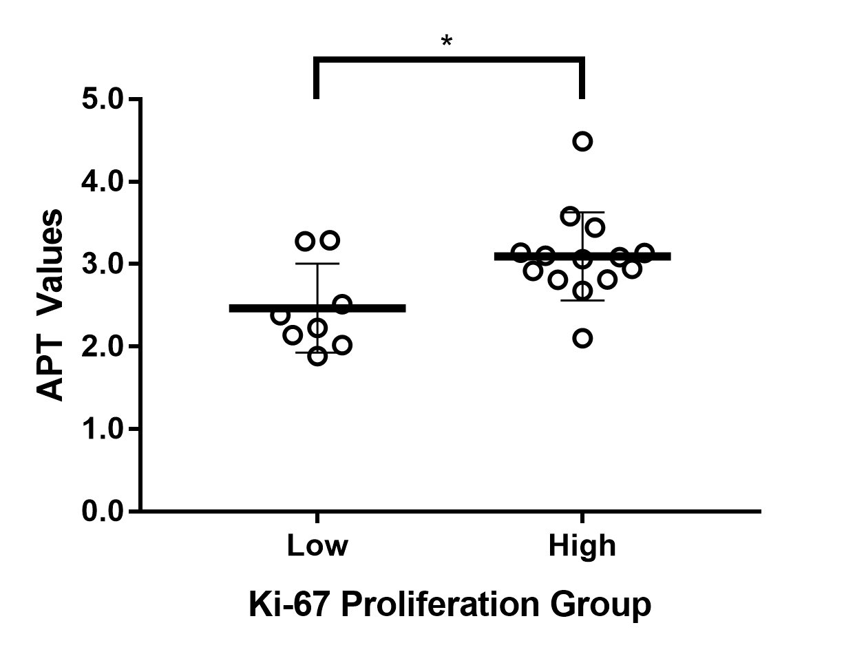
The APT values (in unit of percentage) and Dt, D*, f of all type I endometrial carcinoma were 2.865 ± 0.129, 0.677 ± 0.027 x 10⁻³mm2/s, 31.801 ± 11.492 x 10⁻³mm2/s, 0.179 ± 0.050 with inter-observer ICC 0.996 (95%CI 0.989-0.998), 0.850 (95%CI 0.611-0.942), 0.956 (95%CI 0.885-0.983), 0.995 (95%CI 0.988-0.998) respectively. APT values of Ki-67 low-proliferation group (<30%, n=8) were 2.466 ± 0.191, significantly lower than high-proliferation group (>30%, n=14) with APT values of 3.093 ± 0.142 (p=0.016) (Figure 1, 2). Area under the curve was 0.768. The feasible threshold value was determined as 2.595 with a sensitivity of 92.9% and specificity of 75%. The APT values of type I endometrial carcinoma were moderately positively correlated with Ki- 67 labeling index (r = 0.583, p=0.004). There were no significant difference of Dt (p=0.843), D* (p=0.262), f (p=0.553) between low-proliferation group and high-proliferation group. No correlation was found between IVIM-derived parameters and Ki-67 labeling index (Dt, p=0.717; D* p=0.151; f, p=0.153).DISCUSSION
The present study demonstrates the first attempt of 3D TSE APTw MR imaging for endometrial carcinoma with excellent inter-observer measurement agreement. Our study revealed that the APT values were positively correlated with Ki-67 labelling index, which is consistent with the results of APTw imaging of brain tumors including gliomas and meningioma.7,8 Compared with the previous 2D APT sequence,6 3D spin-echo APT sequence on uterus allowed to cover the whole volume of the tumor during scanning and select the precise slice showing the maximum area during the measuring. In this study, the saturation RF pulses of the APT sequence was maintained for two seconds by interleaving two RF transmitters, and the resulting APTw signal value corresponds to this particular B1 setting (labelling time and strength).CONCLUSION
3D TSE APTw imaging is feasible in type I endometrial carcinoma. APT values positively moderately correlates with Ki-67 proliferation status. However, no correlation was found between IVIM-derived parameters and Ki-67 labeling index.Acknowledgements
NONEReferences
1. Ferlay J, Soerjomataram I, Dikshit R, et al. Cancer incidence and mortality worldwide: sources, methods and major patterns in GLOBOCAN 2012. Int J Cancer. 2015;136(5):E359-386.
2. Murali R, Delair DF, Bean SM, et al. Evolving Roles of Histologic Evaluation and Molecular/Genomic Profiling in the Management of Endometrial Cancer. J Natl Compr Canc Netw. 2018;16(2):201-209.
3. Gulseren V, Kocaer M, Ozdemir IA, et al. Do estrogen, progesterone, P53 and Ki67 receptor ratios determined from curettage materials in endometrioid-type endometrial carcinoma predict lymph node metastasis? Curr Probl Cancer. 2019;10.1016.
4. Liu J, Wan Y, Wang Z, et al. Perfusion and diffusion characteristics of endometrial malignancy based on intravoxel incoherent motion MRI at 3.0 T: comparison with normal endometrium. Acta Radiol. 2016;57(9):1140-1148.
5. Kamimura K, Nakajo M, Yoneyama T, et al. Amide proton transfer imaging of tumors: theory, clinical applications, pitfalls, and future directions. Jpn J Radiol. 2019;37(2):109-116.
6. Takayama Y, Nishie A, Togao O, et al. Amide Proton Transfer MR Imaging of Endometrioid Endometrial Adenocarcinoma: Association with Histologic Grade. Radiology. 2018;286(3):909-917.
7. Suh CH, Park JE, Jung SC, et al. Amide proton transfer-weighted MRI in distinguishing high- and low-grade gliomas: a systematic review and meta-analysis. Neuroradiology. 2019;61(5):525-534.
8. Yu H, Wen X, Wu P, et al. Can amide proton transfer-weighted imaging differentiate tumor grade and predict Ki-67 proliferation status of meningioma? Eur Radiol. 2019;29(10):5298-5306.
Figures