0563
Quad-contrast imaging with quantitative relaxation maps for clinical neuro-evaluation1Electrical and Computer Engineering, Seoul National University, Seoul, Korea, Republic of, 2Biomedical Engineering, Hankuk University of Foreign Studies, Yongin, Korea, Republic of
Synopsis
A 2D quad-contrast sequence is developed to generate four different contrast images commonly used in the clinical patient scan (PDw, T2w, T1w, and FLAIR) and two quantitative maps (T1- and T2- maps) in 4:14 of scan time. The proposed sequence provides comparable tissue contrasts to that of conventional sequences. In particular, native FLAIR contrast is acquired, which does not display hyperintense brain surface that arises from partial volume error during parameter mapping. Psuedo-contrast images with different TE and TI are also synthesized utilizing the quantitative maps, analogous to MAGiC.
Introduction
Fast image acquisition is of great importance for patient comfort and reduction of motion artifacts, particularly for patients with movement disorders such as Parkinson’s disease. A routine clinical scan includes 2D images of various contrasts (i.e. PDw, T2w, T1w, and FLAIR). For clinically feasible scan time, images are often restricted to thick slices (~5mm), and small number of slices (~24). Recently MAGiC, a synthetic MR imaging scheme1 that produces quantitative T1 and T2 maps and synthesize the four different contrast images with ~6min scan time was proposed. However, several studies reported degraded FLAIR images due to artifacts such as hyperintense brain surface2–4. In this study, a 2D quad-contrast sequence that provides the four different contrast images (PDw, T2w, T1w, and FLAIR) and two different quantitative maps (T1- and T2- maps) of the whole brain (4mm, 32 slices) in 4:14 of scan time is proposed.Methods
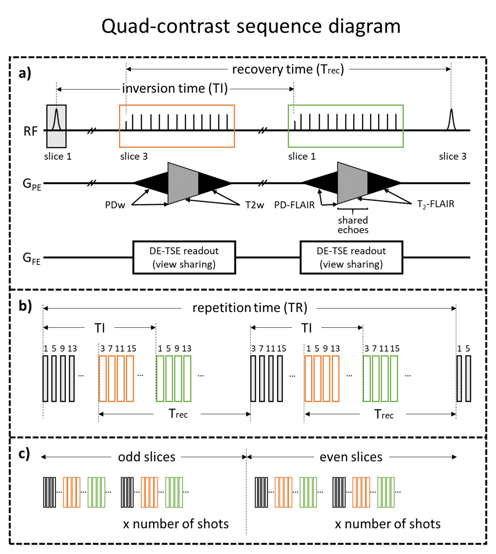
[Quad-contrast sequence] A quad-contrast sequence (Fig. 1) was developed to simultaneously acquire PDw, T2w, PD-FLAIR, and T2-FLAIR contrasts. Three methods were used to reduce total acquisition time.1) Interleaved contrast acquisition: Non-IR-prepped acquisition blocks for PDw and T2w were located between the inversion RF and IR-prepped acquisition blocks for PD-FLAIR and T2-FLAIR. An interleaved multi-slice acquisition scheme was applied (Fig. 1b).
2) View sharing: To reduce acquisition block duration, a view-shared turbo spin-echo (TSE) readout was incorporated. The effective echo train length (ETL) was 8 for each contrast, and 4 echoes were shared between the PDw and T2w; and PD-FLAIR and T2-FLAIR contrasts (Fig. 1a).
3) Parallel acquisition: GRAPPA acceleration factor 4 was used.
Concatenation of 2 was used to enable larger inversion thickness, in order to minimize inflow artifacts (Fig. 1c). Compared with conventional scans with same scan parameters, the total acquisition was 2.4 times accelerated.
[Quantitative mapping and image synthesis] T1w images were generated using the following equation 5.
$$ T1w=\ \frac{PD_{FLAIR}*PD}{{PD_{FLAIR}}^2+{PD}^2} $$
Quantitative T1 and T2 maps were generated by conducting a non-linear least-square fit of the four acquired contrast images to the signal model below.
$$S_{PD}=A\left(1-e^{-\frac{T_{recovery}}{T_1}}\right)$$
$$S_{T_2}=A\left(1-e^{-\frac{T_{recovery}}{T_1}}\right)e^{-ΔTE/T_2}$$
$$S_{PD-FLAIR}=A(1-2e^{-TI/T_1}+e^{-T_{recovery}/T_1}e^{-TI/T_1})\ $$
$$S_{T_2-FLAIR}=A(1-2e^{-TI/T_1}+e^{-T_{recovery}/T_1}e^{-TI/T_1})e^{-ΔTE/T_2}$$
Utilizing the T1 and T2 maps, pseudo-contrast images of IR-prepped and T2w with various TI and TE values were synthesized, analogous to MAGiC. The same exponential model as MAGiC was used for synthesis1.
Pseudo-FLAIR image with TR/TI/TE=9000/2500/90ms was also synthesized and compared with the conventional- and quad-contrast FLAIR.
[Data acquisition] Quad-contrast images were acquired from a healthy subject (IRB approved; 3T, Trio, Siemens). For comparison, conventional PDw, T1w, T2w, and FLAIR contrasts were acquired. The following sequence parameters were common for all acquisitions: FOV=256×256mm2, voxel size=1×1mm2, slice thickness=4mm, number of slices=32, slice gap=25%. Except for the T1w contrast image acquisition, GRAPPA acceleration factor=4, and number of ACS lines=30 was used. For the quad-contrast sequence, TR/TI/short TE/long TE=10500/2100/9.1/109ms and total scan time 4:14 was used. Conventional sequences were acquired with the following parameters. PDw: TSE readout, ETL=12, TR/TE=7000/9.4ms, scan time=0:58. T2w: TSE readout, ETL=12 TR/TE=9000/113ms, scan time=1:14. FLAIR: TSE readout, ETL=12, TR/TE/TI=9000/84/2500ms, scan time=2:26. T1w: GRE readout, TR/TE=250/2.5ms, flip angle=70°, scan time=2:10. Larger ETL than the quad-contrast sequence (=12) was used to match the effective TE for T2w while maintaining readout bandwidth.
Results
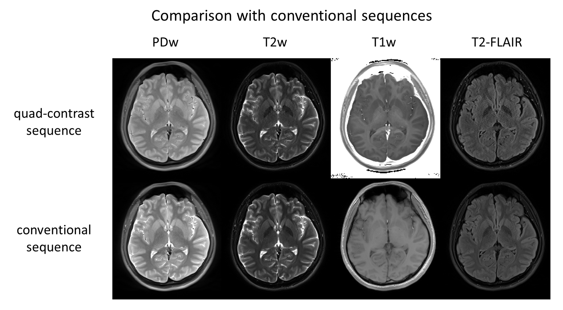
The total scan time of 6:48 for conventional scans was reduced to 4:14 using the quad-contrast sequence, despite of larger ETL in conventional scans. The four routine contrasts acquired by the proposed sequence and the conventional sequences show comparable tissue contrast (Fig. 2).The T1 and T2 maps, and the synthesized images are presented (Fig. 3, video). The T1 and T2 values agree with the literature values (WM: T1=832±10ms, T2=79.6±0.6ms, GM: T1=1331±13ms, T2=110±2ms)6.
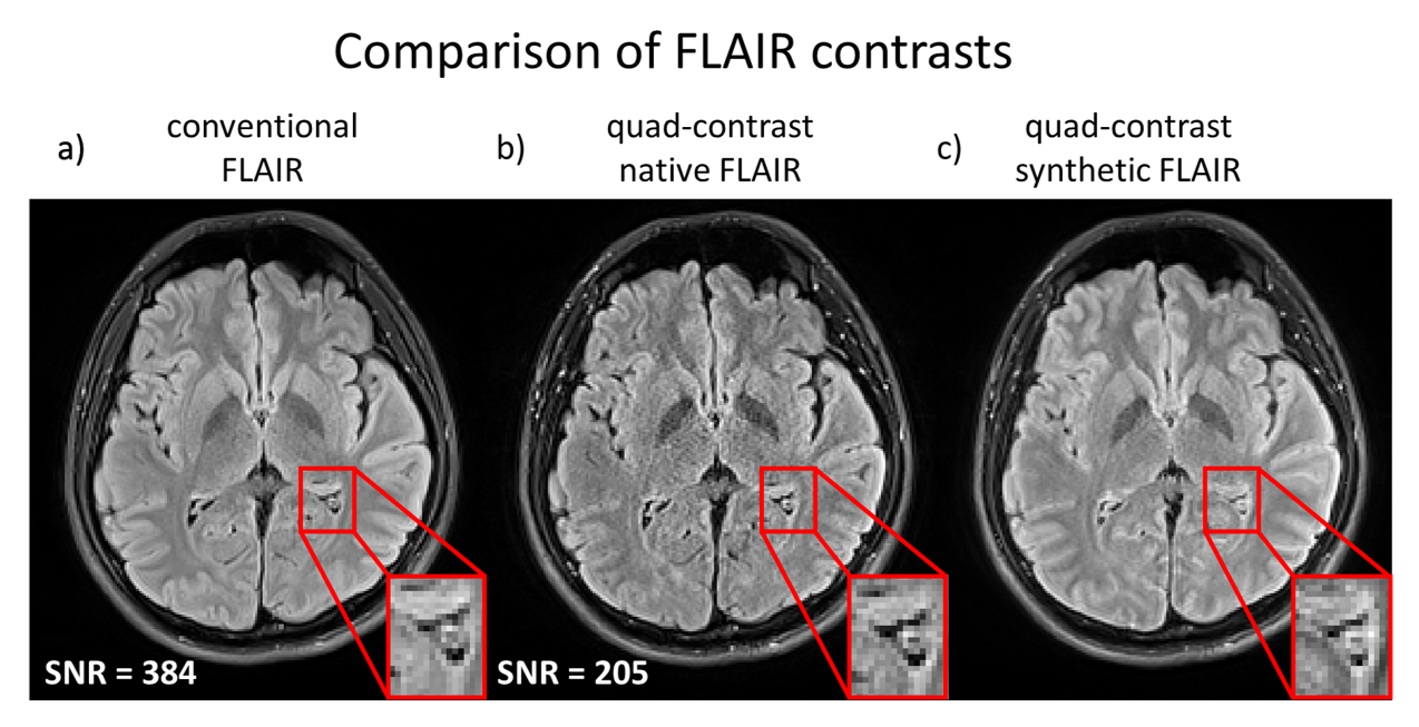
The FLAIR images from three different methods (conventional, quad-contrast, and quad-contrast synthetic) are compared (Fig. 4). The quad-contrast FLAIR has relatively lower SNR (=205) than conventional FLAIR (=384). The synthetic FLAIR appears less noisy (SNR not calculated), comparable to conventional FLAIR. Quad-contrast synthetic FLAIR shows hyperintense brain surface, while the natively acquired contrasts do not (Fig. 4, red box).
Conclusion and discussion
In this study, a 2D quad-contrast sequence was developed to reduce scan time and generate PDw, T2w, T1w and FLAIR contrast images in a single scan. Utilizing an acceleration factor of 4, the total acquisition time of the four contrasts was reduced to ~4min.Because the acquired contrasts are intrinsically aligned together, no further registration is necessary. This may be advantageous in the case where alignment between the images is important.
The FLAIR contrast is natively acquired in the proposed sequence. Hence, the quad-contrast FLAIR contrast is not degraded by hyperintense brain surface, which presumably arises from partial volume errors during parameter mapping7. In contrast, the synthesized FLAIR displays hyperintense brain surface, similar to FLAIR of MAGiC4. The relatively lower SNR of the quad-contrast FLAIR results from the short TI and long TE used. In case SNR of FLAIR is important, synthetic FLAIR can be used. Because T1- and T2- maps are fitted using all four contrasts, the noise level of the synthetic FLAIR is not limited to that of native quad-contrast FLAIR.
In conclusion, the proposed quad-contrast sequence reduces the routine clinical MR scan time to 4:14, with whole-brain coverage (32 slices) and quantitative T1 and T2 maps.
Acknowledgements
This research was supported by the National Research Foundation of Korea(NRF) funded by the MSIT(NRF-2018R1A4A1025891), and the Brain Research Program through the National Research Foundation of Korea(NRF) funded by the Ministry of Science, ICT & Future Planning (NRF-2019M3C7A1031994).References
1. Warntjes, J. B. M., Leinhard, D. O., West, J. & Lundberg, P. Rapid magnetic resonance quantification on the brain: Optimization for clinical usage. Magnet Reson Med 60, 320–329 (2008).
2. Tanenbaum, L. N. et al. Synthetic MRI for Clinical Neuroimaging: Results of the Magnetic Resonance Image Compilation (MAGiC) Prospective, Multicenter, Multireader Trial. Am J Neuroradiol 38, 1103–1110 (2017).
3. Granberg, T. et al. Clinical Feasibility of Synthetic MRI in Multiple Sclerosis: A Diagnostic and Volumetric Validation Study. Am J Neuroradiol 37, 1023–1029 (2016).
4. Hagiwara, A. et al. Synthetic MRI in the Detection of Multiple Sclerosis Plaques. Am J Neuroradiol 38, 257–263 (2017).
5. Marques, J. P. et al. MP2RAGE, a self bias-field corrected sequence for improved segmentation and T1-mapping at high field. Neuroimage 49, 1271–1281 (2010).
6. Wansapura, J. P., Holland, S. K., Dunn, S. R. & Ball, W. S. NMR relaxation times in the human brain at 3.0 tesla. J Magn Reson Imaging 9, 531–538 (1999).
7. Hagiwara, A. et al. SyMRI of the Brain. Invest Radiol 52, 647–657 (2017).
Figures