0561
MP-RAVE: IR-Prepared T1-Weighted Radial Stack-of-Stars 3D GRE Imaging with Retrospective Motion Correction
Eddy Solomon1, Houchun H. Hu2, Kai Tobias Block1, Daniel K. Sodickson1, and Hersh Chandarana1
1Radiology, New York University School of Medicine, New York, NY, United States, 2Radiology, Nationwide Children's Hospital, Columbus, OH, United States
1Radiology, New York University School of Medicine, New York, NY, United States, 2Radiology, Nationwide Children's Hospital, Columbus, OH, United States
Synopsis
Inversion-recovery 3D T1 gradient echo sequences are commonly used in brain examinations for their excellent gray-/white-matter contrast. However, prominent motion artifacts can arise during lengthy Cartesian k-space sampling (typically 5-7 minutes) if the patient is not able to hold still, as is often the case for pediatric or elderly patients. Here, we present an alternative based on radial stack-of-stars imaging and show that comparable image contrast can be achieved, with lower sensitivity to head motion. Moreover, we demonstrate how the radial acquisition scheme can be utilized for additional retrospective motion correction to further improve robustness without increasing acquisition time.
INTRODUCTION
Acquisition of T1-weighted images is an essential component of routine brain MRI. Three-dimensional Magnetization-Prepared Rapidly Acquired Gradient Echo (MP-RAGE) is a commonly used sequence that provides excellent gray-/white-matter contrast at high resolution (1). However, like any sequence using Cartesian phase-encoding, MP-RAGE is prone to ghosting-like motion artifacts that can impair the diagnostic value of the images if the patient cannot hold still during the examination, which poses a particular problem in pediatric patients or elderly patients with tremor. As a result, scans must be repeated or, in the case of pediatric imaging, must be performed during sedation. Recently, it has been demonstrated that non-Cartesian k-space acquisition schemes, such as radial sampling, provide higher inherent robustness to motion (2), and it has been shown that using such sequences provides value for imaging of the head and brain (3,4). The aim of this work was to develop a variant of the MR-RAGE sequence with radial sampling of k-space, named MP-RAVE, and to evaluate its advantages in healthy human volunteers during head motion.METHODS
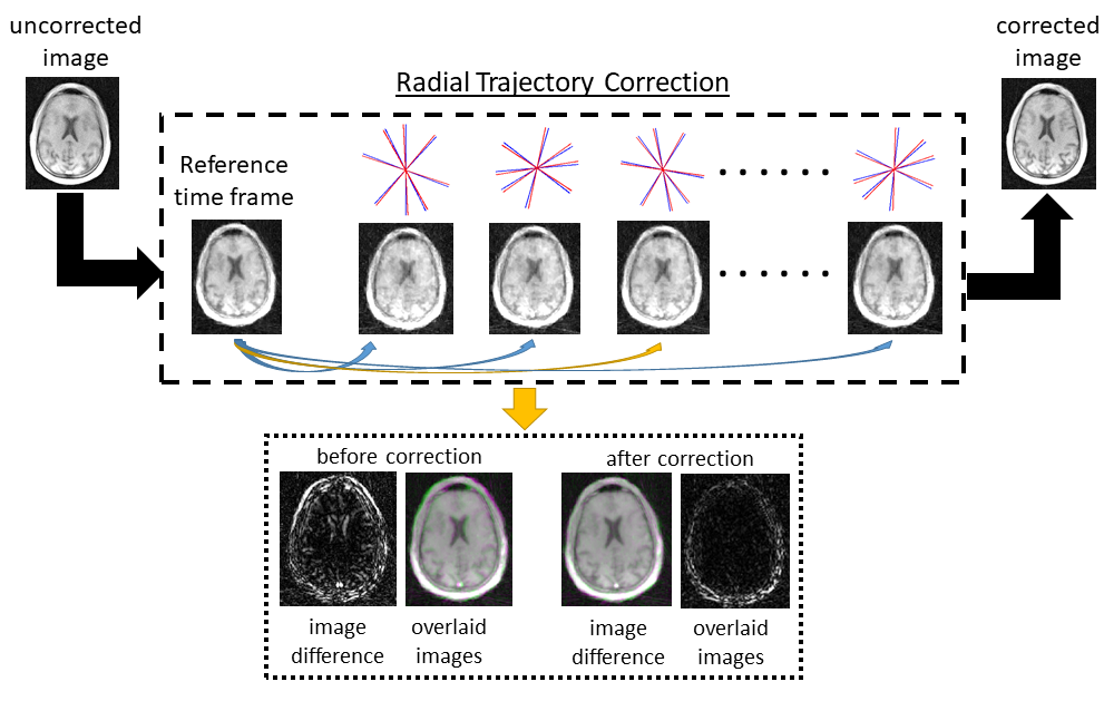
All data were acquired on a 3T Prisma system (Siemens Healthcare, Erlangen, Germany) using a 20-channel head coil array. The study protocol included a conventional T1-weighted MP-RAGE sequence and the radial counterpart MP-RAVE, which has been derived from a previously described radial stack-of-stars 3D GRE sequence (RAVE) (5) by integrating an inversion recovery pulse that is played out once for every stack of radial views with same angle. Common imaging parameters were TR/TI/TE=2200/900/3.2ms, 1.0mm spatial resolution, no fat suppression, and 224 slices. For MP-RAVE, 400 radial views were acquired and a centric acquisition was used along the slice direction. Images were reconstructed by standard gridding (NUFFT). The scan time of MP-RAGE was 4min with 2x GRAPPA acceleration whereas the initial version of MP-RAVE did not use parallel imaging, resulting in double the acquisition time. Both methods were tested in six healthy volunteers.Motion-correction algorithm: While radial data acquisition is inherently more robust to motion and does not show the MR-typical ghosting artifacts, it is not completely immune to motion. However, due to the geometry of the radial trajectory, which covers k-space center with each radial view, it is possible to reconstruct time-resolved subframes from the acquisition using an iterative reconstruction technique like GRASP (6), which utilizes compressed sensing with a temporal total-variation constraint to calculate images from the highly undersampling subframe data. This series of dynamic images can then be used for estimating intra-acquisition head motion via image-based registration, specifically rotation and translation. Once the motion dynamics has been estimated, the k-space trajectory can then be corrected for rigid motion and a final full-resolution motion-corrected image can be reconstructed. To demonstrate this possibility, a volunteer scan was performed under head motion using the RAVE sequence. Each subframe “navigator” image consisted of four radial views, sensing motion every ~0.9 seconds. The motion-correction algorithm scheme is illustrated in Figure 4.
RESULTS AND DISCUSSION
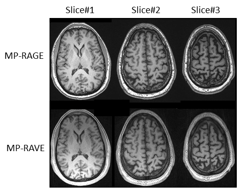
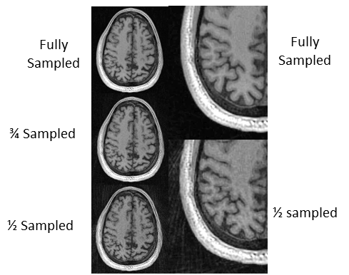
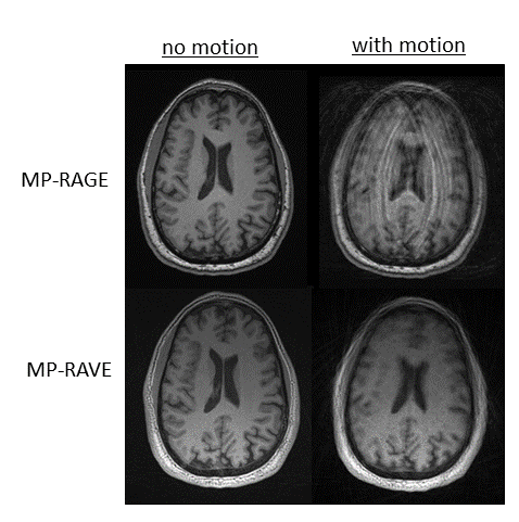
T1-weighted anatomical brain images were acquired by conventional MP-RAGE and the proposed MP-RAVE sequence, showing comparable T1-weighted contrast and image quality (Fig. 1). As a direction to further reduce the relatively longer scan time of MP-RAVE, reconstruction of MP-RAVE data with different undersampling factors is shown in Fig. 2, demonstrating that acceptable image quality is achieved even at 50% undersampling. The higher motion robustness achieved with MP-RAVE compared to MP-RAGE in presence of continuous head motion is shown in Figure 3. None of the sequences are completely immune to motion. However, while MP-RAVE images are somewhat blurred, MP-RAGE images show prominent ghosting artifacts along the phase-encode direction that can potentially obscure pathologies. The latter arise because the phase-encoding scheme is disturbed by motion-induced phase modulation, which is avoided with radial sampling. In addition, MP-RAVE’s higher robustness may also be attributed to oversampling of the k-space center, which has a motion-averaging effect. To overcome residual motion blurring with MP-RAVE, we have tested a motion-correction algorithm (Fig. 4) in a volunteer (Fig. 5). It can be seen that the correction algorithm is able to reduce the amount of blurring and can recover anatomical information in various regions of the brain, allowing for better delineation of specific structures (see yellow arrows).CONCLUSION
This work describes a novel IR-prepared T1-weighted 3D GRE sequence with radial stack-of-stars sampling (MP-RAVE) as a potential alternative to the widely used MP-RAGE sequence. It has been shown that MP-RAVE can generate comparable image quality and contrast to conventional MP-RAGE but with lower sensitivity to head motion. To reduce the scan time, undersampling techniques can be utilized, such as iterative constrained reconstruction. Moreover, to further improve the robustness to motion, retrospective motion correction can be applied without adding to scan time or requiring additional navigator information.Acknowledgements
We acknowledge support from NIH grant P41 EB0171813 and R01 5R01EB018308.References
- Brant-Zawadzki M, Gillan GD, Nitz WR. MP RAGE: a three-dimensional, T1-weighted, gradient-echo sequence--initial experience in the brain. Radiology. 1992 Mar;182(3):769-75.
- Chandarana H, Feng L, Block TK, Rosenkrantz AB, Lim RP, Babb JS, Sodickson DK, Otazo R. Free-breathing contrast-enhanced multiphase MRI of the liver using a combination of compressed sensing, parallel imaging, and golden-angle radial sampling. Invest Radiol. 2013 Jan;48(1):10-6.
- Hu HH, Benkert T, Jones JY, McAllister AS, Rusin JA, Krishnamurthy R, Block KT. 3D T1-weighted contrast-enhanced brain MRI in children using a fat-suppressed golden angle radial acquisition: an alternative to Cartesian inversion-recovery imaging. Clin Imaging. 2019 May - Jun;55:112-118
- Kecskemeti S, Samsonov A, Velikina J, Field AS, Turski P, Rowley H, Lainhart JE, Alexander AL. Robust Motion Correction Strategy for Structural MRI in Unsedated Children Demonstrated with Three-dimensional Radial MPnRAGE. Radiology. 2018 Nov;289(2):509-516.
- Block KT, Chandarana H, Milla S, Bruno M, Mulholland T, Fatterpekar G, Hagiwara M, Grimm R, Geppert C, Kiefer B, Sodickson DK. Towards Routine Clinical Use of Radial Stack-of-Stars 3D Gradient-Echo Sequences for Reducing Motion Sensitivity. Journal of the Korean Society of Magnetic Resonance in Medicine 18 (2), 87-106.
- Feng L, Axel L, Chandarana H, Block KT, Sodickson DK, Otazo R. XD-GRASP: Golden-angle radial MRI with reconstruction of extra motion-state dimensions using compressed sensing. Magn Reson Med. 2016 Feb;75(2):775-88.
Figures
Figure
1. Comparison
of image contrast and quality for three representative brain slices acquired
with MP-RAGE and MP-RAVE
Figure
2. Evaluating
the MP-RAVE reconstruction of a single representative slice under different undersampling. As
can be appreciated in the zoomed images on the right, image quality
and contrast is acceptable even at 50% undersampling.
Figure
3. Behavior
of MP-RAGE and MP-RAVE to head motion. While MP-RAVE images are somewhat
blurred, MP-RAGE images show prominent “ghosting”-like motion artifacts along
the phase-encode direction.
Figure
4. Motion
correction scheme: GRASP is used to reconstruct the uncorrected data into 64
“navigator” frames, each calculated from 4 radial views. Each subframe
image is then registered to a predefined ‘reference frame’ and the trajectory
is corrected accordingly (from blue to red).
The corrected k-space trajectory is used to reconstruct one final
full-resolution motion-corrected image.
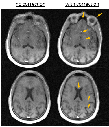
Figure
5. Demonstration
the described
motion-correction scheme for RAVE data acquired during head motion. It can be
seen that the correction algorithm reduces motion blurring, allowing better
delineation of specific anatomical structures.