0560
3D Flow Compensated Interleaved EPI with a Centric Reordering Scheme for Fast High-Resolution Susceptibility-Weighted Imaging1Siemens Shenzhen Magnetic Resonance Ltd., Shenzhen, China
Synopsis
In this study, we implemented a novel centric reordering scheme in a partial flow compensated 3D-iEPI to further reduce the flow effect and assessed its feasibility for a fast high-resolution SWI application. By properly dividing one interleave into two EPI shots sequentially acquired with opposite phase encoding gradient polarities and overlapping one line in the interleave center, we demonstrated that the partial flow compensated 3D-iEPI with such centric reordering scheme can significantly reduce the arterial contamination and obtain comparable contrast and image quality to 3D-GRE, whilst enjoying an approximate 2-fold reduction in acquisition time.
INTRODUCTION
Susceptibility-weighted imaging (SWI) can provide enhanced image contrast for tissues with susceptibility difference by utilizing the phase information of the acquired MR signals, which has been widely used in a variety of clinical setting for evaluations of iron-laden tissues, venous blood vessel, etc1. A conventional technique to acquire SWI images is the fully flow compensated 3D-GRE sequence, which usually requires long scan time. Furthermore, imaging at a high resolution can reduce dephasing across the voxel and allow for a better contrast with longer TE, which is even more time consuming2. Therefore, a 3D interleaved-EPI sequence (3D-iEPI) has been proposed to achieve faster SWI with improved SNR efficiency3-5. By using a short EPI train length, the typical EPI related artifacts (distortion and blurring) are limited and comparable to GRE5. However, the flow effect in EPI is much more complicated than that in GRE. Several studies have analyzed the flow properties in EPI sequence, and a centric reordering scheme has been proposed to reduce flow artifacts in phase encoding direction by reducing the phase encodings before the center of k-space6-7. In our previous work, we demonstrated the feasibility of a partial flow compensated 3D-iEPI for fast SWI8. This time, we implemented a novel centric reordering scheme in that 3D-iEPI sequence to further alleviate the flow effect. It shows that the proposed technique can significantly mitigate the artery contamination in SWI images and provide a better vessel depiction.METHODS
In the proposed centric reordering scheme (Fig. 1), two shots of EPI acquisitions with opposite phase encoding gradient polarities were combined to provide a full interleave coverage. Compared with the previous studies6-7, the center line of the interleave was sequentially acquired in both shots to provide higher SNR and reduce the motion influence. With fully flow compensated in all directions, the repeated acquisitions of the central line can further improve the efficiency of flow compensation. The TE shifting strategy was also adapted for the centric reordering, which can smooth the phase discontinuities from off-resonance effects9. In addition, the prephaser and partition encoding gradients were moved next to the central echo for further reduction of flow effect. All measurements were performed on a commercial 1.5T scanner (MAGNETOM Aera, Siemens Healthcare GmbH, Erlangen, Germany) equipped with a 20-channel head/neck coil. Experimental data was obtained from a healthy volunteer using standard 3D-GRE, a prototype flow compensated 3D-iEPI with linear reordering and centric reordering respectively. The imaging parameters were shown in Table 1. The TR and flip angle were tuned in 3D-iEPI to match the background contrast in 3D-GRE. After data collection, both 3D-GRE and 3D-iEPI data was SWI-processed in the standard way.RESULTS
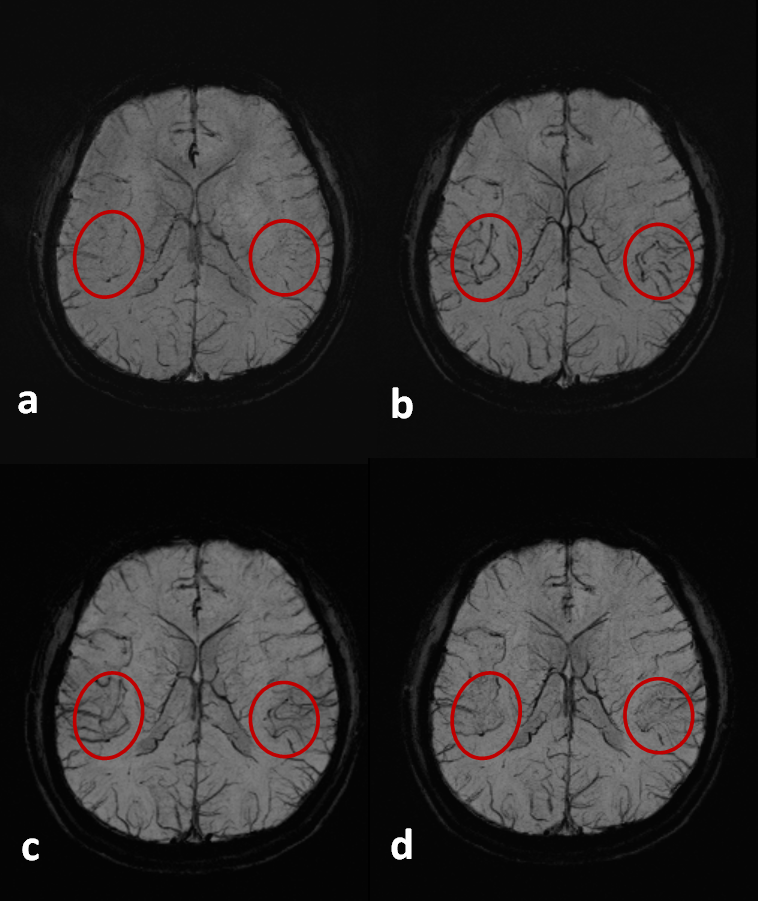
The comparison between the flow compensated 3D-iEPI, with linear and centric reordering schemes, and the conventional 3D-GRE sequence were shown in the Fig. 2. With flow compensation, both 3D-GRE (Fig.2a) and 3D-iEPI (Fig. 2c, Fig. 2d) provide similar image quality with clear vessel depiction. By combining the centric reordering and partial flow compensation in 3D-iEPI, the flow induced arterial contamination can be further mitigated and the vessels will be better depicted. Moreover, more tiny vessels are shown in images for 3D-iEPI (Fig.2c, Fig.2d), which may be due to the increased TE for high frequency signals. Our results show that, the improved 3D-iEPI allows approximately 2-fold reduction in the scan time, with comparable image quality and contrast to the conventional 3D-GRE.DISCUSSION
While the partially flow compensated 3D-iEPI with a centric reordering scheme can provide better suppression of artery signals, the centric reordering scheme inevitably leads to lower SNR efficiency compared to the linear reordering, due to less echoes acquired per shot with the same EPI factor used. However, by sampling the central line of each interleave twice, both SNR and flow effect can be improved. In addition, the motion influence can be mitigated by the sequential acquisition of two partial shots and will benefit the following data splicing. The two shot acquisitions with opposite phase-encoding polarities may also be corrected by a standard reverse-gradients method10, which allows more flexible selection for the echo train length. Eventually, from the in-vivo results, the distortion and blurring artifacts in 3D-iEPI are not noticeable due to the short echo train used.CONCLUSION
Although the flow effect is not fully compensated for all echoes in 3D-iEPI, this study demonstrated that the centric reordering scheme in the 3D-iEPI sequence can significantly reduce the remaining arterial signals and improve the vessel depiction. Furthermore, by taking advantage of high scan efficiency of 3D-iEPI, the proposed method can provide comparable contrast and image quality to 3D-GRE and enjoy an approximate 2-fold reduction in acquisition time, which is promising for high resolution SWI applications.Acknowledgements
No acknowledgement found.References
1. Haacke EM, et al. Susceptibility weighted imaging (SWI), Magn Reson Med. 2004;52:612-618.
2. Akbudak E, et al. Conturo TE. Contrast-agent phase effects: an experimental system for analysis of susceptibility, concentration, and bolus input function kinetics. Magn Reson Med Dec; 1997 38(6):990-1002.
3. Sati P, et al. Ultra-Fast Acquisition of High-Resolution Susceptibility-Weighted-Imaging at 3T. Proc Intl Soc Mag Reson Med. 2011; 19:2364.
4. Poser BA, et al. Three dimensional echo-plannar imaging at 7 Tesla. Neuroimage. 2010; 51:261-266.
5. Zwanenburg JJ, et al. Fast high resolution whole brain T2* weighted imaging using echo planar imaging at 7T. Neuroimage. 2011; 56:1902-1907.
6. Luk Pat GT, et al. Reducing flow artifacts in echo-planar imaging. Magn Reson Med 1997; 37: 436-447.
7. Beck G, et al. Reducing Oblique Flow Effects in Interleaved EPI With a Centric Reordering Technique. Magn Reson Med 2001; 45: 623-629.
8. Liu W, et al. 3D Flow Compensated Interleaved EPI for a Fast High-Resolution Susceptibility-Weighted Imaging at 1.5T. Proc. Intl. Soc. Mag. Reason. Med 2019; 27: 3326.
9. Feinberg DA, et al. Gradient-echo shifting in fast MRI techniques (GRASE imaging) for correction of field inhomogeneity errors and chemical shift. J Magn Reson 1992; 97:177-183.
10. Gallichann D, et al. Reducing Distortions in Diffusion-Weighted Echo Planar Imaging With a Dual Echo Blip-Reversed Seqeucne. Magn Reson Med 2010; 64: 382-390.
Figures
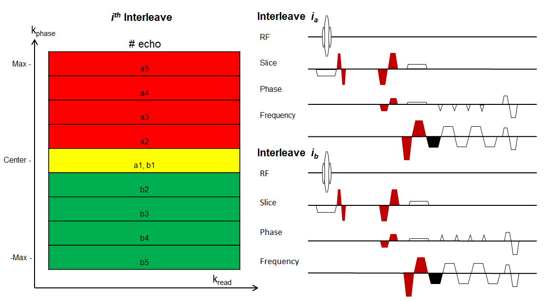
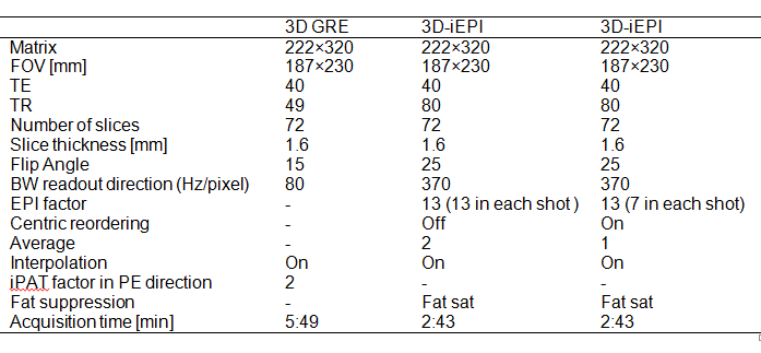
Table 1. Imaging parameters for axial SWI protocols including 3D-GRE, flow compensated 3D-iEPI with linear reordering and centric reordering respectively.