0493
Quantitative Multiple Quantum Filtered Sodium MRI and [18F]-FET-PET: Complementary Imaging Techniques for the Study of Cerebral Gliomas
Wieland A Worthoff1, Aliaksandra Shymanskaya2, Karl-Josef Langen1,3,4, and N. Jon Shah1,2,4,5
1Institute of Neuroscience and Medicine - 4, Forschungszentrum Jülich GmbH, Jülich, Germany, 2Institute of Neuroscience and Medicine - 11, Forschungszentrum Jülich GmbH, Jülich, Germany, 3Department of Nuclear Medicine, RWTH Aachen University, Aachen, Germany, 4Section JARA-Brain, Jülich-Aachen Research Alliance, Aachen, Germany, 5Department of Neurology, RWTH Aachen University, Aachen, Germany
1Institute of Neuroscience and Medicine - 4, Forschungszentrum Jülich GmbH, Jülich, Germany, 2Institute of Neuroscience and Medicine - 11, Forschungszentrum Jülich GmbH, Jülich, Germany, 3Department of Nuclear Medicine, RWTH Aachen University, Aachen, Germany, 4Section JARA-Brain, Jülich-Aachen Research Alliance, Aachen, Germany, 5Department of Neurology, RWTH Aachen University, Aachen, Germany
Synopsis
A cohort of patients with untreated cerebral gliomas underwent consecutive [18F]-FET-PET and sodium MRI exams. It is shown that quantitative results from multiple quantum filtered sodium MRI using the enhanced SISTINA sequence offer access to the metabolic properties of tumours beyond what is observed by [18F]-FET-PET alone, thus presenting the potential to serve as an additional marker in tumour diagnostics.
Introduction
Conventional MRI is a common indicator for the localisation of glioblastomas and is vital in the process of therapy planning. Nevertheless, it is known that tumorous tissue can extend beyond the region seen in such scans1. O-(2-[18F]fluoroethyl)-L-tyrosine ([18F]-FET)-PET is capable of revealing such regions, although not sufficiently enough to completely asses the properties of the tumour. Multiple quantum filtered sodium MRI is capable of indicating the status of the isocitrate dehydrogenase (IDH) mutation2 and possibly further information relating to tumour metabolism. Here we report on additional quantitative information, such as relaxation rates, sodium concentrations, volume and molar fractions, and compare them with the information obtained from amino acid PET.Methods
Ten patients with untreated cerebral gliomas underwent sodium MRI using an enhanced SISTINA sequence3 and [18F]-FET-PET. Quantitative sodium parameters and [18F]-FET uptake in the tumours were compared. After biopsy or resection, histology and the IDH mutational status were determined neuropathologically. Patients were divided into two groups, discriminated by IDH mutational status. Informed consent was granted by all subjects.Sodium MRI
Full brain covering sodium MRI scans were performed on a 4T Siemens scanner with a dual tuned Na/H birdcage coil (Rapid Biomedical, Germany). Enhanced SISTINA was run with a preparatory time of 7ms, a mixing time of 0.04ms and a repetition time of 150ms, yielding a total acquisition time of 8min. Five radially sampled images were acquired after the first pulse, with a base resolution of 22 and a bandwidth of 1kHz/pixel, TE = {0.36; 1.75; 3.14; 4.53; 5.92}ms. A Cartesian readout with 10 mm isotropic resolution was acquired after the third pulse, consisting of 6 echoes sampled with a bandwidth of 120Hz/pixel at TEcart={5.92; 15.10; 25.32; 34.48; 43.64; 52.80}ms. Information on the relaxation of total, restricted and unrestricted sodium was obtained from all images and sodium quantitative parameters were derived. Comparing different quantitative parameters in the region of the gliomas with normal-appearing brain matter in the contralateral tissue (CLT) revealed metabolic changes in the tumorous region.
FET-PET
An ECAT EXACT HR+ scanner (Siemens Medical Systems, Inc.) was used in 3D mode (32 rings; axial field of view of 155mm) up to 50min after injection of approximately 200MBq [18F]-FET tracer to obtain dynamic PET information. The reconstructed dynamic dataset consisted of 16 time frames (5×1min; 5×3min; 6×5min). Three rotating line sources (68Ge/68Ga) were measured in transmission for attenuation correction. Random coincidences, scattered coincidences and dead time were corrected before the reconstruction of 63 images using the OSEM algorithm (16 subsets, 6 iterations). The image resolution is approximately4 5.5mm. Tumour-to-brain ratios (TBR) were calculated on summed [18F]-FET-PET images from 20-40min after injection: mean and maximal ROI value of the lesion were divided by the mean ROI value of normal brain tissue, where a maximum value exceeding a threshold of 1.6 was considered a positive finding.
Results
The mean fast relaxation rate T*2f in white matter is significantly reduced in the tumorous tissue compared to the CLT (with T*2f/ T*2f,CL = 0.64±0.15) in patients suffering from IDH mutated gliomas (IDHmut), while in IDH wildtype gliomas (IDHwt) T*2f remains largely unchanged (T*2f/ T*2f,CL = 0.97±0.20). The slow relaxation rate, T*2s, appears prolonged in both of these groups by approximately a factor of 1.4 compared to the CLT. The mean total sodium concentration is elevated in these gliomas, most dominantly in the IDHmut case, by a factor of 2±0.25 compared to 1.5±0.5 in the case of IDHwt. Mean pseudo intracellular sodium concentration is also increased in IDHwt (factor 1.2±0.21) compared to CLT, whereas it is decreased in IDHmut (factor 0.8±0.26). Mean intracellular molar and volume fractions are reduced significantly in the case of IDHmut (0.26±013 and 0.66±0.08 ). No obvious relationship between the distribution of sodium and [18F]-FET uptake is seen in these groups of subjects. Fig.1 shows enhanced SISTINA data and quantitative sodium parameters of one tumour patient.Conclusions
Quantitative sodium MRI can reveal characteristic metabolic information in brain tumours, making it a promising complement to [18F]-FET-PET. In particular, it is very sensitive to the IDH mutational statusAcknowledgements
The authors wish to express their sincere gratitude to Prof. Dr. Bernd Neumaier, Prof. Dr. Norbert Galldiks, Dr. Johannes Lindemeyer, Dr. Philipp Lohmann, Dr. Gabrielle Stoffels, Elke Bechholz, Petra Engels, Anita Köth and Claire Rick.References
- Lohmann P, Stavrinou P, Lipke K, Bauer EK, Ceccon G, Werner JM, Neumaier B, Fink GR, Shah NJ, Langen KJ, Galldiks N. FET PET reveals considerable spatial differences in tumour burden compared to conventional MRI in newly diagnosed glioblastoma. Eur J Nucl Med Mol Imaging 2019;46(3):591-602.
- Shymanskaya A, Worthoff WA, Stoffels G, Lindemeyer J, Neumaier B, Lohmann P, Galldiks N, Langen KJ, Shah NJ. Comparison of [(18)F]Fluoroethyltyrosine PET and Sodium MRI in Cerebral Gliomas: a Pilot Study. Mol Imaging Biol 2019.
- Worthoff WA, Shymanskaya A, Shah NJ. Relaxometry and quantification in simultaneously acquired single and triple quantum filtered sodium MRI. Magn Reson Med 2019;81(1):303-315.
- Herzog H, Tellmann L, Hocke C, Pietrzyk U, Casey ME, Kuwert T. NEMA NU2-2001 guided performance evaluation of four siemens ECAT PET scanners. Ieee T Nucl Sci 2004;51(5):2662-2669.
Figures
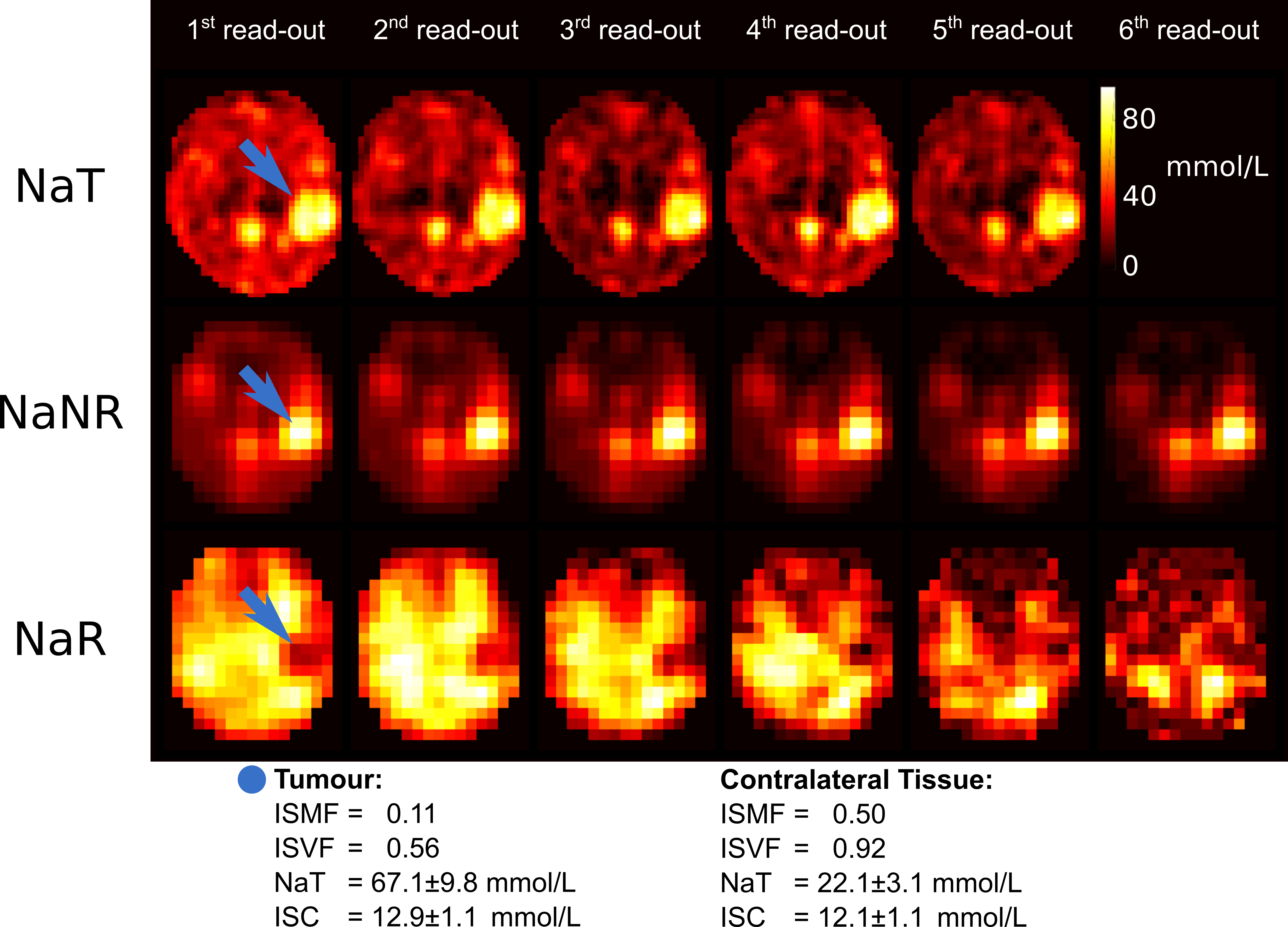
Fig.1: Relaxometry and
quantification of sodium parameters from an enhanced SISTINA sequence in a
patient suffering from a low-grade astrocytoma (blue arrow) with positive IDH
mutation. Sodium parameters are significantly altered in the tumour region
compared to the contralateral tissue.