0491
Simultaneous ultra-short TE-MRS in two voxels using a SPECIAL sequence with Hadamard encoding1McGill University, Montreal, QC, Canada, 2Centre d'Imagerie Cérébrale, Montreal, QC, Canada, 3Johns Hopkins University, Baltimore, MD, United States
Synopsis
The spin echo, full Intensity acquired localized (SPECIAL) sequence consists of a localized spin-echo, preceded by an alternating ISIS pre-inversion for voxel localization. In this study we modified the SPECIAL sequence to simultaneously localize the signal at two different positions. The technique relies on a four-step inversion scheme involving two different inversion positions, followed by a Hadamard encoding reconstruction. Comparing the in vivo performance of dual-SPECIAL sequence to the conventional SPECIAL sequence demonstrated that the dual-Special sequence provides simultaneous metabolite profile from two different regions, reducing the acquisition time by a factor of two, and without any penalty in SNR.
Introduction
The 1H spin echo, full Intensity acquired localized (SPECIAL) sequence1, is one commonly used acquisition technique for localized MR spectroscopy in the brain. Like the STEAM2 sequence, SPECIAL allows acquisition of spectra at ultra-short echo times, but with the additional benefit of a two-fold increase in the signal to noise ratio (SNR). The SPECIAL sequence consists of a localized spin-echo (SE), preceded by an alternating ISIS pre-inversion module. The ISIS subtraction scheme, in which inversion-on scans are subtracted from inversion-off scans, is used for processing the raw SPECIAL data to yields fully localized spectra3. In this study we modified the SPECIAL sequence to simultaneously localize the signal at two different positions. The modified sequence, denoted as dual-SPECIAL, involves a four-step multiplexed inversion scheme with two different inversion positions, followed by a Hadamard reconstruction4 to yield localized short-TE spectra from two voxels in a single acquisition.Methods
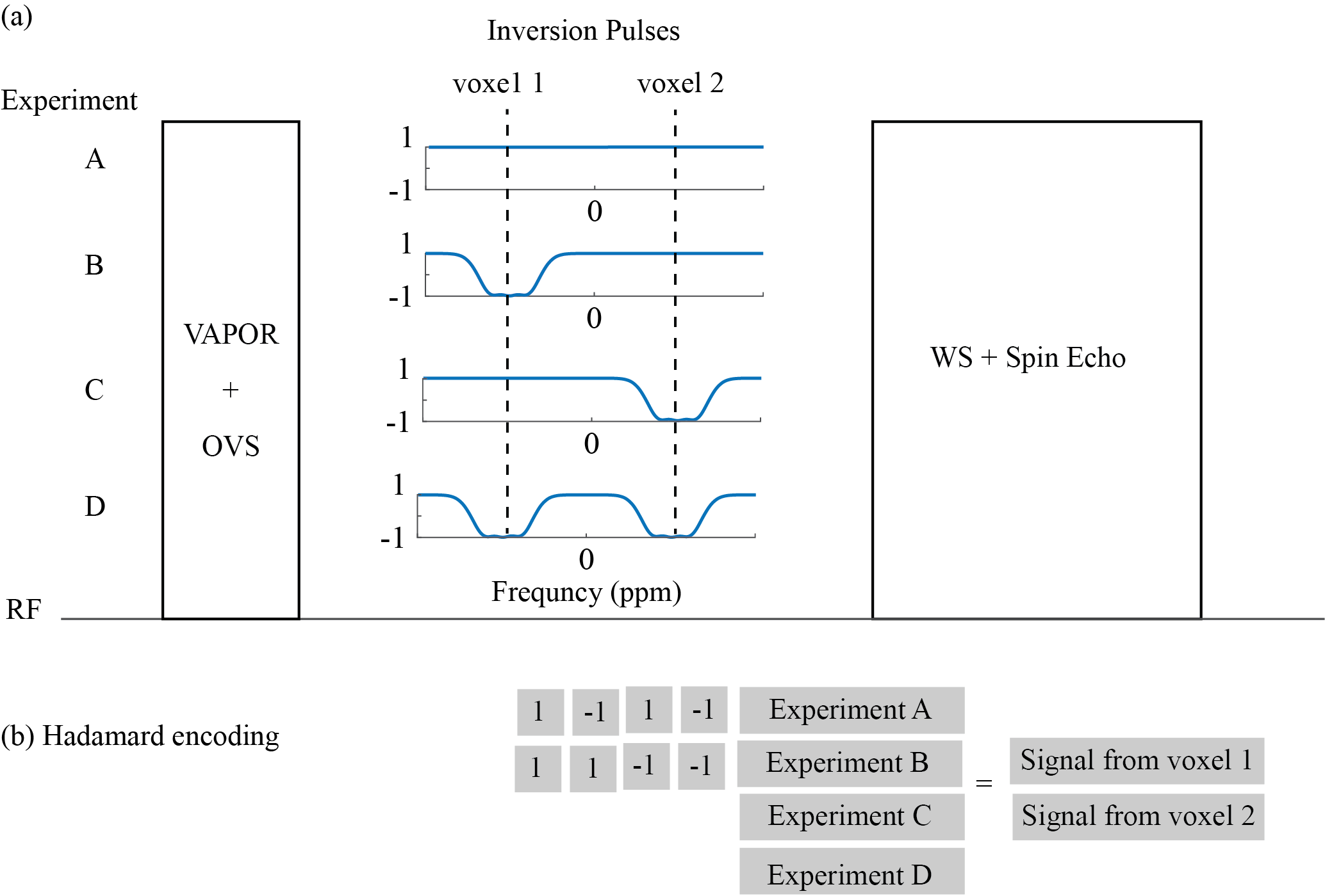
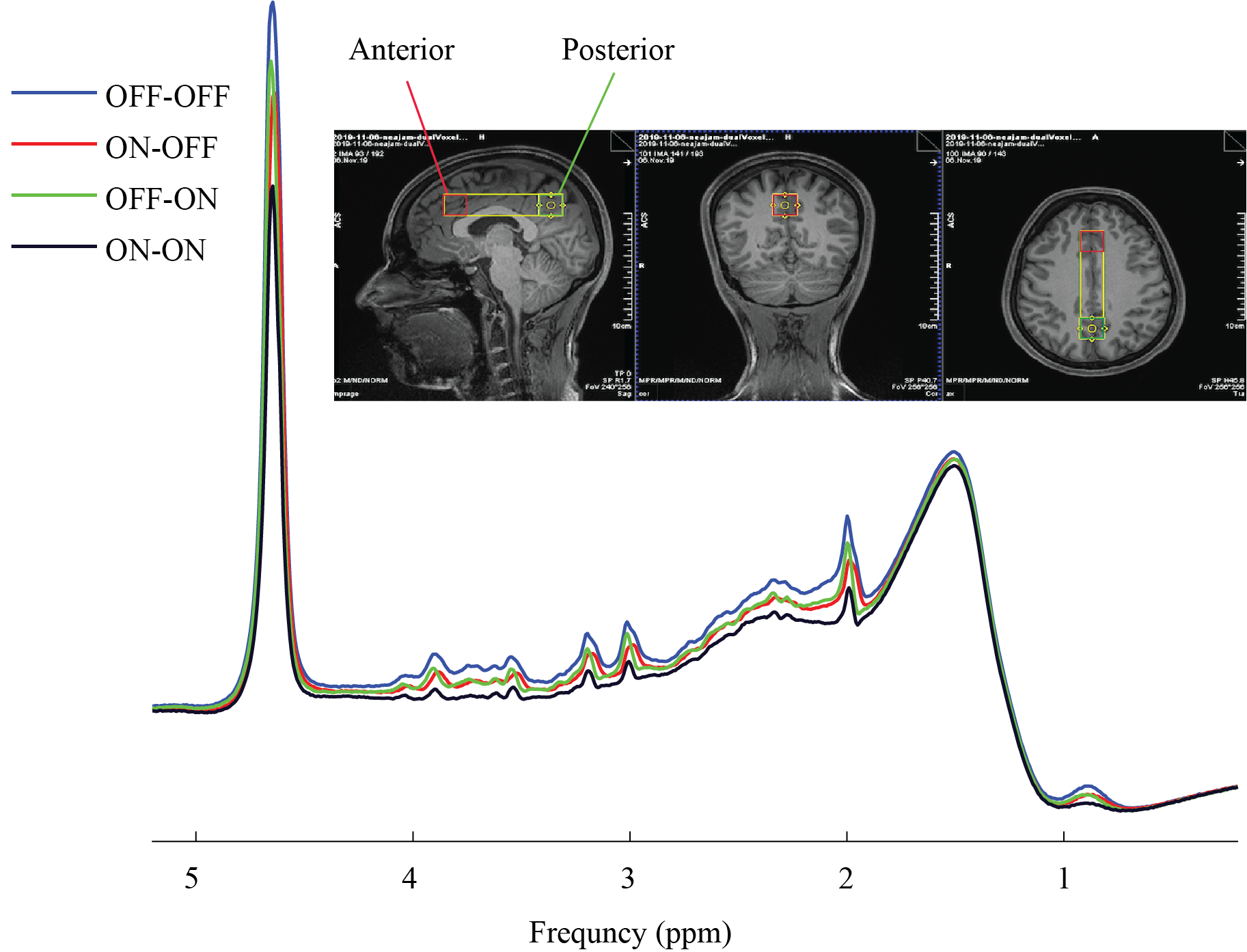
As with the conventional SPECIAL sequence, each acquisition of the dual-SPECIAL sequence consists of a slice-selective SE that is localized to a column of tissue. Localization of two regions was achieved by introducing two different inversion pulses, both perpendicular to the SE column. The two inversion locations correspond to the two voxel positions (voxel 1 and voxel 2) and were arranged in a sequence of four experiments in the dual-SPECIAL sequence (Fig.1). The first scan (OFF/OFF) is acquired from the entire SE column within the field of view of the RF receiver/transmitter coil. The other three experiments incorporated a slice‐selective full‐passage adiabatic inversion pulse followed by a spoiling gradient prior to the SE sequence. Scan ON/OFF is acquired using a single selective inversion pulse centered at the position of first voxel, scan OFF/ON is acquired using a single selective inversion pulse centered at the position of second voxel, scan ON/ON is acquired using a dual selective inversion pulse and two bands centered on the positions of first and second voxels, respectively. The performance of the dual-SPECIAL sequence was investigated in vivo and compared with the conventional SPECIAL sequence. One healthy female volunteer provided informed consent to participate in this study. Experiments were performed on a 3T Siemens Prisma MR scanner with a commercial body transmit-volume coil and 32-channel receive array. The SE column was oriented in the scanner y-direction (anterior-posterior). Bo field inhomogeneities were minimized within a section of the column measuring 2.0 × 10.0 × 2.0 cm3 (encompassing both regions of interest) using the GRE-shim procedure, resulting in a water linewidth of 6.3 Hz and 5.3 in posterior and anterior regions (Fig.2.a). Localized water suppressed 1H spectra were acquired using the dual-SPECIAL sequence and SPECIAL sequence from two equal-size voxels (2.0 × 2.0 × 2.0 cm3), one placed in the medial prefrontal cortex and one placed in the medial posterior cingulate cortex/precuneus posterior regions of VOI and separated by 8.0 cm. All acquisition parameters including TR/TE = 4000/8.5 ms and 128 averages were kept same between the two sequences. Spectral pre-processing steps, including coil combination, phase and frequency correction and averaging were performed in MATLAB using the FID-A toolkit5. To achieve the desired dual localization, the four subspectra from dual-Special sequence were aligned and combined using a Hadamard encoding approach (Fig. 1.b). The signal from posterior and anterior regions is acquired by linear combination of (OFF/OFF)+(ON/OFF)-(ON/ON)-(OFF/ON) and (OFF/OFF)+(OFF/ON)-(ON/ON)-(ON/OFF), respectively. The two subspectra in Special sequence were combined based on the 1D ISIS add‐subtract scheme3.Results
Figure 1 illustrates the four-step inversion scheme and Hadamard‐encoding approach used for combination of subspectra. Figure 2. shows the VOI for shimming along with posterior and anterior voxels selected for spectra acquisition, and the four subspectra acquired using the dual-SPECIAL sequence. Figure 3 shows the localized spectra obtained from posterior and anterior regions by dual-SPECIAL sequence and SPECIAL sequence. In both anterior and posterior regions, the spectral appearance is very similar between the SPECIAL and dual-SPECIAL sequences. Moreover, the SNR of spectra acquired using dual-SPECIAL sequence is similar to the SPECIAL sequence, ~190 for posterior and ~170 for anterior regions.Discussion
Using the described dual-SPECIAL sequence and Hadamard encoding approach, the simultaneous signals from anterior and posterior regions are achieved by coaddition of signal from within the voxels and cancellation of signals from all external regions. Compared to the conventional SPECIAL sequence, the quality of the spectra, linewidth and SNR, in dual-SPECIAL sequence is similar for both anterior and posterior regions while the total measurement time for acquisition of both voxels is reduced by half.Conclusion
In summary, simultaneous measurement of signals from two regions of interest was shown to be feasible using the developed dual-SPECIAL sequence combined with a Hadamard reconstruction. The dual-SPECIAL sequence reduces the total measurement time by half relative to sequential measurements of each voxel using the SPECIAL sequence without any penalty in SNR. These preliminary results suggest that the newly developed dual-SPECIAL sequence can be a potential candidate for simultaneous measuring of metabolites profiles from two different regions in clinical and pre-clinical applications such as functional MRS or 13C MRS.Acknowledgements
This work is supported by the National Engineering and Sciences Research Council (NSERC, RGPIN-2014-06072, J.N.), the Canadian Institutes of Health Research (PJT-165896, J.N.) and the Fonds de recherche du Quebec – Santé (FRQS, J.N.).References
1. Mlynárik V, Gambarota G, Frenkel H, Gruetter R. Localized short-echo-time proton MR spectroscopy with full signal-intensity acquisition. Magnetic Resonance in Medicine. 2006; 56(5):965-970.
2. Frahm J, Merboldt K-D, Hänicke W. Localized proton spectroscopy using stimulated echoes. Journal of Magnetic Resonance. 1987; 72(3):502-508.
3. Ordidge R.J, Connelly A, Lohman J.A.B. Image-selected in Vivo spectroscopy (ISIS). A new technique for spatially selective nmr spectroscopy. Journal of Magnetic Resonance.1986; 66(2):283-294.
4. Hadamard, J.S. Résolution d’une question relative aux determinants. Bulletin des Sciences Mathematiques. 1893; 17: 240-246.
5. Simpson R, Devenyi GA, Jezzard P, Hennessy TJ, Near J. Advanced processing and simulation of MRS data using the FID appliance (FID-A)-An open source, MATLAB-based toolkit. Magnetic Resonance in Medicine. 2017; 77(1):23-33.
Figures