0479
31P-MRSI of the human heart in 2 ½ minutes at 7T using concentric rings (CONCEPT)1Wellcome Centre for Integrative Neuroimaging, NDCN, University of Oxford, Oxford, United Kingdom, 2High-field MR Centre, Department of Biomedical Imaging and Image-guided Therapy, Medical University of Vienna, Vienna, Austria, 3Oxford Centre for Clinical Magnetic Resonance Research, Radcliffe Department of Medicine, University of Oxford, Oxford, United Kingdom, 4Department of Imaging Methods, Institute of Measurement Science, Slovak Academy of Sciences, Bratislava, Slovakia, 5Wolfson Brain Imaging Centre, Department of Clinical Neurosciences, University of Cambridge, Cambridge, United Kingdom
Synopsis
A density-weighted concentric ring trajectory MRSI sequence is implemented for cardiac 31P-MRS at 7T. The sequence is characterised in phantoms and in five healthy participants. Quantitative comparisons are made against a previously implemented acquisition weighted CSI sequence with matched acquisition time and voxel size. The proposed sequence (CONCEPT) was found to robustly measure 3D localised PCr/ATP ratios of the human myocardium 2.59 times faster or with 1.73 times smaller nominal volume than standard acquisition weighted CSI.
Introduction
Phosphorus magnetic resonance spectroscopy (31P‐MRS) allows measurement of the metabolism of the human heart in vivo, specifically the ratio of phosphocreatine to adenosine triphosphate concentrations (PCr/ATP), which is a biomarker of heart failure (1).To date, 3D resolved 31P‐MRS measurements of the human heart have used Cartesian-sampled chemical shift imaging (CSI) or single voxel 3D-ISIS pulse sequences (2,3). While CSI offers optimal SNR per-unit-time, the point-by-point sampling of k-space restricts the minimum scan time at higher spatial resolutions (4).
Concentric ring (CONCEPT) MRSI has recently been shown to be an effective pulse-sequence for high-resolution 1H MRSI of the brain (5,6). In this work we demonstrate a 31P density-weighted CONCEPT sequence for MRSI of the human heart at 7T (7). The proposed sequence is compared against previously published sequences for reduced acquisition durations or increased resolution (8).
Methods
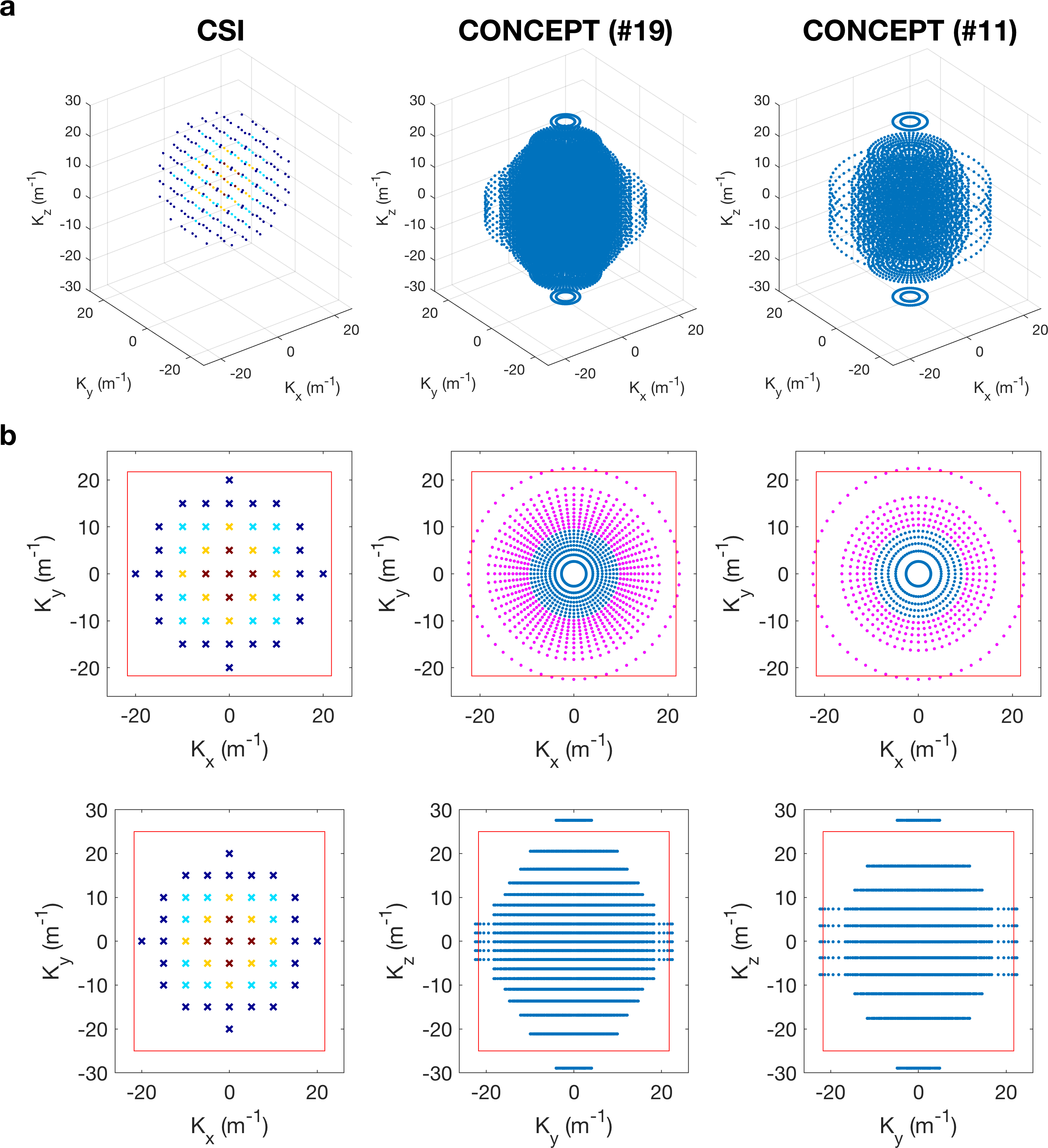
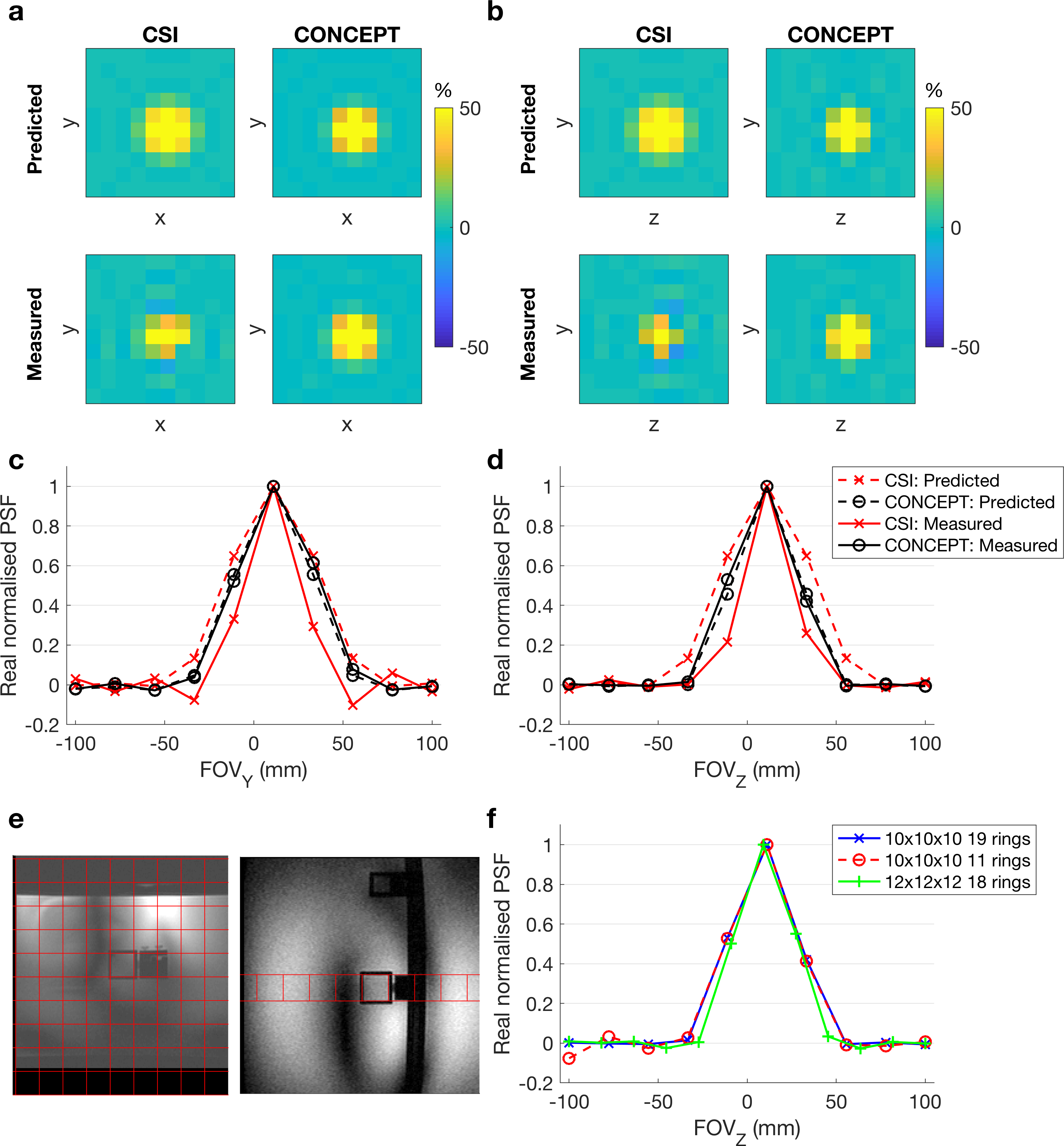
A density-weighted 3D CONCEPT sequence was created from a previously published equidistant ring 3D CONCEPT sequence (9). Density was described as a Hamming Window: by placing rings at particular radii in the kxy-plane, and at particular planes in the kz-direction (Fig 1). The number of rings was attuned to elliptically sample k-space in the kz-direction. The number of points in each ring was constant. Sequence duration was adjusted by varying the number of rings and z-direction partitions whilst keeping the maximum k-space coverage identical. Excitation used a 2.4 ms shaped constant phase pulse designed to uniformly excite metabolites between -3 and 8 ppm (2,3-DPG, PDE, PCr and g-ATP) (3) when centred 270Hz above PCr. A single BISTRO saturation band to suppress the chest wall signal was added to the sequence (10).The sequence SNR and point-spread function was characterised relative to an acquisition weighted CSI sequence on a point-source phantom (11).
Five subjects (4M/1F, 74±11 kg, 30±3 years) were scanned in a supine position using a whole-body Siemens Magnetom 7T scanner (Erlangen, Germany) equipped with a quadrature transmit-receive surface coil positioned over the heart (12). Each subject was scanned using a previously described CSI sequence (8), a matrix size of 8x16x8 (four averages at the centre of k-space, 6:37 min), and with symmetrical matrix size of 10x10x10 (4&1 averages, 6:31&4:21 min). Then the CONCEPT sequence was repeated five times with a matched symmetric matrix size of 10x10x10 (20, 16, 14, 12 and 10 rings/partitions; 6:32, 4:33, 3:19, 2:31 and 1:37 mins) and once with a matrix size of 12x12x12 (19 rings/partitions, 6:55 mins). CSI was run with 240x240x200 mm3 FoV, 1 s TR, 8 kHz bandwidth, and 2048 time-samples. CONCEPT had matched FoV and TR, but 2778 Hz bandwidth with 720 time-samples.
CSI data were reconstructed online and CONCEPT data offline using the non‐uniform FFT (NUFFT) toolbox with min‐max Kaiser‐Bessel kernel interpolation and twofold oversampling (13) in MATLAB (MathWorks, Natick, USA). No density compensation is required. Individual coil data was combined using the WSVD algorithm (14). CSI/MRSI data fitting was performed using the OXSA toolbox (15).
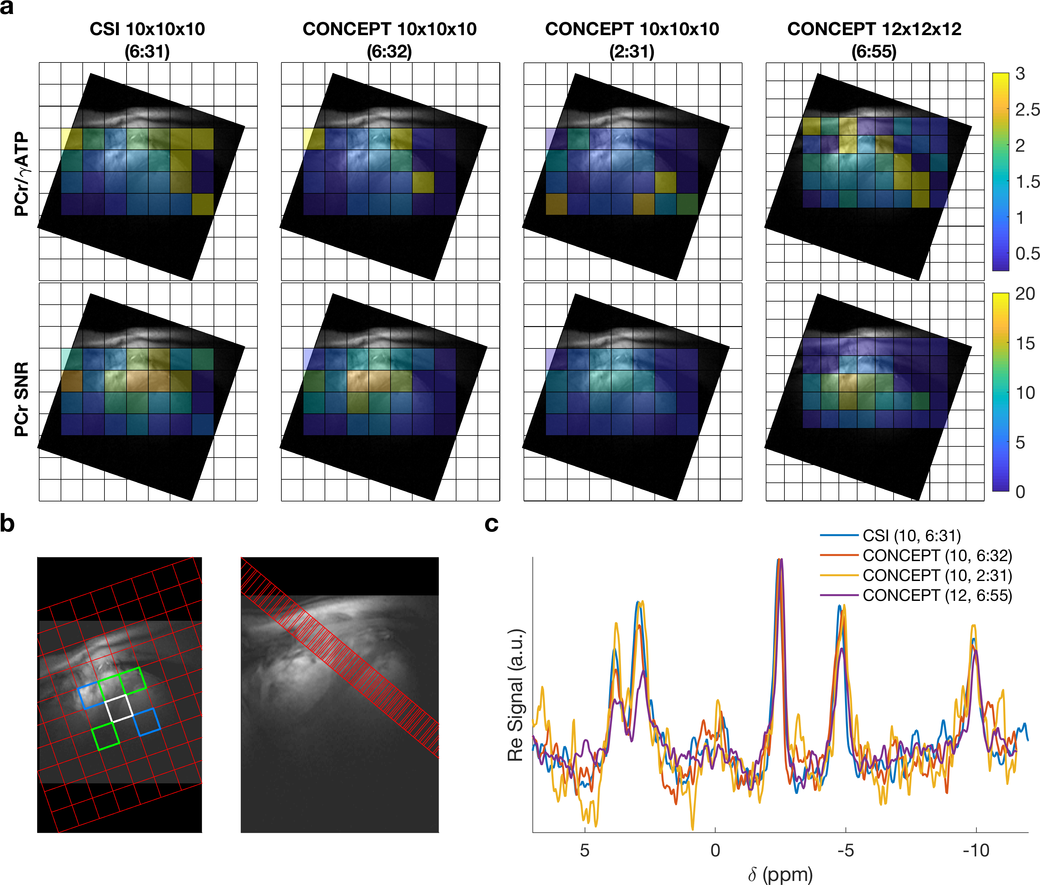
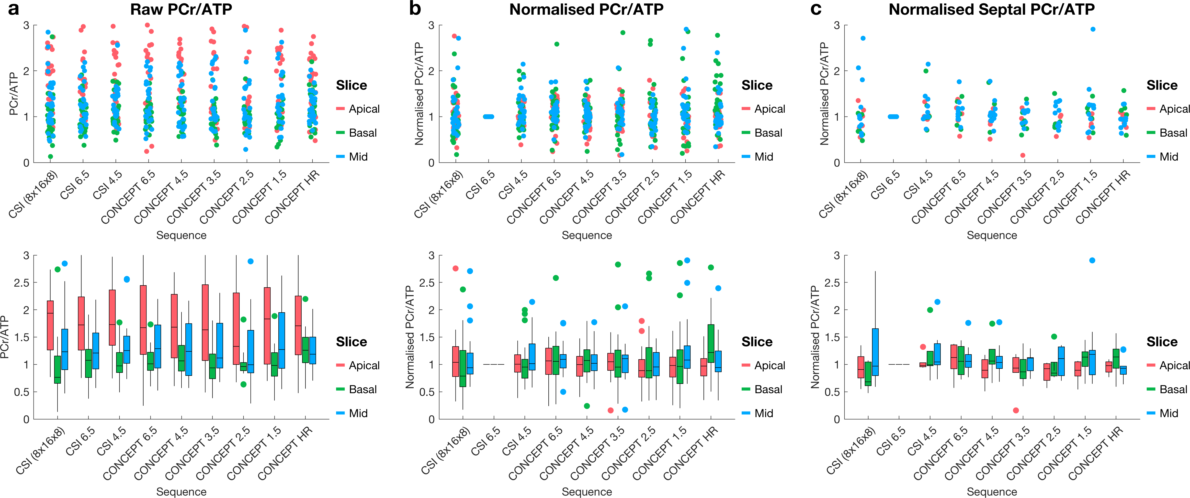
Sequences were compared using PCr/γ‐ATP ratios in anterior-, mid-, and posterior-septal voxels from apical, mid and basal slices of the heart, without correcting for partial saturation or blood contamination.
Results
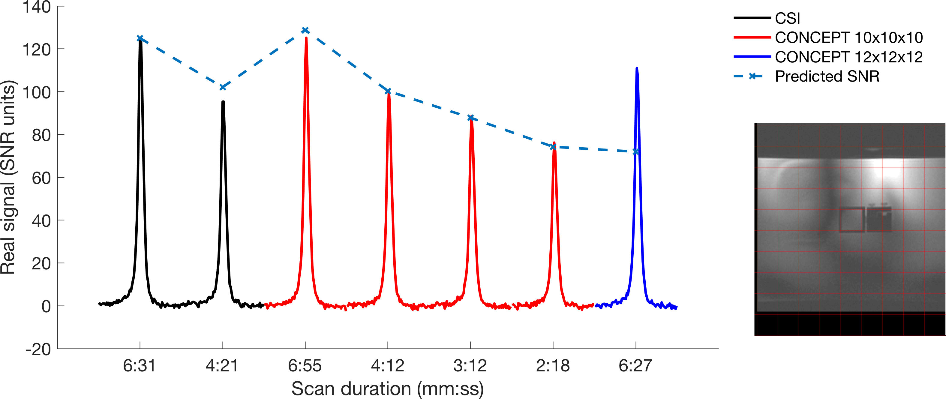
Phantom experiments showed that the measured CONCEPT point-spread-function (PSF) matched the predicted low ripple PSF (Fig 2). Reducing the number of rings to 10 from 19 did not cause significant PSF deterioration. SNR performance of the time-matched sequence was found to match that of the acquisition weighted CSI for acquisitions >4:12 min total and exceed CSI for shorter scans (because post-acquisition reweighting of 1avg CSI causes SNR losses) (Fig 3).In the in vivo experiments (Fig 4), the mean PCr/ATP ratio showed no strong divergence from the time-matched CSI sequences even at 1.5 minutes duration (Fig 5). For every scan duration except the shortest (1:37 min) the mid-septal PCr/ATP was quantified with CRLB < 30% in all subjects.
Discussion
Density-weighted CONCEPT MRSI has been demonstrated for fast (2:31 min) 3D localised cardiac 31P-MRS at 7T. Phantom measurements showed no loss of SNR compared to SNR optimal CSI encoding. In vivo PCr/ATP maps are consistent with maps generated from previously published sequences. This demonstrates a 2.59x and 1.73x reduction in scan time compared to previously described (8) and the shortest feasible Cartesian-sampled CSI sequence at matched TR.Using CONCEPT trajectories has some potential limitations compared to CSI. Incoherent aliasing artefacts could influence measured metabolite ratios, especially in the case of cardiac 31P-MRS with close proximity of potentially contaminating tissues (skeletal muscle and liver) containing the same metabolites at different concentrations. Care must also be taken with the increased susceptibility to off-isocentre distortions caused by gradient non-linearity and strong spatial blurring associated with aliased spectral peaks (16). Neither of these effects are observed in this data, the latter due to the use of a spectrally selective excitation pulse.
Conclusion
The proposed sequence is able to measure the PCr/ATP ratio in the human septal myocardium in 2½ min, which is 2.59 times faster than a standard CSI sequence with the same nominal voxel size of 11.5 mL. Or we can achieve comparable SNR with a nominal voxel size of 6.6 mL in only 6:55 min versus 8:35 min predicted for CSI.Acknowledgements
CTR and LV are funded by a Sir Henry Dale Fellowship from the Wellcome Trust [098436/Z/12/B]. The support of the Slovak Grant Agencies VEGA [2/0001/17] and APVV [#15–0029] is also gratefully acknowledged. The Wellcome Centre for Integrative Neuroimaging is supported by core funding from the Wellcome Trust (203139/Z/16/Z). CTR and LV contributed equally to this work.References
1. Neubauer S. Mechanisms of disease - The failing heart - An engine out of fuel. New Engl J Med 2007;356(11):1140-1151.
2. Lamb HJ, Doornbos J, den Hollander JA, Luyten PR, Beyerbacht HP, van der Wall EE, de Roos A. Reproducibility of human cardiac 31P-NMR spectroscopy. NMR Biomed 1996;9(5):217-227.
3. Rodgers CT, Clarke WT, Snyder C, Vaughan JT, Neubauer S, Robson MD. Human cardiac 31P magnetic resonance spectroscopy at 7 Tesla. Magn Reson Med 2014;72(2):304-315.
4. Pohmann R, von Kienlin M, Haase A. Theoretical evaluation and comparison of fast chemical shift imaging methods. J Magn Reson 1997;129(2):145-160.
5. Chiew M, Jiang W, Burns B, Larson P, Steel A, Jezzard P, Albert Thomas M, Emir UE. Density-weighted concentric rings k-space trajectory for (1) H magnetic resonance spectroscopic imaging at 7 T. NMR Biomed 2018;31(1).
6. Hingerl L, Bogner W, Moser P, Povazan M, Hangel G, Heckova E, Gruber S, Trattnig S, Strasser B. Density-weighted concentric circle trajectories for high resolution brain magnetic resonance spectroscopic imaging at 7T. Magn Reson Med 2018;79(6):2874-2885.
7. Greiser A, von Kienlin M. Efficient k-space sampling by density-weighted phase-encoding. Magn Reson Med 2003;50(6):1266-1275.
8. Ellis J, Valkovic L, Purvis LAB, Clarke WT, Rodgers CT. Reproducibility of human cardiac phosphorus MRS ((31) P-MRS) at 7 T. NMR Biomed 2019;32(6):e4095.
9. Hingerl L, Strasser B, Moser P, Považan M, Hangel G, Heckova E, Gruber S, Trattnig S, Bogner W. Towards Full-Brain FID-MRSI At 7T With 3D Concentric Circle Readout Trajectories. 2018; Proc. Intl. Soc. Mag. Reson. Med. 26, Paris. p 618.
10. Luo Y, de Graaf RA, DelaBarre L, Tannus A, Garwood M. BISTRO: an outer-volume suppression method that tolerates RF field inhomogeneity. Magn Reson Med 2001;45(6):1095-1102.
11. Robson MD, Tyler DJ, Neubauer S. Ultrashort TE chemical shift imaging (UTE-CSI). Magn Reson Med 2005;53(2):267-274.
12. Schaller B, Paritmongkol W, Rodgers CT. Quadrature 31P and single 1H dual‐tune coil for cardiac 31P‐MRS at 7T. 2016; Proc. Intl. Soc. Mag. Reson. Med. 24, Singapore. p 4006.
13. Fessler JA, Sutton BP. Nonuniform fast Fourier transforms using min-max interpolation. Ieee T Signal Proces 2003;51(2):560-574.
14. Rodgers CT, Robson MD. Receive array magnetic resonance spectroscopy: Whitened singular value decomposition (WSVD) gives optimal Bayesian solution. Magn Reson Med 2010;63(4):881-891.
15. Purvis LAB, Clarke WT, Biasiolli L, Valkovic L, Robson MD, Rodgers CT. OXSA: An open-source magnetic resonance spectroscopy analysis toolbox in MATLAB. PLoS One 2017;12(9):e0185356.
16. Mayer D, Levin YS, Hurd RE, Glover GH, Spielman DM. Fast metabolic imaging of systems with sparse spectra: application for hyperpolarized 13C imaging. Magn Reson Med 2006;56(4):932-937.
Figures