0464
Measurement of head motion using a field camera in a 7T scanner1Physics, Sir Peter Mansfield Imaging Centre, University of Nottingham, Nottingham, United Kingdom
Synopsis
In this work, a step towards a non-contact motion correction technique has been made. Measurements of extra-cranial field perturbations made using a 16-channel magnetic field camera have been used to predict head motion parameters with good accuracy. The prediction was performed using both linear (PLS) and non-linear (NARX) methods. The number of field probes used for the prediction was reduced by performing Principal Component Analysis. Magnetic field data was also pre-processed to reduce the unwanted effect of chest movement in respiration. NARX outperformed the PLS approach producing good predictions of head position changes for a wider range of movements.
Introduction
Methods for monitoring head position inside the MR scanner can be used to inform prospective and retrospective motion correction (MoCo) techniques, which ameliorate motion artefacts in MR image data. A variety of methods for measuring head position in the scanner have been developed[1], including optical and field camera [2] based methods, which generally require the rigid attachment of markers to the head and navigator-based methods which necessitate some modification of the MR sequence used for image acquisition. An alternative approach, which does not require attachment of markers to the head, line of site access for an optical camera or significant sequence modification is to use a field camera to monitor the magnetic field perturbations generated by changes in head pose [4]. Here, we extend this approach by comparing the use of linear and non-linear methods for calculating head position from field measurements, with and without additional pre-processing.Methods
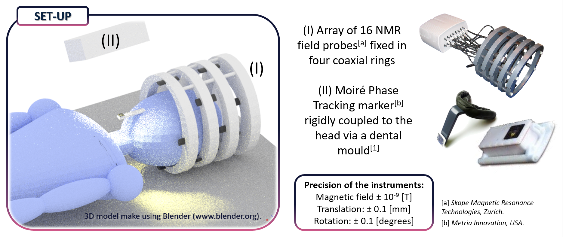
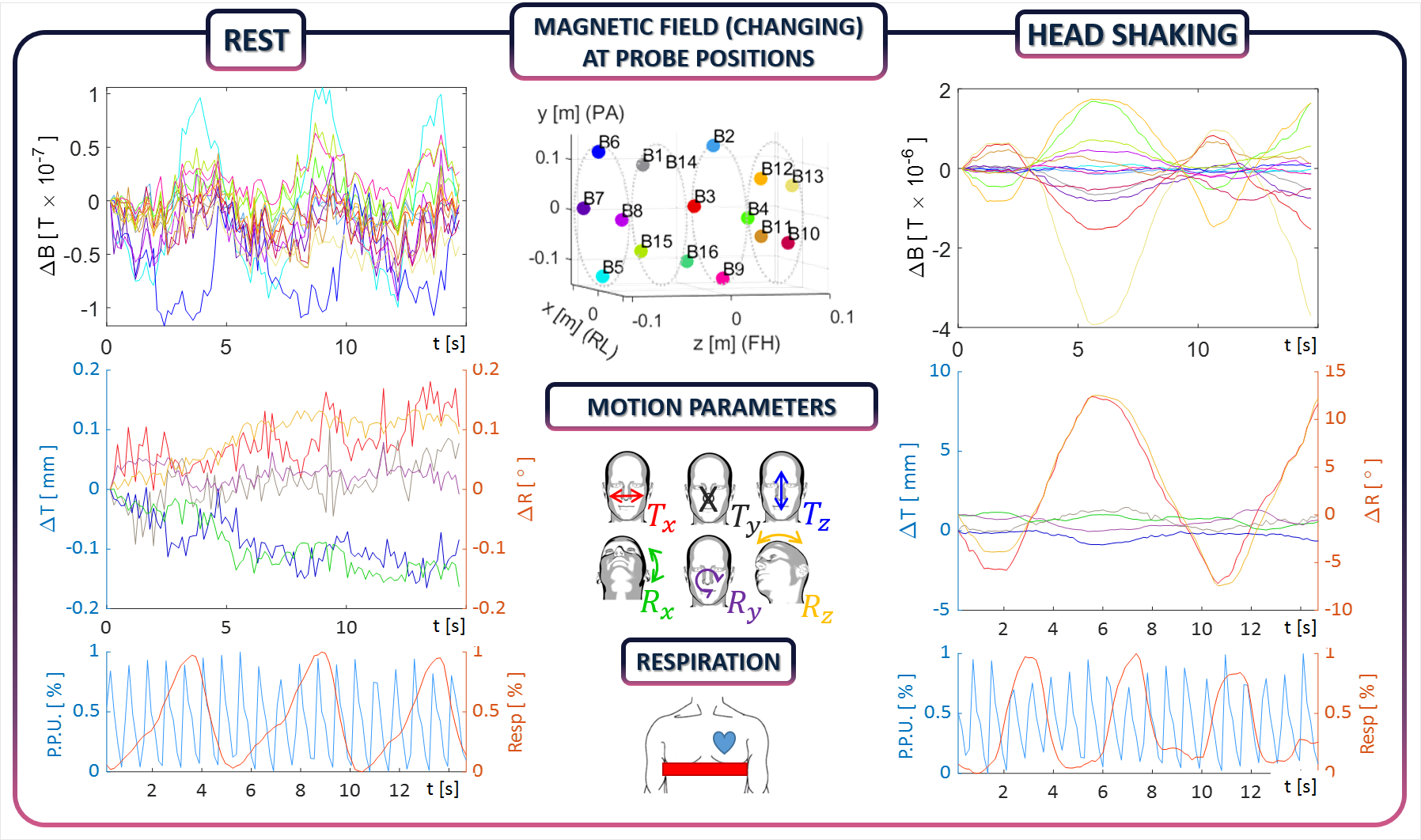
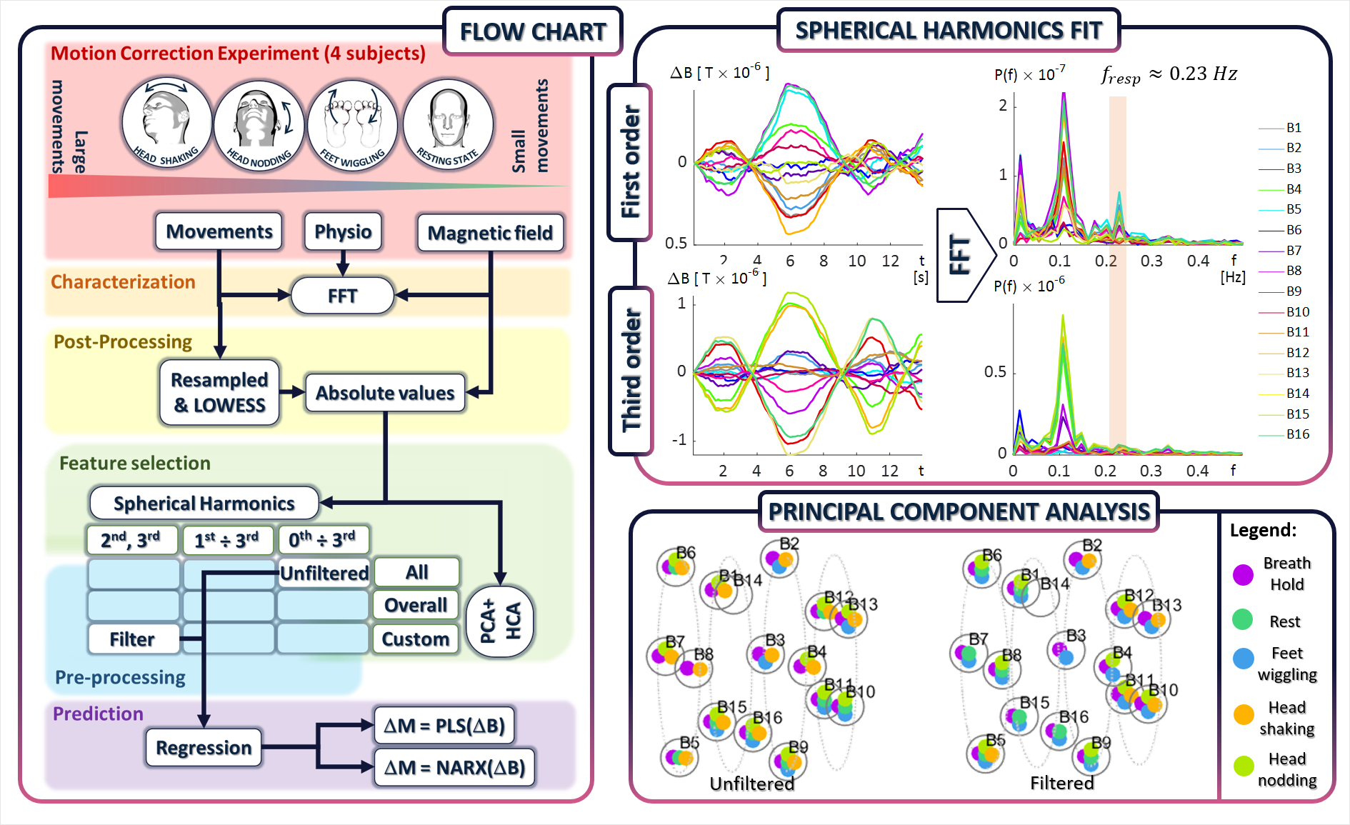
Concurrent measurements of magnetic field variation and head movement, along with cardiac and respiratory cycles were made on 4 subjects executing a range of different head movements in a 7T scanner, using a 16-channel field camera (Skope) and an optical camera (Kineticor), along with a respiratory belt and peripheral pulse unit (see Figure 1).Figure 2 shows example data recorded from one subject for small and large head movements. These show that the magnetic field change (ΔB) varies coherently with change in head position (ΔM). In previous work we demonstrated that the relationship between ΔB and ΔM is linear only for finite range of position changes [4], so that the efficacy of a linear Partial Least Squares (PLS) method in predicting movement from field changes is limited. Here, further analysis has been made using a non-linear autoregressive exogenous model (NARX) [11]. A flowchart of the process used to implement and evaluate the two different regression techniques is reported in Figure 3.
After frequency analysis of the data, a locally weighted polynomial regression method (LOWESS) was used to smooth the motion data, before temporal alignment of the different time series [3]. The regression methods have been applied to both pre-processed and unprocessed, magnetic field data. In the former case, the subset of six or more probes that best described the field variation was selected using Principal Component Analysis (PCA) combined with Hierarchical Cluster Analysis (HCA) [3]. In addition, the ΔB measurements were spatially filtered via spherical harmonic analysis, with only high order harmonics selected so as to reduce the influence of physiological noise [4]. The NARX method was implemented with one hidden layer layer and a 0.9s feedback delay [6]. The best network architecture was automatically selected by evaluating its performance on the experimental data for a range of different network parameters.
Results
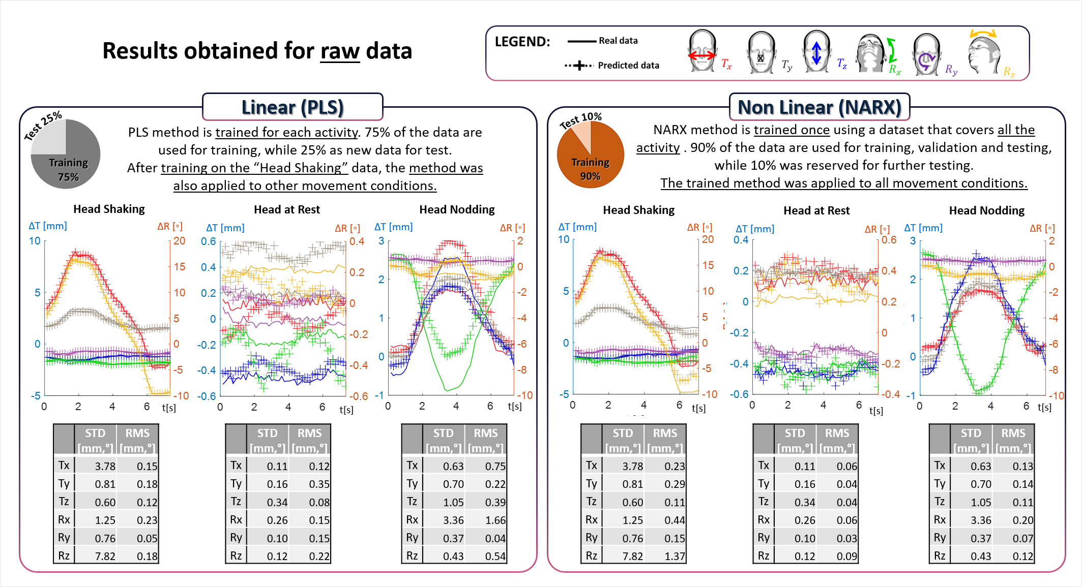
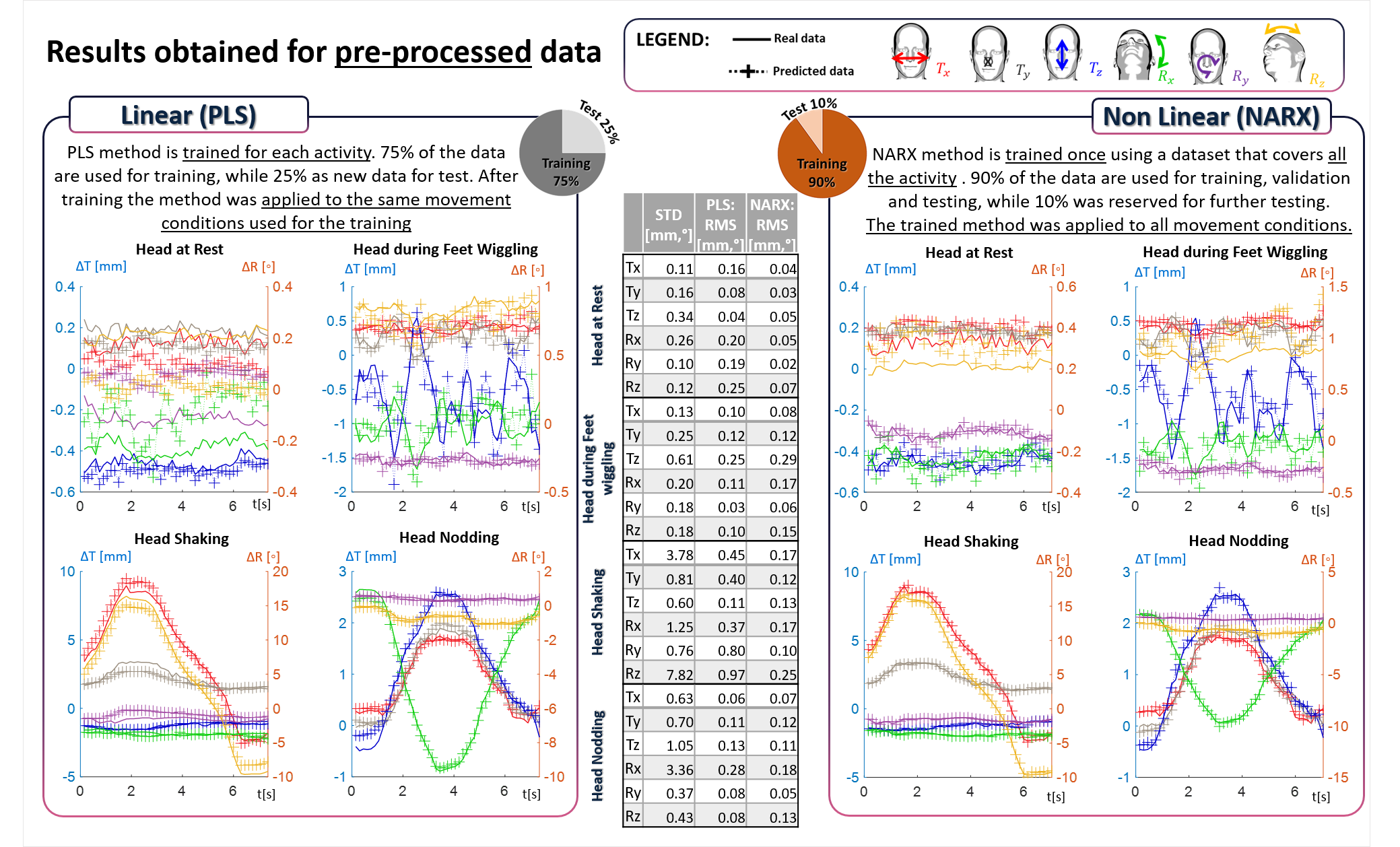
Figures 4 and 5 show results obtained using the PLS and NARX methods applied to field data before and after pre-processing. Comparing the time series plots of the predicted and actual movements, it is evident that the NARX method outperforms the PLS approach, and this is confirmed by the reported RMSE values. In particular, the NARX method works reasonably well for all movement conditions after training on a range of data, while the PLS method only works well when it is trained on, and then applied to, data recorded for a specific type of movement. For both methods the relative error in prediction (RMSE/STD) is best for the dominant movement parameter and for the cases where larger movements occur. Comparing the results obtained from data with and without pre-processing, it is clear that good predictions can be obtained when using data from only a subset of probes and the higher spatial harmonics of the magnetic field. The spatial filtering particularly reduces the RMSE for the rest data, in which the field variation due to chest movement in respiration (which appears mainly in lower spatial harmonics) dominates the effect of small head movements.Discussion and conclusion
The results reported here confirm that measurements of extra-cranial field changes made with a field camera can be used to monitor head position in a 7T MRI scanner. A non-linear (NARX) regression method provides better prediction of movement parameters than a linear (PLS) approach, with the NARX method able to work over a range of different movements. Both these approaches require the acquisition of training data in which head movements are monitored using another approach (here, with an optical camera and an MPT marker attached to a dental mould) at the same time as field variation is measured. In future work, imaging methods could be used in this training phase, to remove the need for the optical camera and mouthpiece. Spatial filtering has been shown to be useful for reducing the confounding effect of field variation due to chest movement in respiration, particularly when predicting small movements. The results also indicate that good predictions can be obtained using a subset of the field probes, which might allow lower cost implementation of this approach. Further work is needed to identify the optimal number and positions of field probes that should be used when the standard receiver coil array is also in place.Acknowledgements
Thanks to James Smith for his long distance support.References
[1] McLaren review J. McLaren Prospective motion correction in brain imaging: A review 2013 March; 69, 621-63
[2] Aranovitch A. et al, “Prospective motion correction with NMR markers using only native sequence elements”, Magn Reson Med. 2018; 79:2046–2056
[3] Bortolotti L., Smith J., Spancer G. et al .Test of multiple sensor set-up for head motion characterization during MRI acquisition. Master Thesis, University of Bologna, Italy, 2017
[4] Bortolotti L. et al, “Non-contact measurement of head movements inside a 7 T Scanner using a 16-channel field camera”, ISMRM2019, Abstract number: 4432.
[5] Bortolotti L. et al, “Simulation of external magnetic field changes due to head motion during 7 Tesla MRI scan”, ISMRM2019; Abstract number: 4431.
[6] Bortolotti L. et al, “Using a field camera to monitor head movement in 7T scanner”, ESMRMB2019, Magn Reson Mater Phy (2019) 32(Suppl 1): 235. https://doi.org/10.1007/s10334-019-00755-1
[7] James Smith, “Motion Correction for MRI using External Tracking Devices”, University of Nottingham 2018.
[8] Robert Odenbach et al, “Remotely controllable phantom rotation system for ultra-high field MRI to improve Cross Calibration”, De Gruyter, Vol 5 issue 1, DOI: https://doi.org/10.1515/cdbme-2019-1570538325
[9] Bortolotti L., Smith J., Gowland P., Bowtell R. . Simulating changes in external magnetic field due to head motion in a 7T scanner. Oxford, UK BCISMRM: PP13. 2018
[10] Andre, Jalal B., et al. "Toward quantifying the prevalence, severity, and cost associated with patient motion during clinical MR examinations.", Journal of the American College of Radiology” 12.7 (2015): 689-695.
[11] Hang Xie et al, “Time series prediction based on NARX neural network: an advanced approach”, 2009 International Conference on Machine Learning and Cybernetics (Hebei, China), DOI:10.1109/ICMLC.2009.5212326
Figures