0454
Motion Compensated Low-Rank(MoCoLoR) constrained reconstruction with application to motion resolved lung MRI1Radiology and Biomedical Imaging, University of California, San Francisco, San Francisco, CA, United States, 2UC Berkeley-UCSF Graduate Program in Bioengineering, University of California, San Francisco and University of California, Berkeley, Berkeley, CA, United States, 3Electrical Engineering, Stanford University, Stanford, CA, United States, 4Electrical Engineering and Computer Sciences, University of California, Berkeley, Berkeley, CA, United States
Synopsis
Respiratory motion is one of the most challenging problems in thoracic and abdominal MRI. Motion resolved reconstruction is introduced to reduce the respiratory motion effects by grouping the data to different motion states, then using compressed sensing techniques to reconstruct different motion states images. Spatio-temporal low-rank constrained reconstruction is one of the widely used techniques. In this work, we proposed a new method incorporating motion compensation into the low-rank model, called MoCoLoR. The proposed method is applied to high resolution free breathing lung MRI, and the results show that MoCoLoR outperforms the standard low-rank constrained reconstruction.
Introduction
Respiratory motion is one of the most challenging problems in thoracic and abdominal MRI, because of the limited encoding speed. Routine clinical protocols require patients to hold breath for single or multiple short duration to reduce respiratory motion. Alternatively, external devices are used to monitor respiratory motion and trigger acquisition at specific motion states to reduce motion artifacts. However, this reduces the scan efficiency and increases the total scan time.To increase the scan efficiency without respiratory motion corruption in free breathing acquisition, one widely-used strategy is to group data into different motion states by the motion signal, then leverage spatial or temporal sparsity to reconstruct motion free images using compressed sensing, called motion resolved reconstruction1,2 . One strategy is to reformulate the motion resolved images to a spatio-temporal matrix, then enforce low-rank in the matrix.However, it has been reported in computer vision applications that the low-rank model breaks down because of spatial misalignment of the images3 . Similarly, in motion resolved MRI reconstruction, low-rank model might also fail because of subject motion. Some recent studies integrate motion information in their models, such as motion adaptive patch-based low-rank4 , and block low-rank sparsity with motion-guidance5 . In this work, we proposed a strategy to directly incorporate motion compensation in the low-rank model, called Motion Compensated Low Rank(MoCoLoR) reconstruction. We apply the proposed technique to 3D free breathing lung MRI.Methods
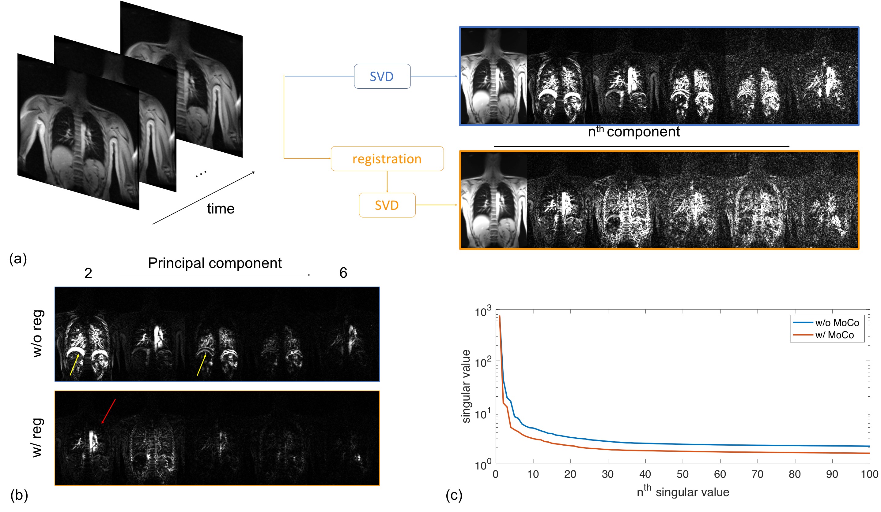
Low-rank with motion compensationA simple experiment is conducted to demonstrate the impact of motion compensation(MoCo) on low-rank model. A 2D coronal slice of chest is continuously acquired under free breathing condition for 1 minute, with a total 150 frames. Without(blue) and with(orange) image based non-rigid registration (MoCo) before singular value decomposition(SVD) are compared in Fig.1. In (b), 2nd to 6th components weighted by singular values are shown. Due to respiratory motion, misaligned diaphragm(yellow arrows) shows up in higher order components. With MoCo, only cardiac perfusion induced intensity fluctuation(red arrow) could be seen in the 2nd component, and higher(>3) order components only has nonstructural residual, while without MoCo, structural information remains in all the shown components. In (c), with MoCo, singular values decrease much faster than without MoCo. That indicates the low-rank property of the spatio-temporal matrix is preserved with MoCo.
Motion Compensated Low-Rank(MoCoLoR) reconstruction
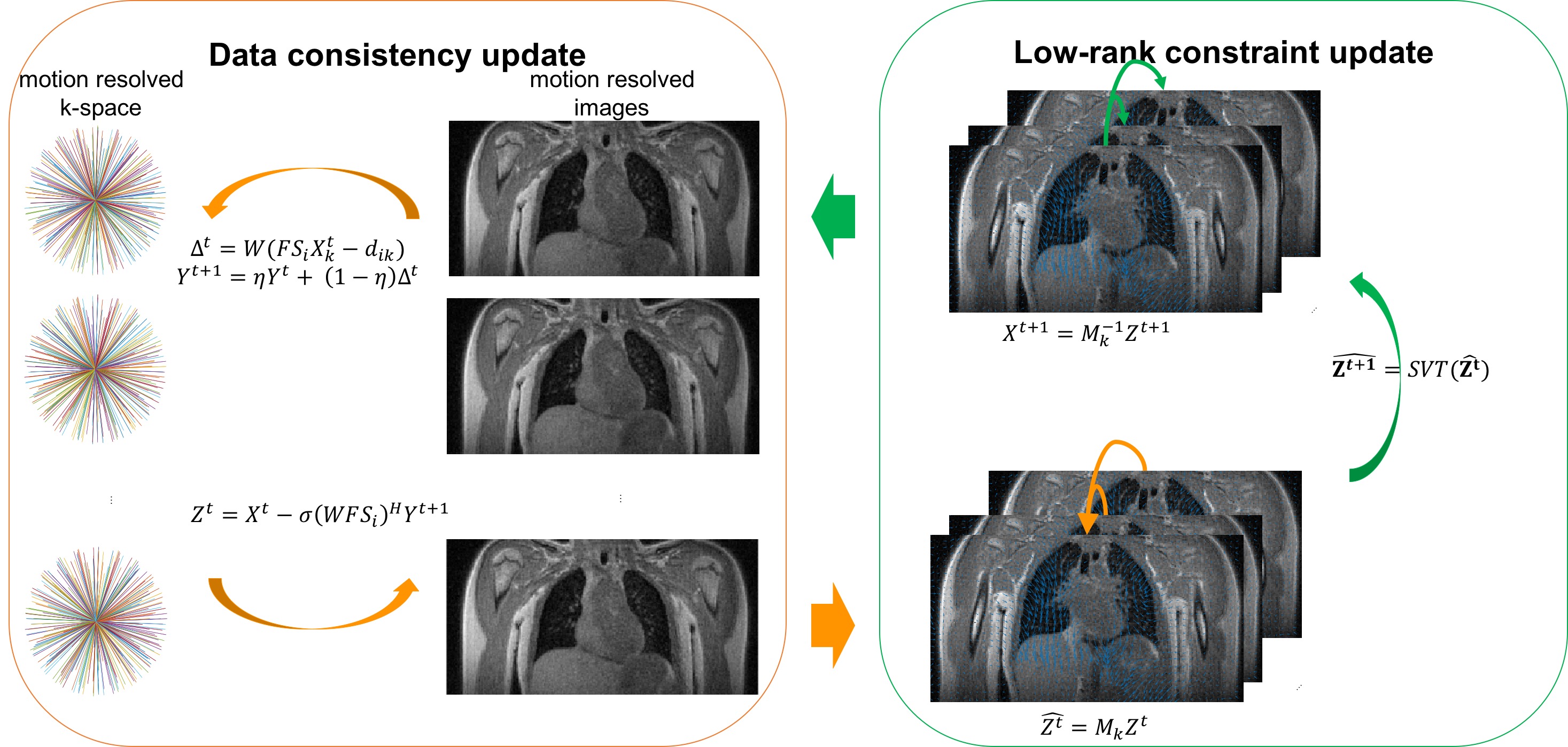
Self-navigation, data binning, and motion field estimation are implemented following the workflow from the previous work, iMoCo-UTE6 . Instead of reconstructing a single state image in iMoCO-UTE, motion resolved images are reconstructed in the proposed MoCoLoR model.
$$ \underset{\bf X}{\operatorname{argmin}} \sum_{i,k}^{N,m}||W(FS_iX_k -d_{i,k})||_2^2+\lambda_L||\textbf{MX}||_* $$
Here, the squared-error data consistency term (left) includes multi-channel sensitivity maps $$$S_i(1,2,...N)$$$, motion states sorted multi-channel data $$$d_{i,k}$$$, $$$W$$$ is sampling density compensation weights, $$$F$$$ is the non-uniform Fourier transform operator, implemented via gridding algorithm, and $$${\bf X}=[X_1,X_2,...X_m]$$$ are the motion-state 3D images, $$${\bf M}=[M_1,M_2,...M_m]$$$ are motion fields corresponding to motion-states images, which are derived by using Demons registration. The iterative reconstruction is summarized in Fig.2.
Lung MRI experiment
An optimized 3D UTE sequence7 with slab selection and variable density readout acquisition was used for pulmonary imaging. A golden angle ordering acquisition scheme was used to randomize undersampling artifacts over time and improve the motion resolved reconstruction and motion estimation. All the studies were performed on 3T MRI clinical scanners (MR750, GE Healthcare, Waukesha, WI, USA). All subjects were scanned with flip angle 4°, 0.9mm isotropic resolution, readout bandwidth 250kHz, TR=2.7~3.1ms, TE=80us, TR increased as the prescribed FOV was reduced. The total scan time was approximately 5 min to 5 min 30 s. Number of total acquired spokes of each scan was approximately 100,000.
Results
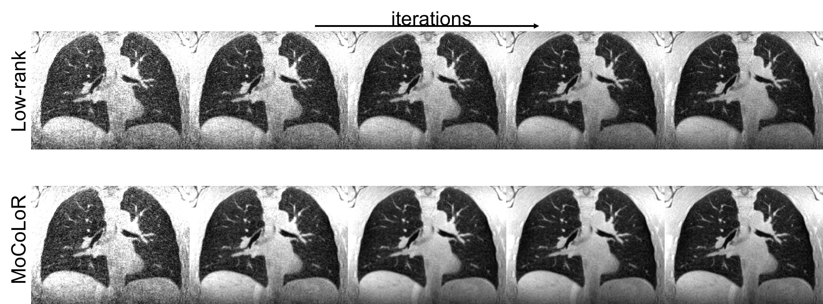
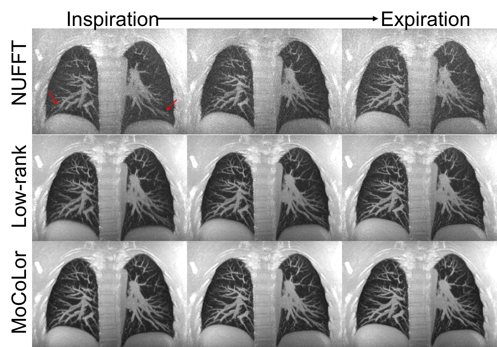
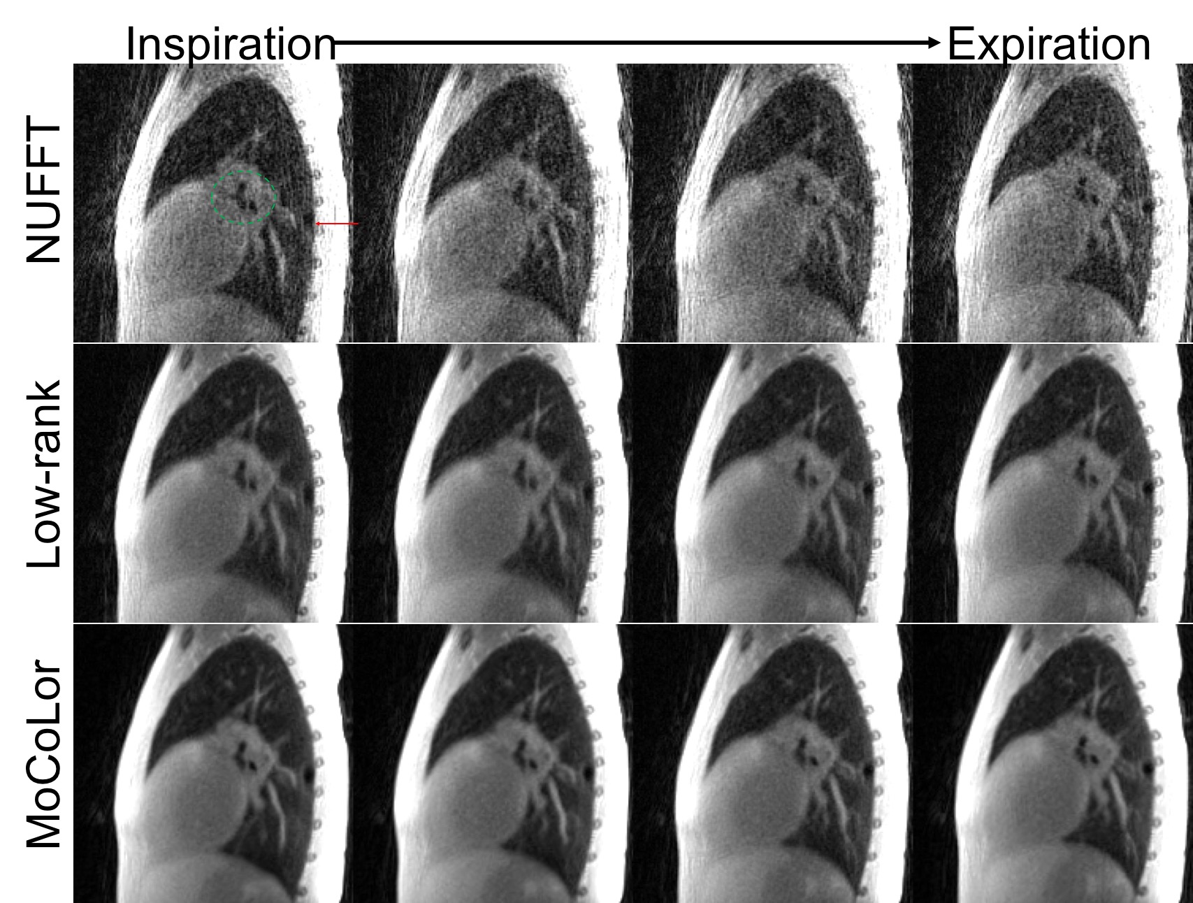
For comparison, directly gridding(NUFFT), low-rank constrained, and proposed MoCoLoR reconstruction are implemented and applied on the same datasets.Firstly, MoCoLoR is applied on an adult volunteer. Fig.3 shows the comparison of intermediate images in the iterative reconstruction. NUFFT images(first row) are used as initial iteration images. Same coronal slice from different reconstruction methods are plotted in each row every 3 iterations. The noise and artifacts in the image are better suppressed with MoCoLoR reconstruction as the number of iterations increases compared to low-rank constrained reconstruction, which indicates that the MoCoLoR formulation better fits the reconstruction problem. 30 coronal slices maximum intensity projection(MIP) images reconstructed by different methods are shown in Fig.4. Overall, MoCoLoR shows sharper vessels and diaphragm than the other two methods, and the improvement of MoCoLoR is more obvious in the areas of largest motion (red arrows).Fig.4 shows the comparison among the methods in an 8 year-old pediatric patient. The patient had surfactant protein C deficiency which caused small pneumatoceles (cysts) in the lung. One air-filled cyst could be found in the posterior part of the right lung(red arrow). Due to sliding motion of the cyst, the low-rank constrained reconstruction blurred the cyst, especially in the inspiratory state. In contrast, images reconstructed with MoCoLoR better delineate the shape and edge of the cyst.Conclusion
In this work, we proposed a new motion resolved reconstruction method by incorporating motion compensation in the low-rank constrained reconstruction model, called MoCoLoR. The proposed method outperforms the standard low-rank constrained reconstruction in both volunteer and patient studies.Acknowledgements
This work is supported by NIH grant R01 HL136965.References
[1] Feng, L., Axel, L., Chandarana, H., Block, K. T., Sodickson, D. K., & Otazo, R. (2016). XD‐GRASP: golden‐angle radial MRI with reconstruction of extra motion‐state dimensions using compressed sensing. Magnetic resonance in medicine, 75(2), 775-788.
[2] Jiang, W., Ong, F., Johnson, K. M., Nagle, S. K., Hope, T. A., Lustig, M., & Larson, P. E. (2018). Motion robust high resolution 3D free‐breathing pulmonary MRI using dynamic 3D image self‐navigator. Magnetic resonance in medicine, 79(6), 2954-2967.
[3] Peng, Y., Ganesh, A., Wright, J., Xu, W., & Ma, Y. (2012). RASL: Robust alignment by sparse and low-rank decomposition for linearly correlated images. IEEE transactions on pattern analysis and machine intelligence, 34(11), 2233-2246.
[4] Yoon, H., Kim, K. S., Kim, D., Bresler, Y., & Ye, J. C. (2014). Motion adaptive patch-based low-rank approach for compressed sensing cardiac cine MRI. IEEE transactions on medical imaging, 33(11), 2069-2085.
[5] Chen, X., Salerno, M., Yang, Y., & Epstein, F. H. (2014). Motion‐compensated compressed sensing for dynamic contrast‐enhanced MRI using regional spatiotemporal sparsity and region tracking: Block low‐rank sparsity with motion‐guidance (BLOSM). Magnetic resonance in medicine, 72(4), 1028-1038.
[6] Zhu, X., Chan, M., Lustig, M., Johnson, K. M., & Larson, P. E. (2019). Iterative motion‐compensation reconstruction ultra‐short TE (iMoCo UTE) for high‐resolution free‐breathing pulmonary MRI. Magnetic Resonance in Medicine.
[7] Johnson, K. M., Fain, S. B., Schiebler, M. L., & Nagle, S. (2013). Optimized 3D ultrashort echo time pulmonary MRI. Magnetic resonance in medicine, 70(5), 1241-1250.
Figures