0367
Robust Outer Volume Suppression Utilizing Elliptical Pulsed Second Order Fields (ECLIPSE) for Human Brain Proton MRSI1Radiology and Biomedical Imaging, Yale University, New Haven, CT, United States
Synopsis
Extracranial lipid contaminants impede the reliable and accurate metabolite quantification with human brain MRSI. Elliptical localization with pulsed second order fields (ECLIPSE) was previously demonstrated for MRSI with inner volume selection (IVS), providing robust lipid suppression with improved elliptical brain coverage relative to a cubical ROI. In this work, alternative ECLIPSE-based OVS and IVS sequences were developed for human brain MRSI at 4T. Both ECLIPSE methods provide > 100-fold mean lipid suppression for short-TE MRSI. In addition, ECLIPSE-OVS consumes 30% of the power required by a traditional 8-slice OVS method, making ECLIPSE-OVS attractive for high field MRSI.
Introduction
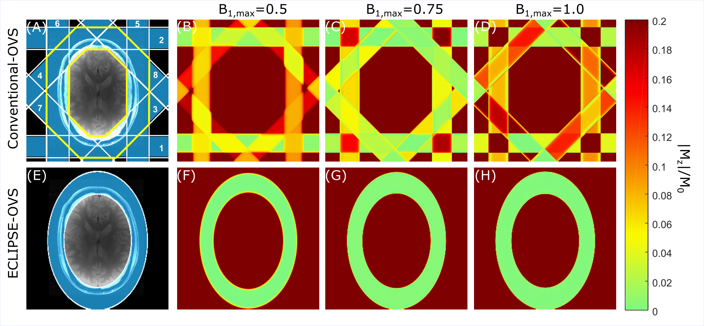
Proton Magnetic Resonance Spectroscopic Imaging (MRSI) is a powerful technique that can map the metabolic profile in the human brain, non-invasively. Extracranial lipid contaminants is a challenge for MRSI, that impedes reliable and accurate metabolite quantification in the brain regardless of magnetic field strength. The 8-slice OVS (hereafter called the conventional-OVS) method, in which eight saturation bands are placed around the skull, is an attractive alternative to cubical IVS methods (see Figure 1A) for improved brain coverage. However, due to overlapping of saturation-slices, and finite time requirements for RF pulses and spoiler gradients, spatially differential relaxation of lipids is unavoidable with conventional-OVS. Previous studies1-2 have shown improvements in the performance of conventional-OVS (mean lipid suppression of ~25-fold) by optimizing inter-pulse delays and RF pulse powers. However, a 16-RF pulse (8 x 2) based OVS module becomes SAR intensive and can be prohibitive moving towards ultra-high fields. Elliptical localization with pulsed second order fields (ECLIPSE) for MRSI was previously demonstrated3 with IVS of an elliptical ROI, providing improved brain coverage relative to a cubical ROI and reduced number of RF pulses relative to conventional-OVS. The objectives of this work were to develop ECLIPSE-based OVS and inversion recovery (IR) sequences with highly effective and robust lipid suppression combined with low SAR.Methods
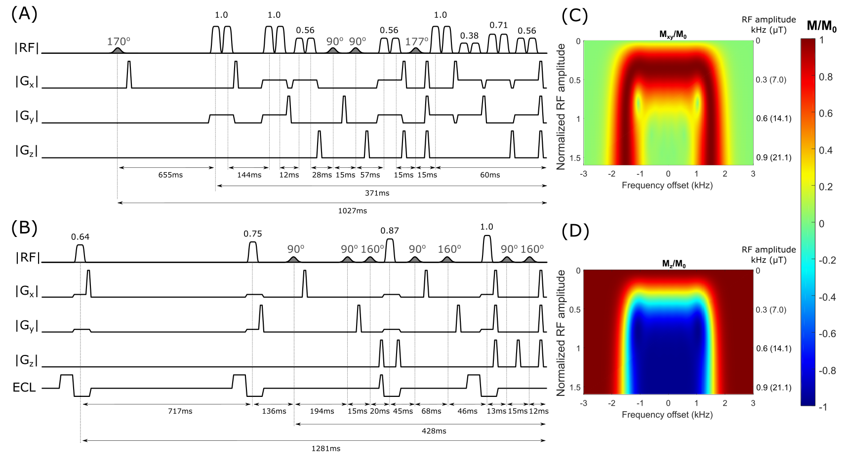
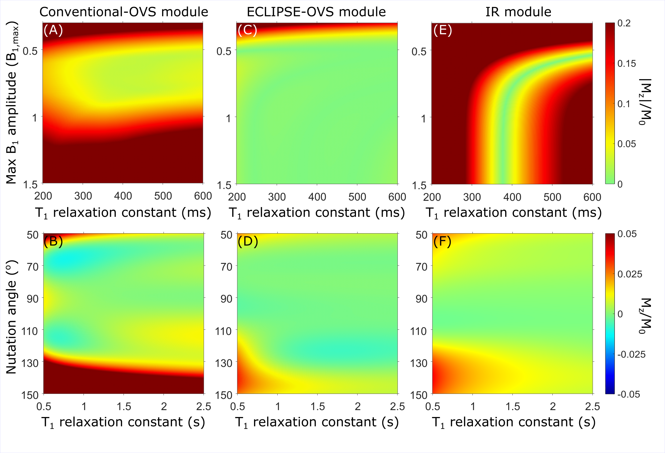
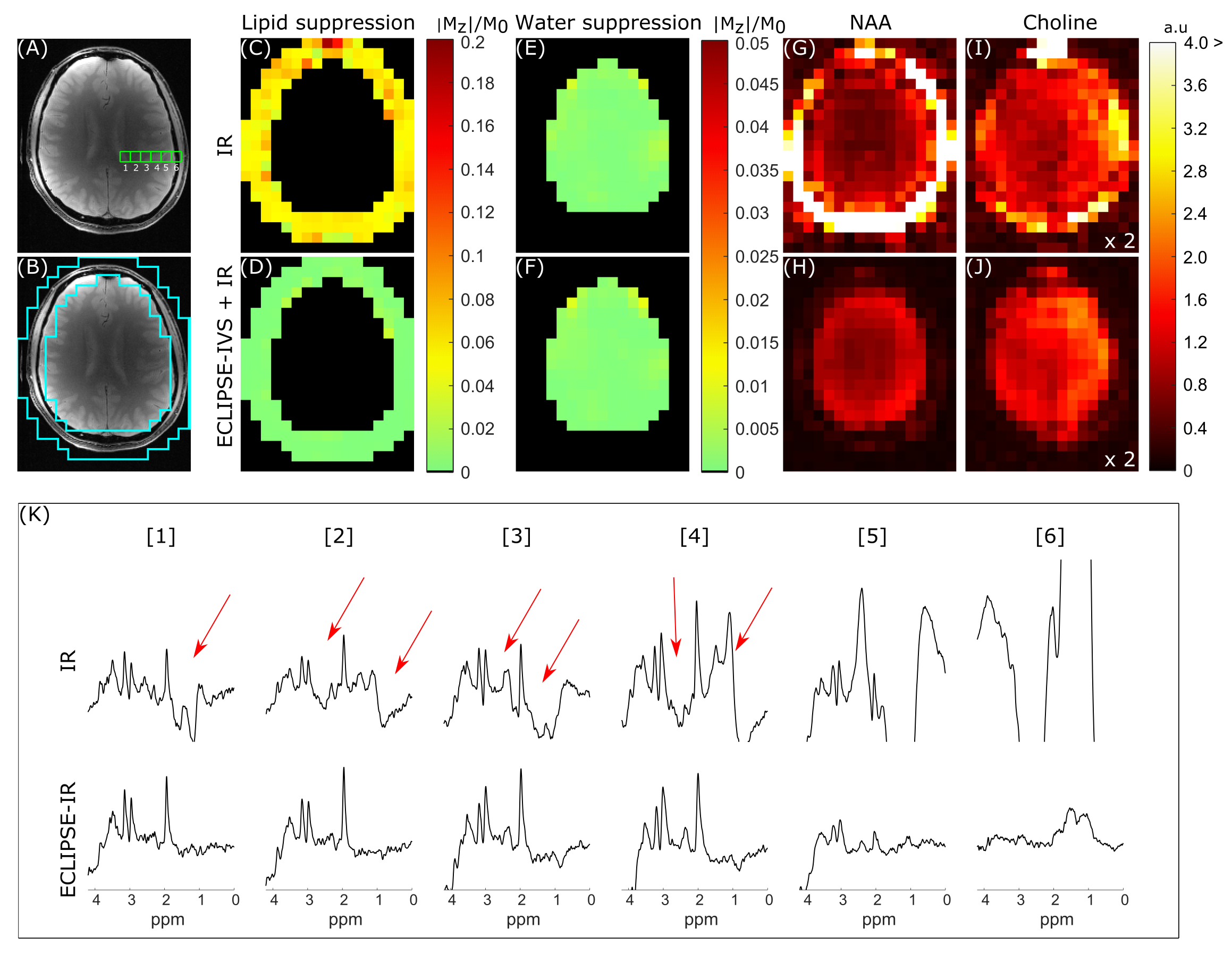
All MR experiments were performed on a 4T magnet (Magnex Scientific Ltd.) interfaced to a Bruker Avance III HD spectrometer running ParaVision 6 (Bruker, Billerica, MA, USA). The MR system contains actively shielded gradients capable of switching 30mT/m in 1150μs, and up to third order shimming. A within-brain B1+ optimized, 8-element Tx/Rx volume coil was used in a fixed phase configuration with ± 30% and ± 60% B1+ variation within the brain and extracranial region, respectively. The ECLIPSE system3 is a home-built, unshielded gradient insert consisting of Z2, X2Y2, and XY second order spherical harmonic magnetic fields with efficiencies of 5.48, 2.58 and 2.76 Hz/cm2/A, respectively, driven by 100A Techron 7780 current amplifiers (Techron, Elkhart, IN, USA) interfaced to a home-built multi-channel gradient controller4. Switching the ECLIPSE gradient coil resulted in small eddy currents in terms of time-varying B0 field variations that were virtually eliminated by an empirically determined gradient pre-pulse applied with opposite polarity. No post-processing eddy current corrections were required.A conventional-OVS module as shown in Figure 1A was optimized with two cycles and used as the reference against an ECLIPSE-based OVS module. The global T1 value of extracranial lipids was determined to be ~380 ms at 4T in vivo. The conventional-OVS module optimization was carried out over a T1 range spanning from 370-390 ms, and a B1 range spanning ± 40% with four allowed RF power settings to be allocated among the 16 RF pulses. ECLIPSE based OVS is achieved by repeatedly exciting and dephasing an elliptical ROI as illustrated in Figure 1E. Since each ECLIPSE based OVS pulse uniformly affects the entire in-plane extracranial lipid region, spatially differential relaxation as associated with conventional-OVS is avoided. A four RF pulse-based ECLIPSE-OVS module capable of being immune to a ± 60% variation in B1, while covering T1 species from 300-400 ms, was optimized and implemented. Extracranial lipid suppression with a global inversion recovery (IR) was implemented utilizing an AFP inversion pulse. To improve extracranial lipid suppression of the global IR sequence, the spin-echo excitation pulse was modified with ECLIPSE magnetic fields, thereby selecting an elliptical ROI for IVS. This sequence utilizes a global IR, with an ECLIPSE-IVS excitation pulse, is here after termed the ECLIPSE-IVS + IR module. Three healthy volunteers participated in the study to compare ECLIPSE-OVS and conventional-OVS, and the same three volunteers participated in a separate session to compare the IR and ECLIPSE-IVS + IR methods.
Results
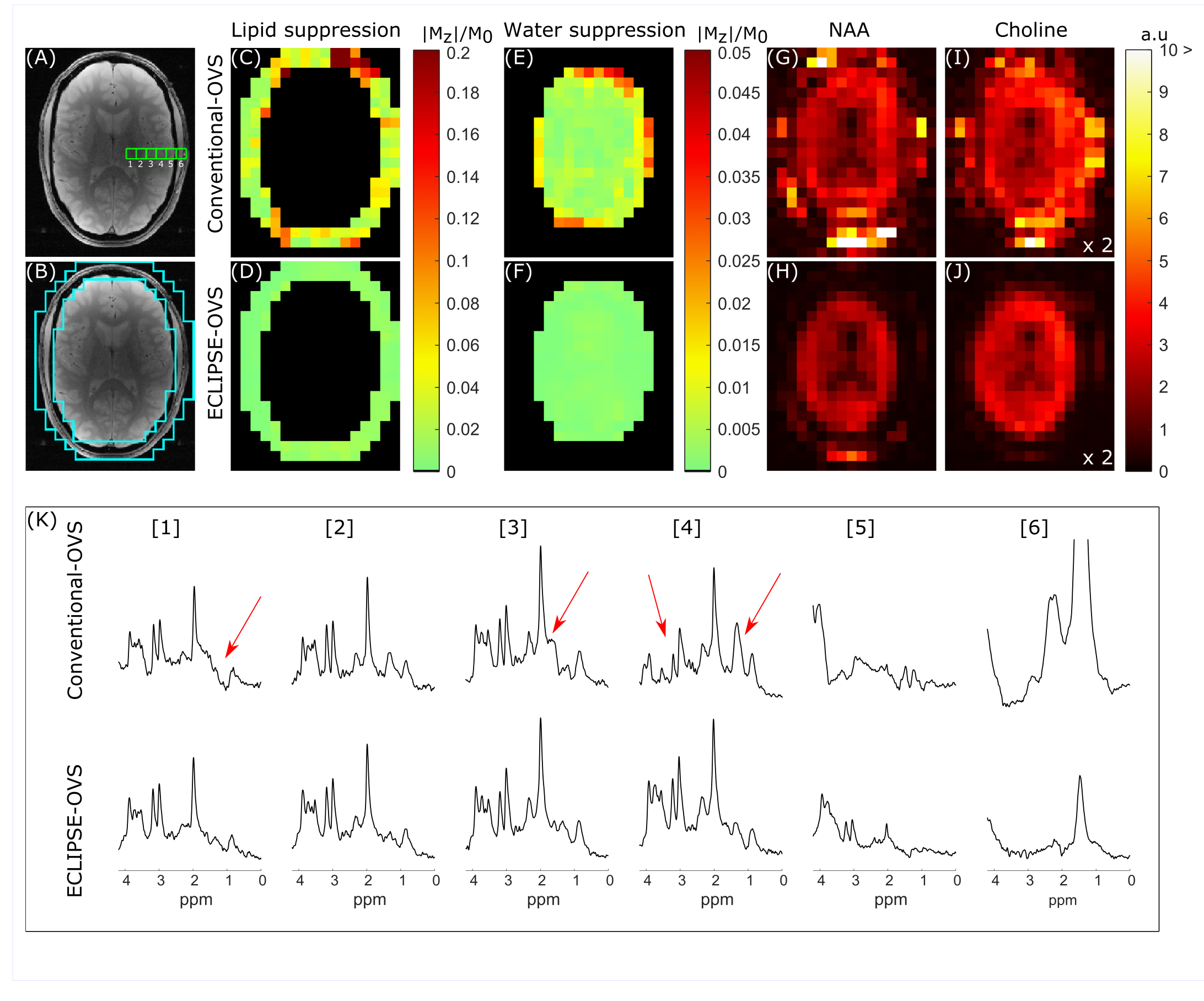
Lipid and water suppression performance in simulation for the conventional-OVS module (see Figure 2A), the optimized ECLIPSE-OVS module (see Figure 2B), and the IR module (not shown) are summarized in Figure 3 A-B, C-D, and E-F, respectively. Lipid and water suppression performance, and the overall MRSI data quality from one volunteer for conventional-OVS and ECLIPSE-OVS are summarized in Figure 4. The mean lipid suppression factor for this volunteer was 18-fold for conventional-OVS and ~135-fold for ECLIPSE-OVS. The IR based MRSI sequence was compared against the ECLIPSE-IVS + IR sequence as summarized in Figure 5 with an identical layout to the OVS comparison in Figure 4. The mean lipid suppression factor for this volunteer was ~18-fold with IR, and ~257-fold with ECLIPSE-IVS + IR (Figure 5D). In addition to the highly robust lipid suppression provided by ECLIPSE, the results are also achieved with only 30% of the power required by conventional-OVS.Conclusions
Both ECLIPSE methods provide > 100-fold mean lipid suppression for robust and short-TE MRSI of the human brain. The low power requirements in combination with insensitivity to B1 and T1, makes ECLIPSE-based OVS particularly attractive for high field MRSI.Acknowledgements
This research was supported by NIH grant R01- EB014861.References
[1] Henning A, Schar M, Schulte RF, Wilm B, Pruessmann KP, Boesiger P. SELOVS: brain MRSI localization based on highly selective T1- and B1- insensitive outer-volume suppression at 3T. Magn Reson Med 2008;59(1):40-51.
[2] Henning A, Fuchs A, Murdoch JB, Boesiger P. Slice-selective FID acquisition, localized by outer volume suppression (FIDLOVS) for (1)H-MRSI of the human brain at 7 T with minimal signal loss. NMR in biomedicine 2009;22(7):683-696.
[3] de Graaf RA, Brown PB, De Feyter HM, McIntyre S, Nixon TW. Elliptical localization with pulsed second-order fields (ECLIPSE) for robust lipid suppression in proton MRSI. NMR in biomedicine 2018;31(9):e3949.
[4] Nixon TW, McIntyre S, de Graaf RA. The design and implementation of a 64 channel arbitrary gradient waveform controller. Proc Int Soc Magn Reson Med. 2017;25:969.
Figures