0365
Intra-session and inter-subject variability of 3D-FID-MRSI using single-echo volumetric EPI navigators at 3T1High Field MR Centre, Department of Biomedical Imaging and Image-guided Therapy, Medical University of Vienna, Vienna, Austria, 2Athinoula A. Martinos Center for Biomedical Imaging, Department of Radiology, Harvard Medical School, Massachusetts General Hospital, Vienna, MA, United States, 3Athinoula A. Martinos Center for Biomedical Imaging, Department of Radiology,, Harvard Medical School, Massachusetts General Hospital, Boston, MA, United States, 4Christian Doppler Laboratory for Clinical Molecular MR Imaging, Vienna, Austria
Synopsis
We demonstrate the combination of 3D free induction decay proton MR spectroscopic imaging and spatial encoding via concentric-ring trajectories at 3T. To improve the reliability a well as the temporal stability, single-echo, imaging-based volumetric navigators for real-time motion/shim-correction were additionally integrated. All intra-subject coefficients of variation and most of the inter-subject coefficients of variation obtained with motion/shim-correction were lower (i.e., better) than without and resulted in higher SNRs and lower CRLBs.
Introduction
We demonstrate the combination of 3D free induction decay (FID)[1] proton MR spectroscopic imaging (MRSI) and spatial encoding via concentric-ring trajectories (CRT) at 3T[2-4]. FID-MRSI[1] has many benefits including high detection sensitivity, in particular for J-coupled metabolites like glutamate (Glu) or glutamine (Gln). This makes it highly attractive not only for clinical, but also for, potentially, functional MRSI[5]. To improve the reliability a well as the temporal stability, single-echo, imaging-based volumetric navigators (se-vNavs) for real-time motion/shim-correction (SHMOCO) were additionally integrated but still preserve ultra-short TR acquisitions[6].Methods
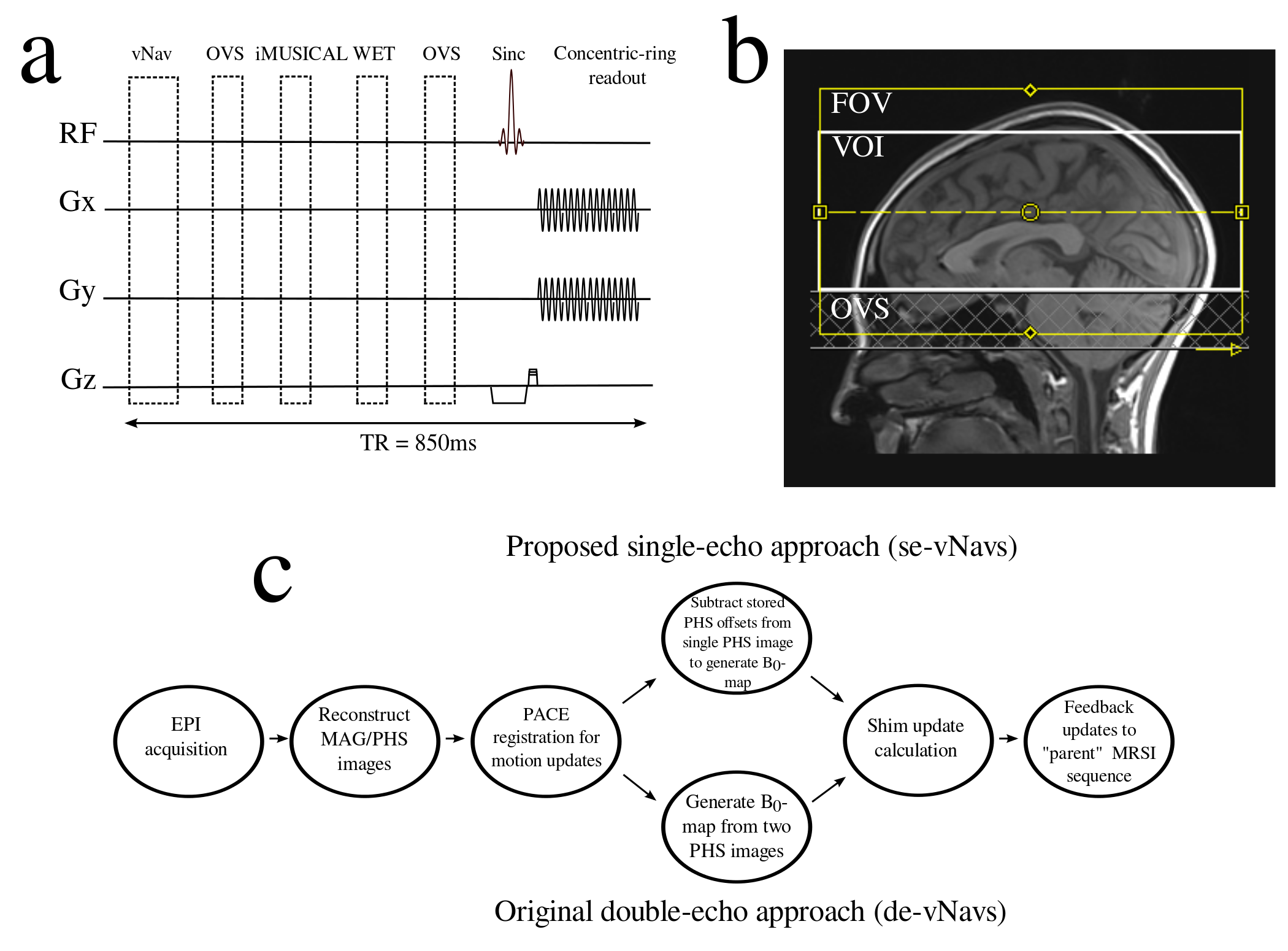
This study was performed on a 3T Prisma MR scanner with a 64-channel receive-only head coil (all Siemens Healthineers) in six healthy volunteers (male/female,4/2; age,28.8±5.4y). Institutional Review Board approval and written, informed consent were obtained prior to the MR examinations. The tracking accuracy (position and B0-field) of our proposed se-vNavs was compared to the original double-echo volumetric navigators (de-vNavs)[6] in phantoms (rest and translation) and in vivo (voluntary head rotation). While motion updates remain unchanged for the se-vNavs compared to de-vNavs, B0-maps were created from the single phase images after subtracting pre-stored, coil-dependent phase offsets [7]. These are calculated using ASPIRE[8] from a dual-echo EPI reference prescan whose parameters matched those of the se-vNavs listed below, except for the dual-echo TE1/TE2 of 7/14ms.The parameters for se-vNavs were: TR, 17ms; TE, 7ms (de-vNavs: TE1/TE2, 7/9.4ms); matrix, 32×32; slices, 18; FOV,256×256×144mm3; bandwidth,4734Hz/pixel; flip angle, 4°;echo train length, 32;water excitation only; slice partial Fourier, 6/8. Using our se-vNavs, the total navigator block requires only ~360ms.
The 3D-FID-MRSI sequence (Figure 1) with CRT readout used the following settings: TR, 850ms; acquisition delay, 0.8ms; flip angle, 70°; 600µs sinc excitation pulse; B1, 13.2µT; VOI, 220×220×76mm3; field-of-view, 220×220×126mm3; in vivo matrix size, 50×50×21 (in phantoms, 32×32×21); complex spectral data points, 360; acquisition bandwidth, 1030Hz; no temporal interleaving; acquisition window, 350ms; averages, 1; spherical k-space coverage; WET water suppression; outer-volume saturation (OVS) band (30mm thick) below VOI covering nasal cavity and skull base; TA, 5:40min.The CRT data reconstruction was performed as described previously[2].
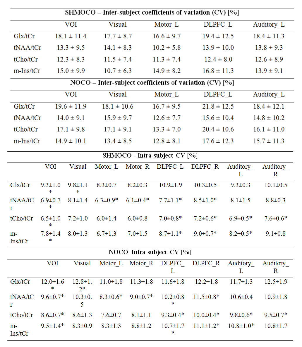
We investigated the intra-session stability of a 5:40min 3D-FID-MRSI scan with SHMOCO and no correction (NOCO) in five resting subjects which were scanned each within one session and without repositioning with a total of eight 3D-FID-MRSI scans: four no correction (NOCO) and four shim/motion-correction(SHMOCO) in an interleaved fashion. Intra/inter-subject coefficients of variation (CV) of major metabolites over the whole 3D volume and in selected regions of interest (ROI) were assessed.
Results
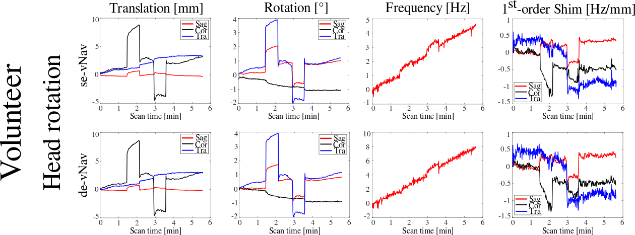
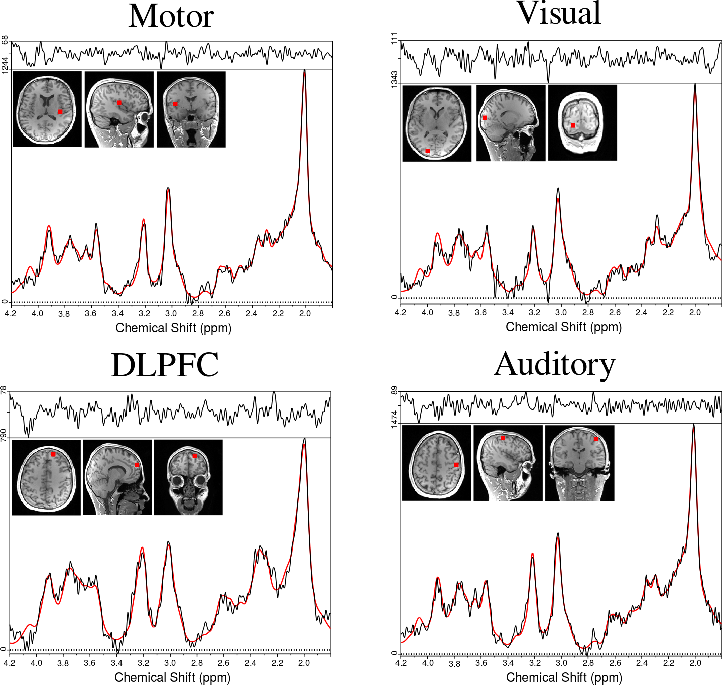
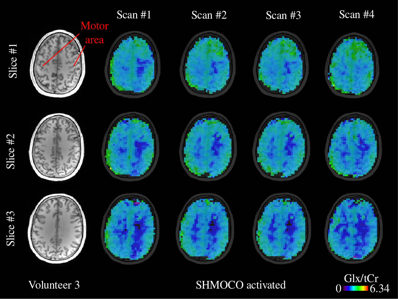
Figure 2 shows motion and B0-field logs from the phantom and in vivo (volunteer 1) measurements. Phantom and in vivo scans showed highly consistent tracking performance for se-vNavs compared to the original de-vNavs, but lower frequency drift. Sample spectra for volunteer 2 are shown in Figure 3. Figure 4 depicts the four Glx/tCr (Glx=glutamate+glutamin, tCr=total creatine) metabolic ratio maps with SHMOCO for volunteer 3 in three adjacent slices. The average maximum translations from the NOCO (0.9±0.4mm) and SHMOCO (1.2±0.4mm) scans were statistically non-significant (p=0.1). Table 1 summarizes the means and standard deviations of the intra/inter-subject CVs obtained for the different metabolites ratios and brain regions averaged over all five volunteers. All intra-subject CVs and most of the inter-subject CVs obtained with SHMOCO were lower (i.e., better) compared to NOCO, with improvements of up to ~30%. Intra-subject CVs of 9.3%, 6.9%, 6.5%, and 7.8% were obtained across the full VOI for Glx/tCr, tNAA/tCr (tNAA=N-acetyl-aspartate+N-acetyl-aspartyl glutamate), tCho/tCr (tCho=total choline) and m-Ins/tCr (m-Ins=myo-inositol) with SHMOCO. For Glx/tCr, values of 9.8% and 8.2% were obtained in visual and motor cortex. Significant differences were found between NOCO and SHMOCO over the whole VOI (p<0.001), but no moderation effect (p=0.67) with the metabolites, i.e., no evidence that the difference between NOCO and SHMOCO was more pronounced for certain metabolites. No moderation effect was also found with the ROIs and metabolites, i.e., no significant evidence that SHMOCO performed better for certain metabolites in certain ROIs (p=0.92). Using SHMOCO led to moderately higher SNR (VOI SNR: 16.5±5.3, FWHM 8.2±2.8 Hz) and lower CRLBs (tNAA 4.1±1.3%) compared to NOCO (VOI SNR 15.9±5.3, FWHM 8.4±2.5 Hz, tNAA CRLB 4.2±1.5%), while the FWHMs were little affected.Discussion and Conclusion
We presented the first use of CRTs in a 3D-FID-MRSI sequence at 3T to generate high-resolution metabolic maps in 5:40 min. The sequence was further equipped with a two times shorter, single-echo volumetric EPI navigator for real-time motion/shim correction. Our results confirm that the use of SHMOCO gives overall significantly better intra-subject CVs than NOCO, which mainly reflects SHMOCO’s ability to correct for temporal frequency drifts and involuntary subject movements. Concerning possible applications, functional MRS studies have reported changes in glutamate [5] range from only subtle increases of 2-4% after visual or motor stimuli to more pronounced changes (up to 22%) after pain stimuli. While functional MRSI has not yet been conducted so far, this report contributes to paving the way to functional MRSI by assessing the stability of 3D-FID-MRSI which makes it possible to judge if small metabolite changes can actually be resolved on a single-subject basis or on a group level.Acknowledgements
This study was supported by the Austrian Science Fund (FWF): KLI 718, P 30701, P 31452 and J 4124, and the Christian Doppler Laboratory for Clinical Molecular MR Imaging.References
1 Bogner W, Gruber S, Trattnig S, Chmelik M. High-resolution mapping of human brain metabolites by free induction decay 1H MRSI at 7 T. NMR Biomed. [Internet] 2012;25:873–882. doi: 10.1002/nbm.1805.
2. Hingerl L, Bogner W, Moser P, Považan M, Hangel G, Heckova E, Gruber S, Trattnig S, Strasser B. Density-weighted concentric circle trajectories for high resolution brain magnetic resonance spectroscopic imaging at 7T. Magn. Reson. Med. [Internet] 2017. doi: 10.1002/mrm.26987.
3. Furuyama JK, Wilson NE, Thomas MA. Spectroscopic imaging using concentrically circular echo-planar trajectories in vivo. Magn. Reson. Med. [Internet] 2012;67:1515–1522. doi: 10.1002/mrm.23184.
4. Steel A, Chiew M, Jezzard P, Voets NL, Plaha P, Thomas MA, Stagg CJ, Emir UE. Metabolite-cycled density-weighted concentric rings k-space trajectory (DW-CRT) enables high-resolution 1H magnetic resonance spectroscopic imaging at 3-Tesla. Sci. Rep. [Internet] 2018;8:7792. doi: 10.1038/S41598-018-26096-Y.
5. Stanley JA, Raz N. Functional Magnetic Resonance Spectroscopy: The “New” MRS for Cognitive Neuroscience and Psychiatry Research. Front. Psychiatry [Internet] 2018;9. doi: 10.3389/fpsyt.2018.00076.
6. Hess AT, Dylan Tisdall M, Andronesi OC, Meintjes EM, Van Der Kouwe AJW. Real-time motion and B0 corrected single voxel spectroscopy using volumetric navigators. Magn. Reson. Med. 2011;66:314–323. doi: 10.1002/mrm.22805.
7. Dymerska B, Poser BA, Barth M, Trattnig S, Robinson SD. A method for the dynamic correction of B0-related distortions in single-echo EPI at 7 T. Neuroimage [Internet] 2018;168:321–331. doi: 10.1016/j.neuroimage.2016.07.009.
8. Eckstein K, Dymerska B, Bachrata B, Bogner W, Poljanc K, Trattnig S, Robinson SD. Computationally Efficient Combination of Multi-channel Phase Data From Multi-echo Acquisitions (ASPIRE). Magn. Reson. Med. [Internet] 2018;79:2996–3006. doi: 10.1002/mrm.26963.
Figures