0363
Calibration-free regional RF shims for localised MR spectroscopy1Sir Peter Mansfield Imaging Centre, School of Physics and Astronomy, University of Nottingham, Nottingham, United Kingdom, 2Russel H. Morgan Department of Radiology and Radiological Science, Johns Hopkins University School Of Medicine, Baltimore, MD, United States
Synopsis
RF shimming can increase B1+ availability, which is critical for robust localised MR spectroscopy at ultra-high field. Shim calibration is performed on a region-wise basis and is, therefore, time consuming. Additionally, B1 distributions become difficult to predict. Recent work has shown that ‘universal’ pulses can be generated offline – avoiding the need for calibration. Here, we determine static calibration-free RF shims, optimised over 5 heads, for 3 different brain regions. B1+ availability using calibration-free shims was significantly higher than quadrature and comparable to tailored shimming. High quality spectra were also obtained from 3 regions with the calibration-free shims.
Aim
To develop robust, calibration-free, fixed RF shim settings for localised single-voxel MR spectroscopy in different brain regions at ultra-high field.Introduction
Inhomogeneity in the transmit field (B1+) is a considerable challenge for MRS at ultra-high field (≥ 7 T). Low B1+ availability and flip angle variation across voxels leads to poor water suppression and localisation errors.1 Parallel transmission (pTx) allows for RF shimming of channel phases and amplitudes to improve B1+ availability in desired regions.2,3 However, calculating subject-specific shims is time-consuming when using channel cycling for B1-mapping and when several ROIs are required in one session (~10 mins). Approaches to avoid calibration include using fixed settings4 or predicted shims using machine-learning.5,6 Recently, Gras et al. generated a set of ‘universal’ k-T points pulses by jointly optimising over a database of B1+ maps and these could be applied to any head without calibration.7,8 A similar approach was shown for spatially-selective excitation pulses.9,10 Given the similarity in B1+ profile across heads, we propose a calibration-free regional RF shim method based on offline ROI registration and database optimisation for predefined locations.Methods
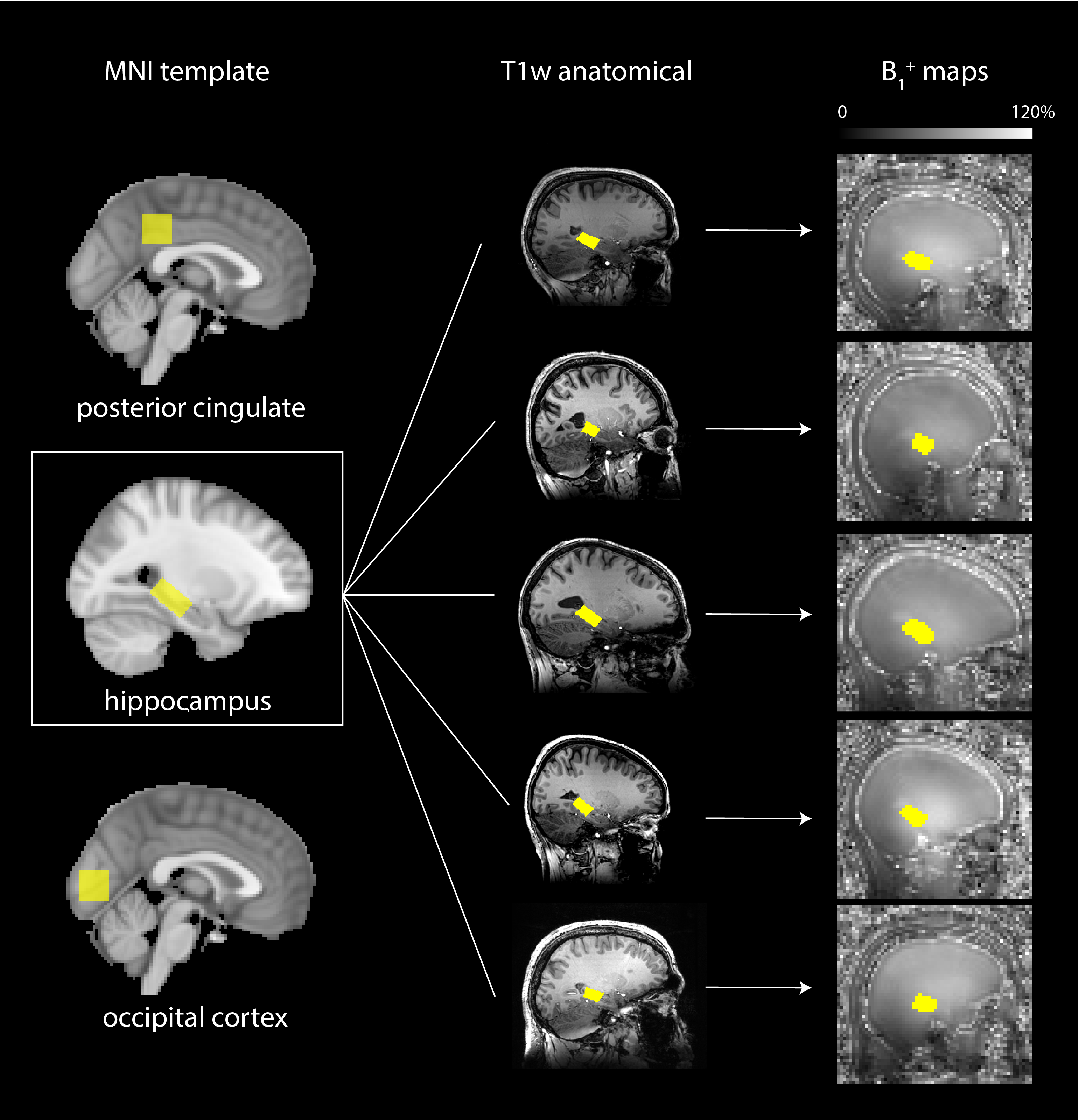
A database of B1 maps was generated from 5 volunteers (P1-5, mean age = 28±5 years, 2F) scanned on a 7 T Philips Achieva MR system with pTx head coil (8Tx/32Rx, Nova Medical). B1+ maps for each channel were obtained by combining whole-brain B1 maps acquired with a DREAM11 sequence (TA=9s) and gradient-echo images acquired on each channel (TA=3m 51s). ROIs for 3 commonly measured regions were defined in MNI space12, namely, occipital cortex (OCC, 20x20x20mm3), hippocampus (Hippo., 30x15x12mm3) and posterior cingulate cortex (PCC, 20x20x20mm3) (see Fig. 1). Each ROI was registered to the B1+ maps using non-linear transformation from MNI-space to T1-weighted images (FNIRT, FSL13) followed by an affine transform.Tailored RF phase-shimming was performed on each subject by minimising the mean least-square error between the predicted B1+ and a target value of 1 (or 100%) over all $$$N_{ROI}$$$ voxels in the ROI. Calibration-free shims were found by finding the shim, $$$w$$$, which, when applied to each subject, $$$k$$$, in the database, minimises this error for the worst-case subject in a minimax7 approach such that,
$$ \min_{w} \max_{k} \left( \frac{1}{N_{ROI,k}} \sum_{i=1}^{N_{ROI,k}} (A_{i,k}w -1)^2 \right) $$
where $$$A_{i,k}$$$ is the fractional B1+ on each channel in the $$$i^{\textrm{th}}$$$ voxel of the ROI for the $$$k^{\textrm{th}}$$$ subject.
Calibration-free shims were applied in 3 test volunteers (mean age = 29±5 years, 1F) not included in the database, and MRS was acquired using STEAM (TE=14ms/TR=3-4s, NT=64, VAPOR water suppression). RF shimming and optimisation was carried out with an in-house tool written in MATLAB (The MathWorks, Natick, MA). Data were compared to results produced using quadrature mode. Forward power through each channel was fixed across shim conditions. Statistical differences were assessed using a two-way ANOVA with significance threshold, $$$\alpha$$$=0.05. SNR was measured as NAA peak height divided by RMS noise from 12-13 ppm.
Results
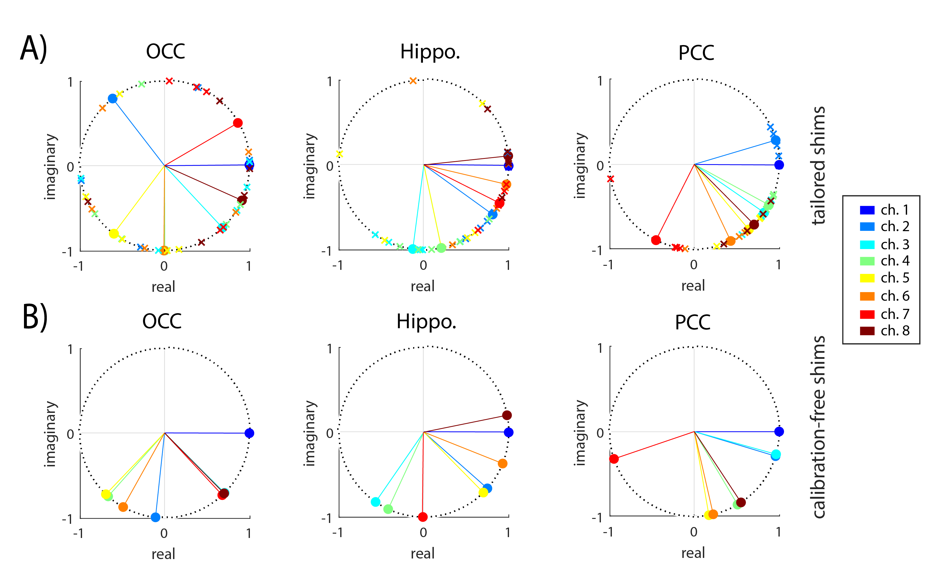
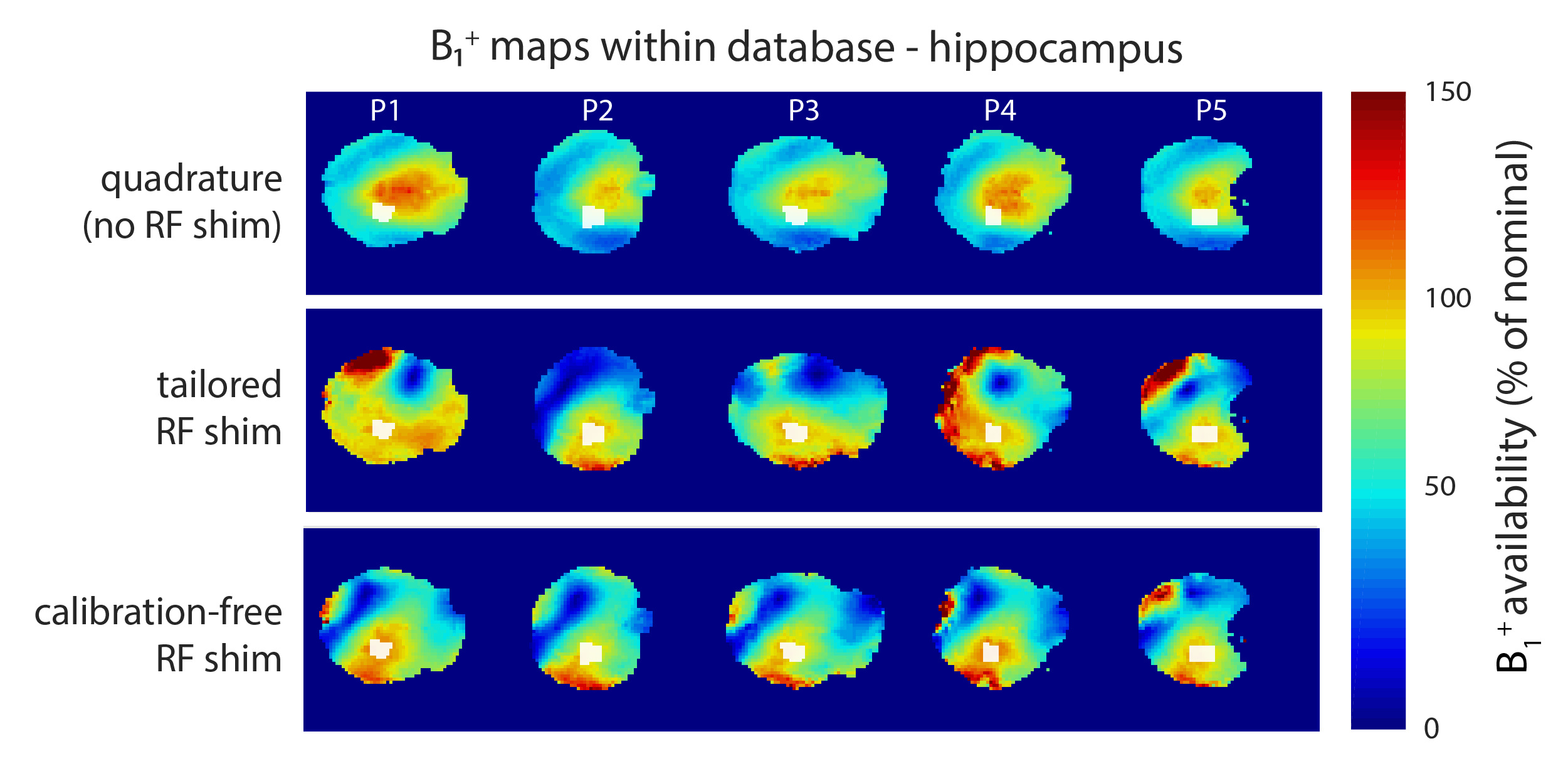
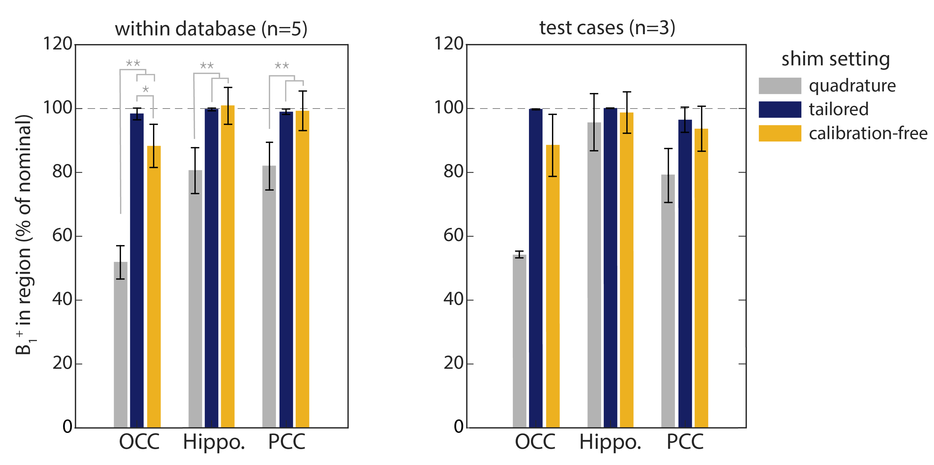
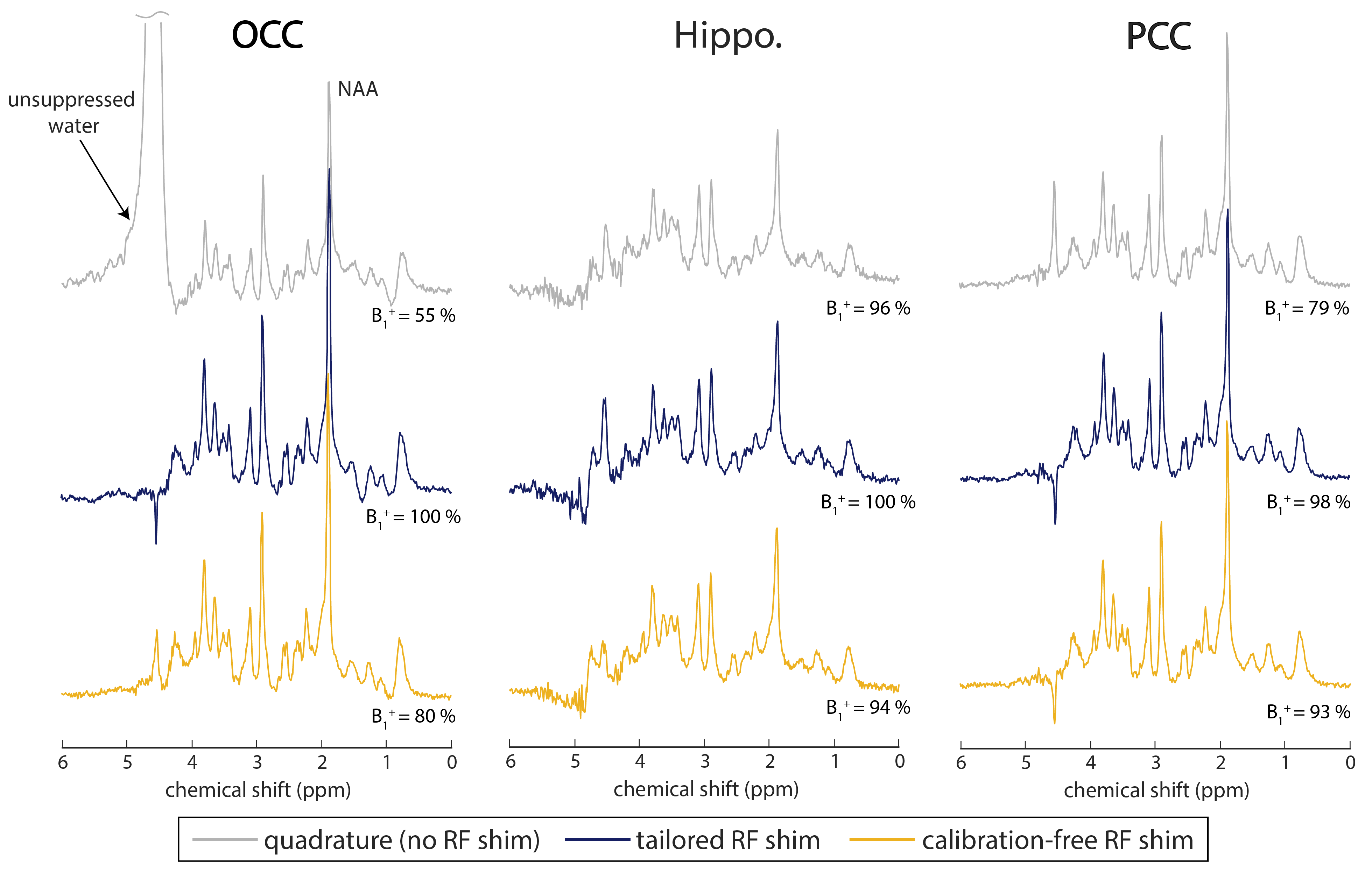
Fig. 2 shows the shims calculated for each ROI using tailored- and calibration-free approaches. Channel phases for calibration-free shims (Fig. 2B) in hippocampus and PCC are similar to the mean of the tailored shims (Fig. 2A). Greater variability in phases was observed for OCC. Predicted B1+ distributions are shown in Fig. 3 for the hippocampal ROI. Tailored shimming led to a greater variation in B1+ outside of the ROI, whereas calibration-free shims produced more consistent profiles across datasets. In all ROIs, the mean available B1+ was significantly higher after shimming than when operating in quadrature mode (p<0.0005) (Fig 4). No statistical differences were found in mean B1+ when using calibration-free shims compared to tailored shimming, apart from in OCC which was (10.0±9.9)% lower with the calibration-free shims (p=0.046). Similar trends in B1+ availability after calibration-free shimming were observed in the 3 test volunteers. Fig. 5 shows spectra obtained for one test volunteer. Spectra using calibration-free shims were of high quality and similar to those produced using tailored shimming. In OCC, there was large residual water in quadrature mode (B1+=55%). For calibration-free shims, residual water was lower than the NAA signal across all subjects and ROIs. Across the 3 test cases, mean SNR was similar (279±35 vs. 245±25) in OCC, (94±8 vs. 87±12) in hippocampus and (202±8 vs. 196±34) in PCC for tailored vs. calibration-free shims.Discussion
A method is demonstrated to generate static RF shims applicable across different heads in localised regions, using a set of measured B1 maps and anatomical scans. Applying these calibration-free shims on subjects within, and outside of, the database revealed a highly similar B1+ availability compared to tailored shimming, similar to previous findings using universal pulses.7 As expected, B1+ availability was significantly higher in all ROIs than in quadrature mode. The difference in B1+ in OCC may be attributable to larger variation in that region across head positions and sizes (Fig 2A). Spectra obtained using calibration-free shims were of similar SNR and residual water compared to tailored shimming. Further work will explore the effect of database size and target functions. Advantages of the calibration-free method are the elimination of time spent shimming, as well as more predictable SAR distributions, which may allow for increases in transmit power.Acknowledgements
The authors would like to acknowledge the Precision Imaging Beacon, University of Nottingham.References
1. Kreis R. Issues of spectral quality in clinical 1H-magnetic resonance spectroscopy and a gallery of artifacts. NMR Biomed. 2004;17(6):361-381. doi:10.1002/nbm.891
2. Metzger GJ, Snyder C, Akgun C, Vaughan T, Ugurbil K, Van de Moortele P-F. Local B1+ shimming for prostate imaging with transceiver arrays at 7T based on subject-dependent transmit phase measurements. Magn Reson Med. 2008;59(2):396-409. doi:10.1002/mrm.21476
3. Emir UE, Auerbach EJ, Moortele P-F Van De, et al. Regional neurochemical profiles in the human brain measured by 1H MRS at 7 T using local B1 shimming. NMR Biomed. 2012;25(1):152-160. doi:10.1002/nbm.1727
4. Nistler J, Diehl J, Renz W, Eberler L. Homogeneity improvement using a 2 port birdcage coil. In: Proceedings of the 15th Annual Meeting of the ISMRM, Berlin. ; 2007:1063.
5. Ianni JD, Cao Z, Grissom WA. Machine learning RF shimming: Prediction by iteratively projected ridge regression. Magn Reson Med. 2018;80(5):1871-1881. doi:10.1002/mrm.27192
6. Tomi-Tricot R, Gras V, Thirion B, et al. SmartPulse, a machine learning approach for calibration-free dynamic RF shimming: Preliminary study in a clinical environment. Magn Reson Med. 2019;(December 2018):2016-2031. doi:10.1002/mrm.27870
7. Gras V, Vignaud A, Amadon A, Le Bihan D, Boulant N. Universal pulses: A new concept for calibration-free parallel transmission. Magn Reson Med. 2017;77(2):635-643. doi:10.1002/mrm.26148
8. Gras V, Mauconduit F, Vignaud A, et al. Design of universal parallel-transmit refocusing k T -point pulses and application to 3D T 2 -weighted imaging at 7T. Magn Reson Med. 2018;80(1):53-65. doi:10.1002/mrm.27001
9. Geldschläger O, Shao T, Henning A. Universal Parallel Transmit Pulse Design for Local Excitation. In: Proceedings of the Joint Annual Meeting ISMRM-ESMRMB, Paris. ; 2018:3395.
10. Geldschläger O, Shao T, Henning A. Universal Parallel Transmit Pulse Design for 3-Dimensional Local-Excitation: A 9.4T Simulation Study. In: Proceedings of the 27th Annual Meeting of the ISMRM, Montreal. Montreal; 2019:4639.
11. Nehrke K, Börnert P. DREAM-a novel approach for robust, ultrafast, multislice B1 mapping. Magn Reson Med. 2012;68(5):1517-1526. doi:10.1002/mrm.24158
12. Park YW, Deelchand DK, Joers JM, et al. AutoVOI: real-time automatic prescription of volume-of-interest for single voxel spectroscopy. Magn Reson Med. 2018;80(5):1787-1798. doi:10.1002/mrm.27203
13. Jenkinson M, Beckmann CF, Behrens TEJ,
Woolrich MW, Smith SM. FSL. Neuroimage. 2012;62(2):782-790.
doi:10.1016/j.neuroimage.2011.09.015
Figures