0362
Compositional and Morphological Characterization of Knee Articular Cartilage in Collegiate Basketball Players using Multiparametric MRI1Department of Radiology and Biomedical Imaging, University of California, San Francisco, San Francisco, CA, United States, 2Department of Radiology, Stanford University, Stanford, CA, United States, 3Department of Radiology and Imaging, Hospital for Special Surgery, New York, NY, United States
Synopsis
Magnetic resonance imaging (MRI) is commonly used to evaluate the morphology of athletes with high knee impact; however, the biochemical composition of their cartilage is not as well understood. In this study, we utilized voxel-based relaxometry (VBR), a fully automatic registration technique, to compare local distribution of knee articular cartilage T1ρ and T2 relaxation times between high knee impact athletes (basketball players) and non-knee impact athletes (swimmers). Statistical analysis revealed laminar differences near the patella, with basketball players having prolonged values in the deep layer. These findings, amongst others, related well to morphological evaluation of the image set.
Introduction
The knee is the joint most vulnerable to articular cartilage degeneration and injury, particularly in jumping athletes who exert high compressive and torsional forces during practice and competitive play. Morphological changes are consistently prevalent in knee cartilage of jumping and impact athletes using magnetic resonance imaging (MRI).1-3 Recent advanced compositional MRI techniques, such as simultaneous T1ρ- and T2-weighted acquisition, can quantify biochemical changes of macromolecules that initiate cartilage degeneration prior to the manifestation of visible morphological changes.4-6 Quantitatively determining early changes in matrix biochemistry would advance understanding of how impact sports affect cartilage health and overall risk of degenerative disease. In this study, we complement morphological evaluation with voxel-based relaxometry (VBR),7 a fully automatic registration technique that aligns images to a single reference template, to compare the local distribution of knee articular cartilage T1ρ and T2 relaxation times between high knee impact athletes (basketball players) and non-knee impact athletes (swimmers).Methods
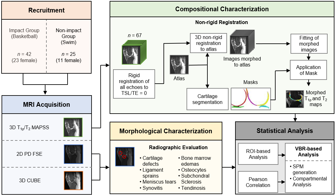
This was a multicenter cross-sectional study performed in accordance to the rules and procedures approved by the Institutional Review Boards of the 3 participating sites. All participants provided informed written consent. Two cohorts of NCAA collegiate-level athletes were imaged on 3.0T MRI prior to their competitive seasons: 42 basketball players (23 female); and 25 swimmers (11 female). The MRI protocol included 2D proton-density-weighted (PD) fast spin echo (FSE), 3D CUBE, and a 3D combined T1ρ/T2 magnetization-prepared angle-modulated portioned k-space spoiled gradient echo snapshots (MAPSS) sequence.4 For morphological characterization, a board-certified musculoskeletal radiologist with 25 years of experience evaluated the MR images. Cartilage lesions were graded in a blinded fashion using the Modified Noyes Score.8 Cases with cartilage lesions (modified Noyes ≥ 1) were not considered for VBR to focus compositional analysis on pre-structural abnormalities. For each case, sagittal MAPSS images in all echoes were rigidly registered to the first time of spin-lock/echo time (0 msec) and nonrigidly registered to an atlas to morph all images to a common reference space. T1ρ and T2 maps were then created using Levenberg-Marquardt mono-exponential fitting. In VBR-based analysis, statistical parametric maps (SPMs) were generated voxel-by-voxel to assess local group differences after controlling for sex, body mass index, and site of acquisition. A classic region of interest (ROI)-based technique, consisting of automatic compartmental segmentation of the cartilage and averaging of T1ρ and T2 values across the entire thickness of each compartment, was performed for comparison.7,9 Unpaired t-test and Pearson Correlation were used to evaluate group differences in ROI- and VBR-analysis, respectively.Results
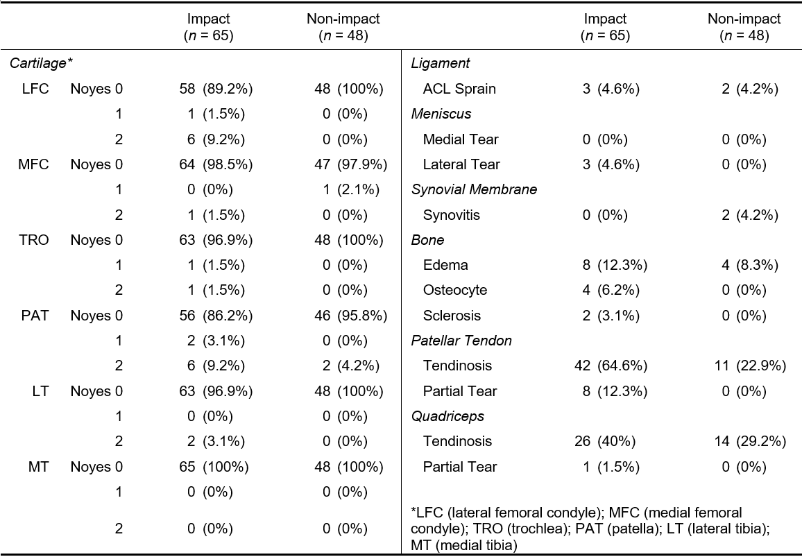
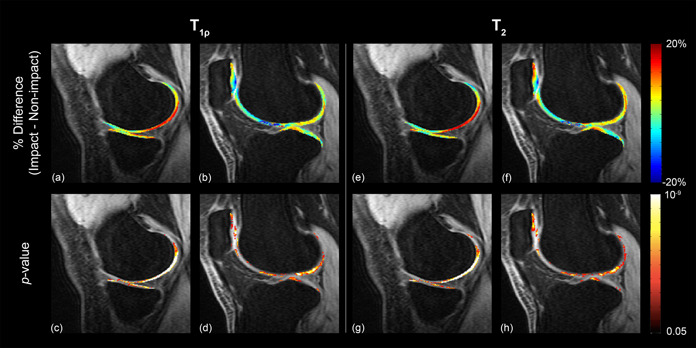
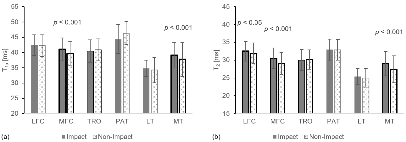
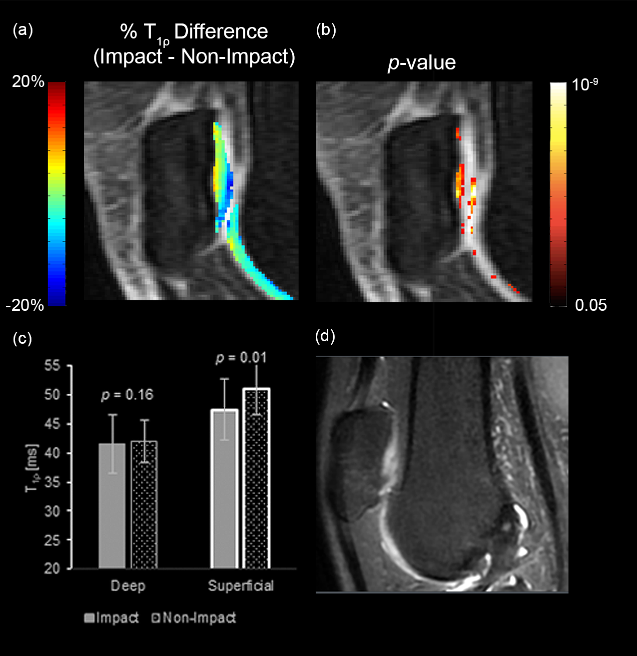
By morphologic evaluation, the occurrence of structural abnormalities was significantly higher in the impact group for all categories evaluated, except synovitis (Table 1). Articular cartilage defects accounted for 17% of all defects and occurred in 18% of basketball players and 4% of swimmers. From VBR-based analysis, the average SPMs in both impact and non-impact groups displayed prolonged values near the trochlear groove and areas of tibio-femoral articulation, and shorter values anteriorly and posteriorly. Comparison of the two groups demonstrated significant differences by sport, with basketball players generally having higher values, particularly in femoral condyle cartilage (lateral: 12.63% average % difference, 42.7% significant voxels; medial: 3.48% average % difference, 29.2% significant voxels) (Fig. 2). Group analysis also revealed differences through the depth of the articular cartilage: basketball players had higher T1ρ and T2 values in the deep layer of cartilage while swimmers had higher values in the superficial layer. This is most notable in the patellofemoral joint. Amongst the ROI-based results, basketball players had significantly elevated T1ρ values in the medial femoral and tibial compartments (Fig. 3) and no significant differences were detected in the patellofemoral compartment. Further division of the patellofemoral ROI into deep and superficial layers showed significant laminar differences (Fig. 4).Discussion
This study used multiparametric MRI to explore morphological and compositional differences between high knee impact athletes (basketball players) and low knee impact athletes (swimmers). Our results demonstrate that a fully automatic, voxel-by-voxel quantification of MR T1ρ/T2 relaxation times can provide information about the local biochemical composition of the cartilage matrix that complements structural findings. Disparity between the deep and superficial articular layers could be attributed to differences in the frequency and magnitude of loading forces in jumping and running exercises practiced in basketball, with more forceful compressive loads imparted to the basilar cartilage layers and subchondral bone. While the laminar VBR analysis may be limited by the in-plane resolution (0.6 mm x 1.2 mm), the differences detected in the patellar cartilage relate well to the morphological findings being predominantly in the patellofemoral joint. This contrast also illustrates the disadvantages of the commonly used ROI-based results, where laminar differences may be averaged out within the traditional divisions of cartilage compartments without closer investigation. Future studies will include post-season imaging to quantify effects of a season of play and longitudinal data to investigate the long-term effects of impact sports.Conclusion
To the best of our knowledge, VBR-based analysis has not been used in combination with morphological grading to characterize cartilage health in impact athletes. The application of this technique expands on our understanding of local degenerative patterns in this population, allowing improvements to be made in areas of clinical assessment, injury prevention, and rehabilitation.Acknowledgements
This work was supported by the NBA and GE Healthcare Orthopedics and Sports Medicine Collaboration.References
1. Kaplan LD, Schurhoff MR, Selesnick H, Thorpe M, Uribe JW. Magnetic resonance imaging of the knee in asymptomatic professional basketball players. Arthroscopy. 2005 May;21(5):557-61.
2. Walczak BE, Mculloch PC, Kang RW, Zelazny A, Tedeschi F, Cole BJ. Abnormal findings on knee magnetic resonance imaging in asymptomatic NBA players. J Knee Surg. 2008 Jan;21(1):27-33.
3. Pappas GP, Vogelsong MA, Staroswiecki E, Gold GE, Safran MR. Magnetic Resonance Imaging of Asymptomatic Knees in Collegiate Basketball Players: The Effect of One Season of Play. Clin J Sport Med. 2016 Nov;26(6):483-489.
4. Li X, Wyatt C, Rivoire J, Han E, Chen W, Schooler J, Liang F, Shet K, Souza R, Majumdar S. Simultaneous acquisition of T1ρ and T2 quantification in knee cartilage – reproducibility and diurnal variation. J Magn Reson Imaging. 2014;39(5):1287-1293.
5. Wang L, Chang G, Bencardino J, Babb JS, Krasnokutsky S, Abramson S, Regatte RR. T1rho MRI of Menisci in Patients with Osteoarthritis at 3T: A Preliminary Study. J Magn Reson Imaging. 2014;40(3):588-595.
6. Taylor C, Carballido-Gamio J, Majumdar S, Li X. Comparison of quantitative imaging of cartilage for osteoarthritis: T2, T1rho, dGEMRIC and contrast-enhanced computed tomography. Magn Reson Imaging. 2009 Jul;27(6):779-84.
7. Pedoia V, Li X, Su F, Calixto N, Majumdar S. Fully automatic analysis of the knee articular cartilage T1ρ relaxation time using voxel-based relaxometry. J Magn Reson Imaging. 2016 Apr;43(4):970-80.
8. Gold GE, Chen CA, Koo S, Hargreaves BA, Bangerter NK. Recent Advances in MRI of Articular Cartilage. AJR Am J Roentgenol. 2009;193(3):628-638.
9. Russell C, Pedoia V, Amano K, Potter H, Majumdar S. Baseline cartilage quality is associated with voxel-based T1ρ and T2 following ACL reconstruction: A multicenter pilot study. J Orthop Res. 2017. Mar;35(3):688-698.
10. Paranjape CS, Cutcliffe HC, Grambow SC, Utturkar GM, Collins AT, Garrett WE, Spritzer CE, DeFrate LE. A New Stress Test for Knee Joint Cartilage. Sci Rep. 2019 Feb 19;9(1):2283.
Figures