0295
THE LONG ROAD FROM INVENTION TO IMPLEMENTATION: A PAN-EUROPEAN NEURORADIOLOGICAL SURVEY ON QUANTITATIVE MRI TECHNIQUES IN CLINICAL PRACTICE1Neuroradiology, University Hospital Bonn, Bonn, Germany, 2Department of Radiology and Nuclear Medicine (Ne-515), Erasmus MC, Rotterdam, Netherlands, 3Lysholm Department of Neuroradiology, National Hospital for Neurology and Neurosurgery Queen Square, London, United Kingdom, 4Department of Brain Rehabilitation and Repair, UCL Institute of Neurology Queen Square, London, United Kingdom, 5Institute of Radiopharmaceutical Cancer Research, Helmholtz-Zentrum Dresden-Rossendorf, Dresden, Germany, 6Universita Politecnica delle Marche. Facolta di Medicina e Chirurgia, Ancona, Italy
Synopsis
This pan-European online survey study revealed that clinically working Neuroradiologists appreciate the additional diagnostic accuracy rendered by quantitative MRI techniques. However, the clinical implementation of many techniques is hampered by a lack of knowledge on how to acquire, post-process and interpret results of multiple quantitative MRI techniques including ASL, CEST/APT, IVIM and others. With exception of DSC and DWI in tumor imaging and stroke, the number of indications is also still limited especially regarding head/neck Radiology and neurodegenerative diseases.
INTRODUCTION
The majority of today’s ground-breaking innovations in magnetic resonance imaging (MRI) research are sequence approaches that contain quantifiable image data and are frequently summarized as “quantitative MRI techniques”.Quantitative MRI methods already showed benefits on innumerable levels of diagnostic imaging e.g. in Neuroradiology whether it be perfusion evaluation in stroke or brain tumours, relaxometric changes in neurodegenerative diseases or differential diagnostics of infectious and inflammatory brain lesions only to present very few examples. Based on the long-standing research efforts and more user-friendly post-processing software evolving. One should thus expect a broad application of advanced MRI techniques in clinical practice. Also, some standardisation of applications may be assumed since several of these techniques already have been initially proposed several decades ago (e.g dynamic susceptibility-weighted MRI (DSC) 19901, dynamic contrast-enhanced MRI (DCE) 19892, intravoxel-incoherent motion MRI (IVIM) 19863, arterial spin labelling (ASL) 19924, relaxometric mapping beginning in 19715).
In the long term, the routine implementation of innovative MR techniques is pivotal to justify future research funding.
This pan-European survey was set up to illuminate which, how and to what extent quantitative MRI techniques are used in Europe in 2019. The in-depth analysis also focuses on reasons for avoidance of application and general knowledge of quantitative MRI.
METHODS
The online survey was structured in 13 sections ranging from multiple choice, single choice and free text answers in GoogleForms and was emailed to Neuroradiologists in all European countries. Mailing lists were obtained through National Health System lists, publicly available emails from department websites (603/1444 Neuroradiologists in 10 countries including Turkey and Russia) and by asking National Neuroradiological Societies to distribute the survey to members if direct contact was not achievable (all other European countries including Israel). Departments of all sizes including (private) practices were included. A balance of large cities and small settlements as well as country-wise population-size balance was attempted. We surveyed basic demographic information about the respondents and their institutions as well as their experience with quantitative MRI techniques, usage frequency, reasons for/against application and knowledge of the institutions QIBA (Quantitative Imaging Biomarker Alliance) and EIBALL (European Imaging Biomarkers Alliance). The MRI techniques surveyed are listed in table 1. Analyses were performed with Excel and in GoogleForms.RESULTS
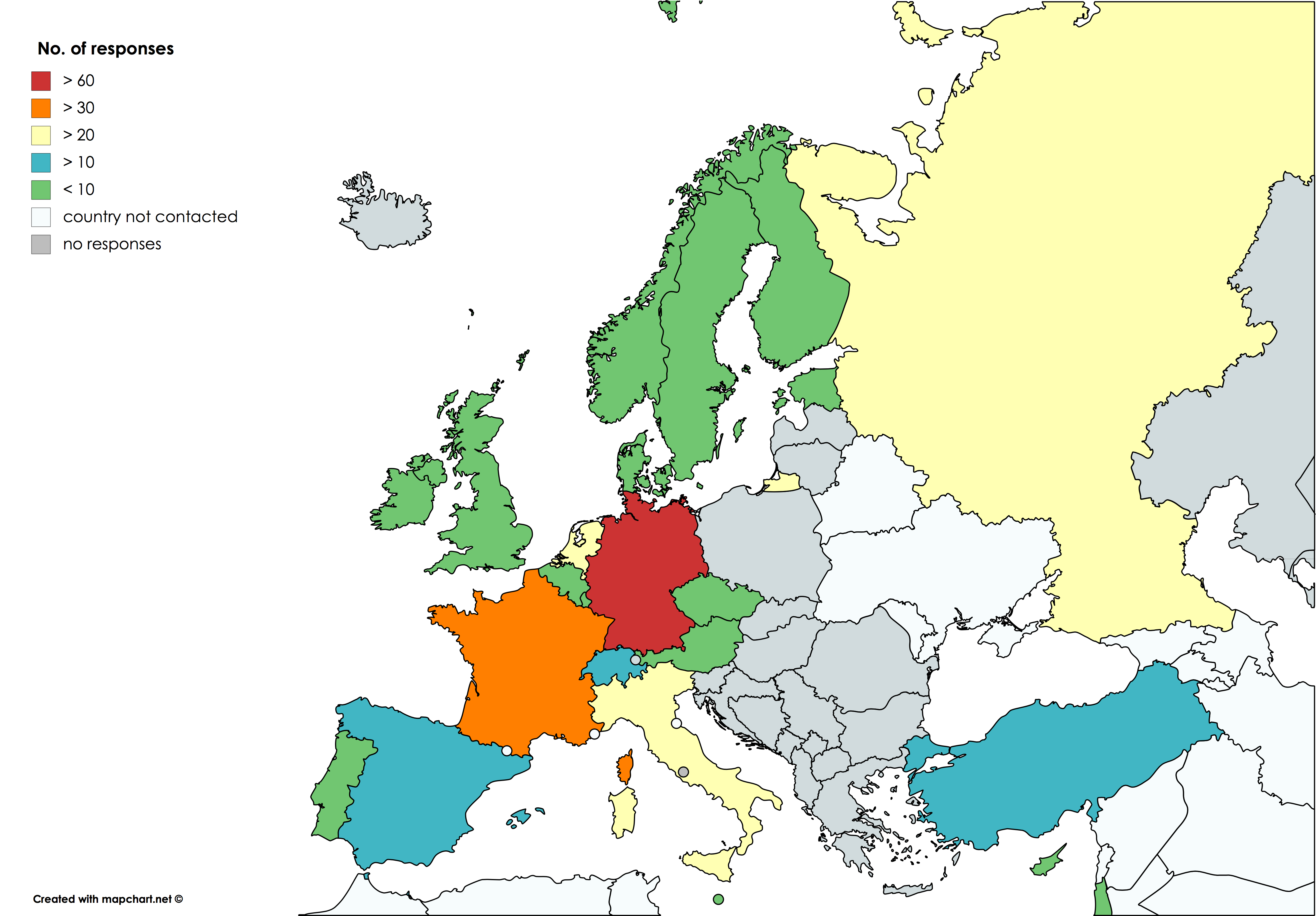
272 professionals/institutions (57.35% university hospitals) in 23 European countries completed the survey (table 2; fig. 1).The most frequently used sequence was DWI (81.99%), followed by DSC (67.28%) and MR-spectroscopy (64.34%). DCE, BOLD-based techniques and ASL still had an intermediate usage (43.38%, 42.65% and 37.50% respectively). All other techniques however were only marginally used with kurtosis imaging being the least used (3.31%).
Usage of techniques differed greatly between countries, e.g. ASL was applied by 77.50% of French institutions, but only by 20.34% of German institutions. Some form of quantitative MRI technique was used in the brain by 94.85% of institutions, while only 31.25% applied quantitative MRI sequences in head/neck MRI.
Furthermore, the most relevant indication found for a specific technique was the use of DSC (73.16%) and spectroscopy (54.78%) in glioma. Tissue differentiation and oncological monitoring were the most common reasons to apply a quantitative technique (82.35% and 72.79%), while stroke (58.82%) was a far less common indication and only a minority of institutions used quantitative MRI for neurodegenerative diseases (26.10%) or multiple sclerosis (22.79%).
Only a minority of responding Neuroradiologists (4.04%) did not see advantages in using other quantitative MRI techniques besides DWI. Neuroradiologists especially endorsed the usage of quantitative MRI because of an improved diagnostic accuracy (89.34%). However, reasons for omitting advanced imaging modalities included time constraints (38.97%), lack of availability of MR sequences/software for processing (29.41%) and insufficient compensation of the extra effort by insurers (12.87%). In consequence, in order to be motivated to use more quantitative MR sequences one in two Neuroradiologists replied he/she would need technical support in the handling of a sequence, one in three would need extra training for the medical staff/technicians as well as more exchange with scientists/colleagues.
EIBALL and QIBA were both not well known to the respondents (12.50% and 11.02%).
DISCUSSION AND CONCLUSION
This survey attempted to estimate the use of quantitative MRI in Neuroradiological institutions across Europe.While wide usage of DWI, DSC and MRS was confirmed for some indications such as glioma6, it also revealed that other techniques are either hardly used, show variable use by country or are only used for limited indications mostly omitting neurodegenerative diseases.
Our data suggests that the biggest obstacle to implement quantitative MRI sequences is not a lack of acceptance by clinicians, but indeed a multi-level lack of skill. Clinicians demanded better knowledge transfer as a motivation for implementation. Here not only scientists, but also vendors are in demand to act through hands-on teachings.
This key finding of our study is important, since Neuroradiologists working frequently with quantitative techniques were likely more willing to answer the survey. Also university centres were highly over-represented implicitly indicating that smaller centres were not interested to answer and possibly use quantitative techniques even less.
In conclusion, usage of quantitative MRI techniques in Neuroradiology is still not a standard and should be promoted clinically through improved networking between clinicians and scientists.
Acknowledgements
No acknowledgement found.References
- Edelman RR, Mattle HP, Atkinson DJ, et al. Cerebral blood flow: assessment with dynamic contrast-enhanced T2*-weighted MR imaging at 1.5 T. Radiology 1990;176(1):211-220.
- Tofts PS, Kermode AG. Blood brain barrier permeability in multiple sclerosis using labelled DTPA with PET, CT and MRI. J Neurol Neurosurg Psychiatry 1989;52(8):1019-1020.
- Le Bihan D, Breton E, Lallemand D, Grenier P, Cabanis E, Laval-Jeantet M. MR imaging of intravoxel incoherent motions: application to diffusion and perfusion in neurologic disorders. Radiology 1986;161(2):401-407.
- Williams DS, Detre JA, Leigh JS, Koretsky AP. Magnetic resonance imaging of perfusion using spin inversion of arterial water. Proc Natl Acad Sci U S A 1992;89(1):212-216.
- Damadian R. Tumor detection by nuclear magnetic resonance. Science 1971;171(3976):1151-1153.
- Thust SC, Heiland S, Falini A, et al. Glioma imaging in Europe: A survey of 220 centres and recommendations for best clinical practice. Eur Radiol 2018;28(8):3306-3317.
Figures
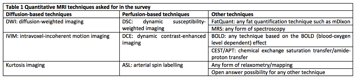
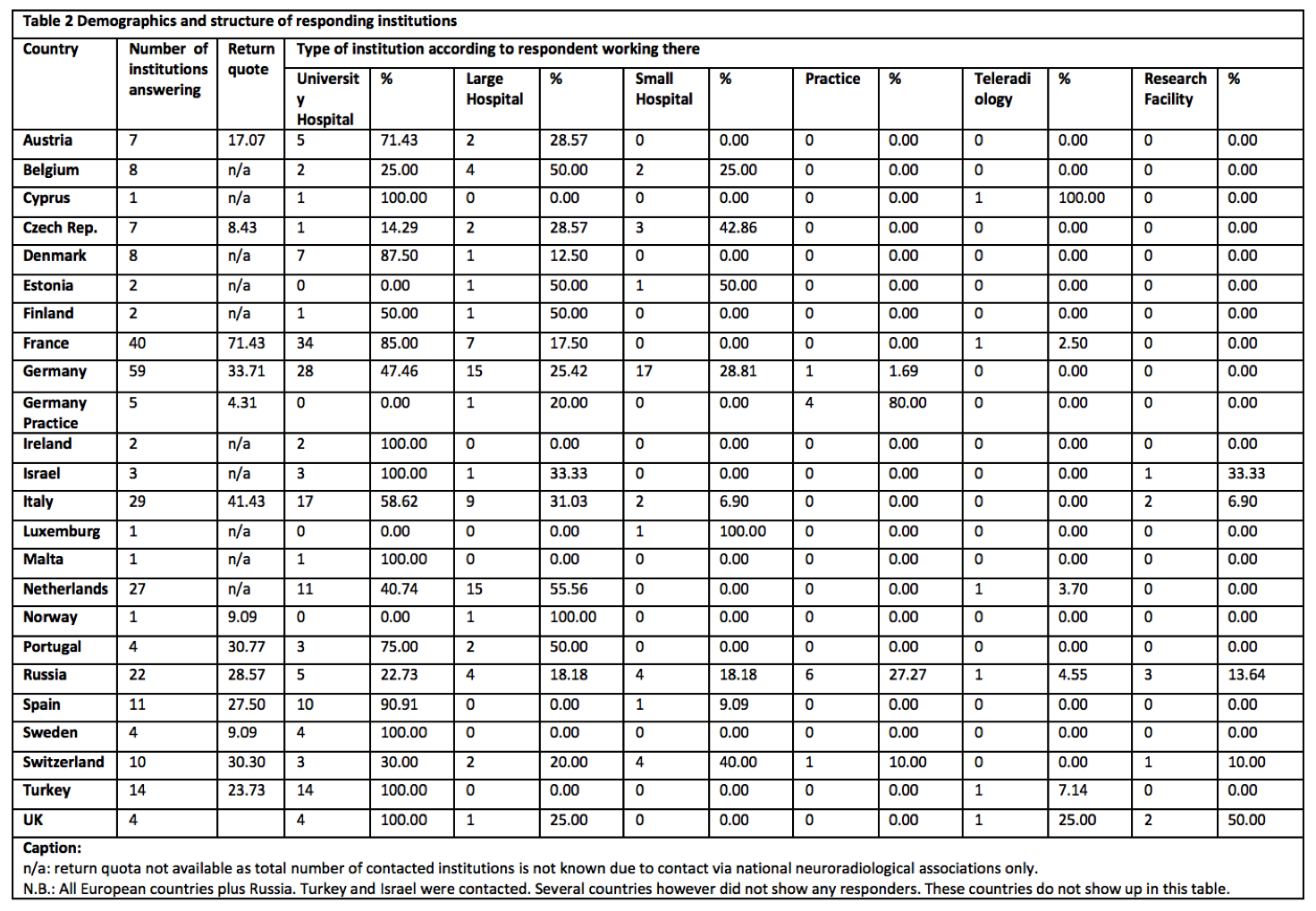
Table 2 Demographics of responding institutions
n/a: return quota not available as total number of contacted institutions is not known due to contact via national neuroradiological associations only.N.B.: All European countries plus Russia. Turkey and Israel were contacted. Several countries however did not show any responders. These countries are not listed in this table.