0263
Triaging dense breast screening MR images using a dilated convolutional neural network
Angelo Zuffianò1, Bob de Vos1, Jorrit Glastra1, Pim Moeskops1, Valerio Fortunati1, Ivana Išgum2, Tim Leiner3, Carla van Gils3, and Wouter Veldhuis3
1Quantib, Utrecht, Netherlands, 2Biomedical Engineering and Physics, Radiology and Nuclear Medicine, Amsterdam University Medical Center, Amsterdam, Netherlands, 3Radiology, Utrecht University Medical Center, Utrecht, Netherlands
1Quantib, Utrecht, Netherlands, 2Biomedical Engineering and Physics, Radiology and Nuclear Medicine, Amsterdam University Medical Center, Amsterdam, Netherlands, 3Radiology, Utrecht University Medical Center, Utrecht, Netherlands
Synopsis
Dynamic contrast enhanced (DCE) MRI is the key series to analyze for the detection of breast cancer in women with extremely dense breasts. Given the increasing number of women receiving dense breast MRI screening we aimed to reduce radiologist workload without reducing the high sensitivity of MRI. We developed a convolutional neural network (CNN) based method able to defer 8.1% of the workload by identifying non-enhancing scans with a sensitivity of 96.3%.
Introduction
Dynamic contrast enhanced (DCE) MRI has the potential to become a standard screening modality in women with extremely dense breast tissue. The goal of this study is to investigate the feasibility of using a convolutional neural network (CNN) to recognize MR image series with a negligible risk of containing lesions, with the aim of supporting the radiologist in prioritizing the screening workload.Methods
A total of 2211 MR studies of subjects with extremely dense breast tissue (Volpara Density Grade 4) and a negative result at screening mammography (BIRADS category 1 or 2)1 were analyzed. Fast-DCE images (352 × 352 × 60 voxels with spacing 0.966 × 0.966 × 3.000 mm3) were acquired on a 3.0T Achieva or Ingenia Philips system at 16 timepoints with an interval of 5 seconds, and within 90 seconds after contrast injection1. All the acquisitions were performed in the axial plane with bilateral anatomic coverage, and a 0.1 mL dose of gadobutrol per kilogram of body weight injected at a rate of 1 mL/sec.Manual classification into non-enhancing images, symmetric enhancing images, and images containing possible lesions was performed by a single expert radiologist (WV) with 14 years experience reading breast MRIs. The classification was based on an axial maximum intensity projection (MIP) of the subtraction images between the last timepoint and the pre-contrast image.
A dilated2 2D CNN with a receptive field of 131 voxels followed by a global pooling layer was trained to distinguish non-enhancing scans (negative class) from enhancing scans, i.e. symmetric enhancement or possible lesion (positive class). The model at the convergence point of the training loss was chosen for the final evaluation. The performance was evaluated on the test set by performing receiver operating characteristic (ROC) curve analyses.
Results
Automatic classification of the breast MR images between the non-enhancing class (negative class) and all other classes (positive class) was achieved with an average inference time below 1 second on a GPU. ROC analysis showed an area under the curve (AUC) of 0.80. This allows 8.1% of the workload to be triaged as non-enhancing with a sensitivity of 96.3% when comparing to the radiology expert labels. Of those studies which were incorrectly classified as non-enhancing none were found to contain relevant lesions for cancer detection according to the expert radiologist.Discussion and conclusion
We have presented a method to quickly triage breast MRIs with a negligible risk of containing any enhancing lesions relevant to cancer detection, as a proof of principle for CNN-based screening workload reduction. The CNN can subsequently be extended to further subclassify the enhancing class by including information from high spatial resolution and diffusion-weighted images that are acquired in the protocol as well1.Acknowledgements
We would like to acknowledge the DENSE Study Group. No funding was obtained for this study, but the data was acquired with funding from The Netherlands Organization for Health Research and Development (ZonMW-200320002-UMCU, ZonMW-50-53125-98-014), Bayer Pharmaceuticals (BSP-DENSE), and the Dutch Cancer Society (KWF-UU-2009-4348).References
- Emaus, Marleen J., et al. "MR imaging as an additional screening modality for the detection of breast cancer in women aged 50–75 years with extremely dense breasts: the DENSE trial study design." Radiology 277.2 (2015): 527-537.
- Fisher Yu and Vladlen Koltun "Multi-Scale Context Aggregation by Dilated Convolutions", 2015.
Figures

Subject characteristics. Physical data are
expressed as a range or average; frequency data are expressed as subject count
(relative frequency within set).
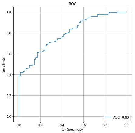
Receiver operating characteristic (ROC) curve
of the trained model.
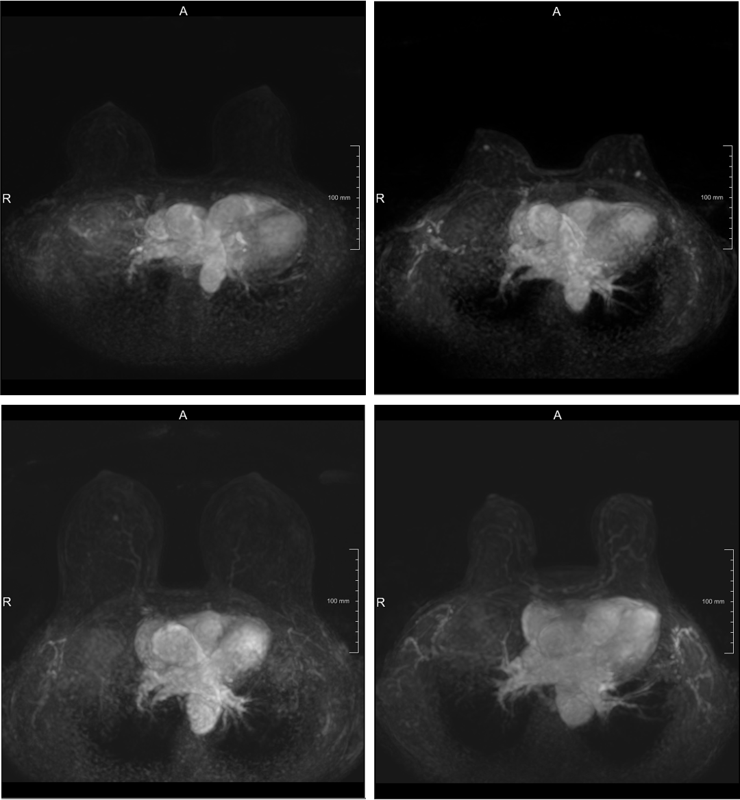
Sample image correctly classified as
“non-enhancing” (Top Left), correctly classified as “enhancing” (Top Right),
incorrectly classified as “non-enhancing” (Bottom Left), incorrectly classified
as “enhancing” (Bottom Right).