0206
Association of ApoE gene polymorphism and caudate functional connectivity in mild cognitive impairment of Parkinson’s disease1Department of Radiology, Nanjing First Hospital, Nanjing Medical University, Nanjing, China, 2GE Healthcare, MR Research China, Beijing, China, 3Northern Jiangsu People’s Hospital, Yangzhou, China
Synopsis
In this study, we aimed to investigate the association of Apolipoprotein E (ApoE) gene polymorphism and caudate functional connectivity in mild cognitive impairment of Parkinson’s disease (PD-MCI), using resting-state functional magnetic resonance imaging (rs-fMRI) and genotyping. Our findings revealed that gene-brain-behavior associations involve alterations of caudate activity with posterior cortical, thereby provide potential imaging-based markers that contribute to the early diagnosis and monitoring of PD-MCI.
Introduction
The early presence of mild cognitive impairment (MCI) of Parkinson’s disease (PD) would markedly increase the incidence of PD dementia. Neurochemical alterations induce impairment of neuronal processing in caudate nucleus, which plays a vital role in cognitive changes of PD1. Recent studies have reported that ApoE is a vulnerability genetic biomarker linked to cognitive decline in PD1, 2. Interestingly, resting state functional MRI (rs-fMRI) is particularly appealing since it has shown the ability to combine genetic polymorphism information with MR images, characterizing the genetic effects on brain activity. However, whether an interaction, which contributes to cognitive impairment in PD-MCI between ApoE and caudate, has still been rarely studied. Thus, in the present study, the possible association between different ApoE genotypes and caudate functional connectivity (FC) in PD-MCI was investigated, and we hypothesized that this association is involved in the pathogenesis of cognitive impairment in PD patients.Materials and Methods
95 PD-MCI patients (Male 54, Female 41, 63.83±7.39 years) and 99 matched healthy controls (Male 55, Female 44, 62.75±10.82 years) were recruited. Global cognitive function of all subjects was scored using the MoCa. The impairments in cognitive domains were assessed using a neuropsychological battery. MR image acquisition was performed at a 3T MR scanner (Discovery MR750w, GE, USA). Functional images were acquired with the following parameters: TR 2000ms, TE 30ms, flip angle 90°, slice thickness 4 mm without slice gap, FOV 240mm×240mm and matrix 64×64; voxel size 4.0mm×4.0mm×4.0 mm, number of phases 240, each frame included 35 continuous slices that covered the whole brain volume. The slices were parallel to the anterior/posterior commissure. The scan time was 6 minutes 20 seconds. The functional MRI data were preprocessed using the DPARSF 4.3. Genomic DNA of all participants was isolated from peripheral blood samples using QIAamp DNA Blood kits. Two single nucleotide polymorphisms were genotyped in order to differentiate ε2, ε3, and ε4 carrier status: rs7412 and rs429358. The Mann-Whitney U test/ Student’s t-test and χ2 test were utilized to investigate the differences in demographic data and genetic data in SPSS 19.0 software. Full factorial analysis was conducted to analyze the main effect of diagnosis, genotype, and genotype-by-disease interactions in SPM (functional data) and SPSS (neuropsychological performances). Correlation between these Z-transformed FC strengths of regions and neuropsychological performances were investigated separately using the Pearson’s correlation analysis in SPSS software. The statistical threshold for significance was defined as P < 0.05, corrected for age, gender and education.Results
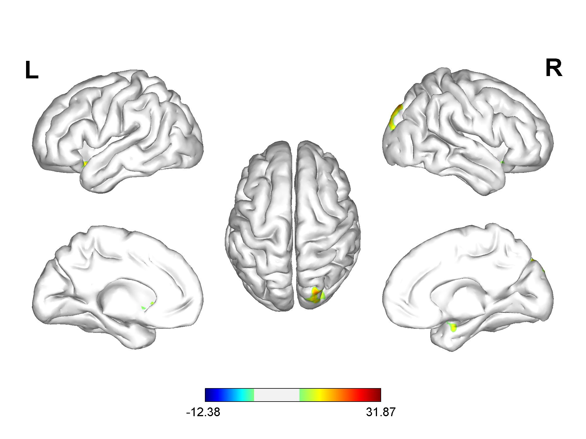
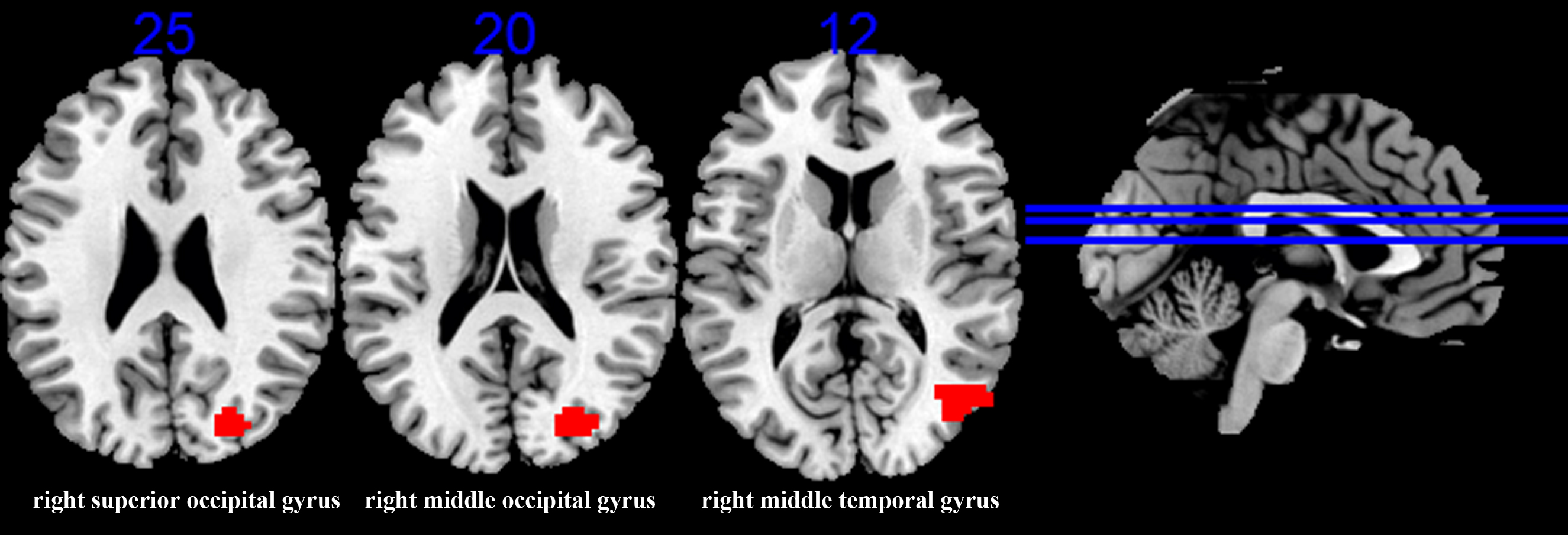
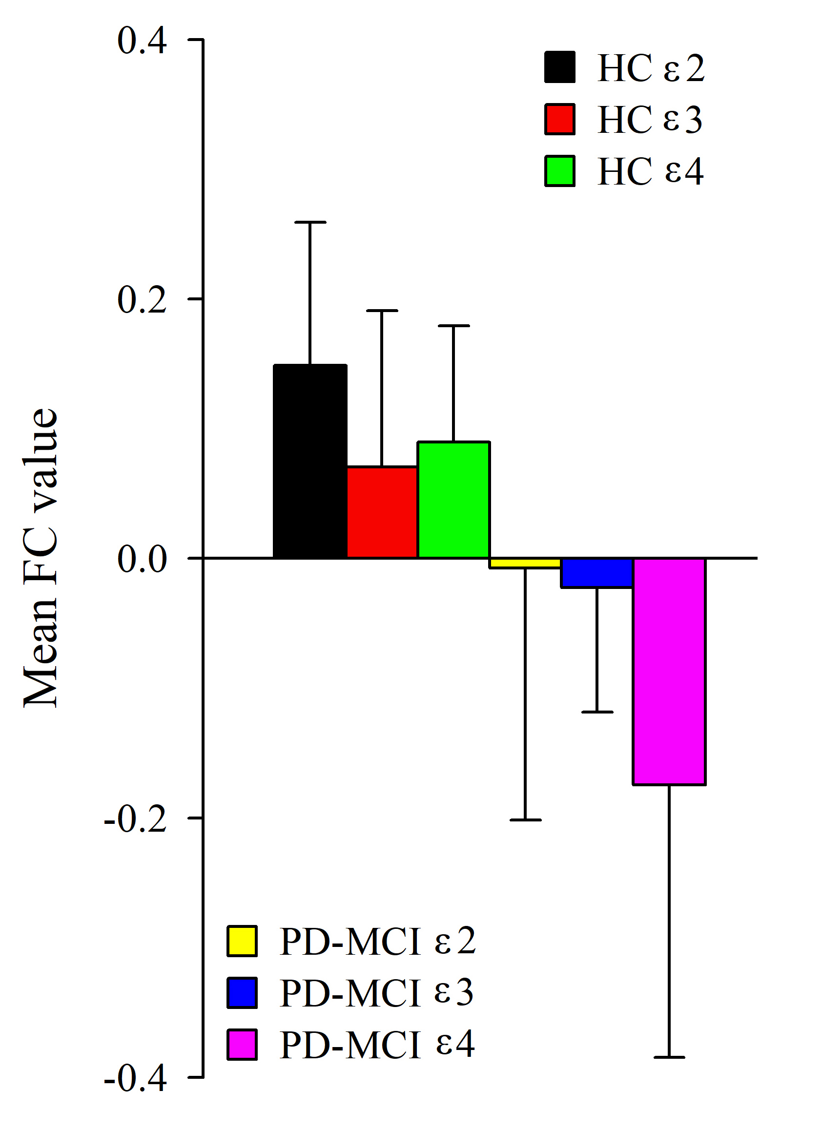
The distribution of ApoE genotype in PD-MCI group was equal to that of NC group (P = 0.20, χ2 = 3.23). A significant main effect of diagnosis was observed in general cognition, executive, memory, language, and visuospatial function. However, there was no significant effect of gene or interaction found in all the cognitive domains. A significant main effect of diagnosis was discovered in decreased FC from bilateral caudate to bilateral inferior orbit frontal gyrus, bilateral superior occipital gyrus (SOG), and right middle occipital gyrus (MOG) (Fig.1). There was no significant difference found in the main effect of genotype. Significant main effects of interaction were discovered in right MOG, right middle temporal gyrus (MTG), and right SOG (Fig. 2). The FC value of ε4 carriers was significantly lower than the other carriers in PD-MCI group (P=0.028, P=0.010, Fig. 3). There was no significant difference observed between ε2 carriers and ε3 carriers of PD-MCI, and neither among NC group (P>0.05, Fig. 3). In PD-MCI group, we found a significantly positive correlation between JoLo score (visuospatial function) and FC values to right MOG (P=0.003, r=0.31).Discussion and conclusion
Our findings included that the caudate nucleus is organized into cortical-striatal-thalamo loops connecting to prefrontal cortex, which mediates some forms of executive functions 3, 4. Furthermore, we revealed that ApoE gene polymorphism has the potential to influence caudate FC to the occipital cortex, binding with cholinergic dysfunction, while ε4 was the most risky isoform among the other isoforms (ε2, ε3). Thus, we could indicate that the impairment of visuospatial function is not simply the effect of PD, but rather an interaction effect with ApoE gene polymorphism. In conclusion, for the first time, by using FC approach and genotyping, we provided promising evidences to support that ε4 genotype in ApoE gene may be influencing the caudate activity to posterior cortical, and associated with underlying pathology of visuospatial impairment in an interactive manner. Gene-based imaging approaches might strengthen the credibility in imaging genetic associations, and provide new powerful insights into the neural mechanisms underlying PD-MCI.Acknowledgements
No acknowledgement found.References
1. Tropea TF, Xie SX, Rick J et al APOE, thought disorder, and SPARE-AD predict cognitive decline in established Parkinson's disease. Mov Disord 2018;33:289-297
2. Sun R, Yang S, Zheng B et al Apolipoprotein E Polymorphisms and Parkinson Disease With or Without Dementia: A Meta-Analysis Including 6453 Participants. J Geriatr Psychiatry Neurol 2019;32:3-15
3. Zhang S, Ide JS, Li CS Resting-state functional connectivity of the medial superior frontal cortex. Cereb Cortex 2012;22:99-111
4. Manza P, Zhang S, Hu S et al The effects of age on resting state functional connectivity of the basal ganglia from young to middle adulthood. Neuroimage 2015;107:311-322
Figures