0198
Development of a spatio-temporally consistent longitudinal structural template of the older adult brain1Biomedical Engineering, Illinois Institute of Technology, Chicago, IL, United States, 2Rush Alzheimer's Disease center, Rush University Medical Center, Chicago, IL, United States
Synopsis
One of the major challenges in constructing a longitudinal structural template of the older adult brain is to ensure spatio-temporal consistency. In this work, a new method was introduced to construct a spatio-temporally consistent longitudinal structural template of the older adult brain based on high quality cross-sectional older adult data from a large cohort. The new template was compared to templates generated with previously published methods in terms of spatio-temporal consistency, image quality, and representativeness of age-related brain changes, and was shown to have superior performance.
Introduction
Longitudinal studies of structural brain MRI changes in aging, and investigations tracking progression of age-related diseases or treatment outcomes could benefit from a longitudinal structural brain template that is representative of brain aging and of high quality. One of the major challenges in constructing such a template is to ensure spatio-temporal consistency. This challenge is relatively simple to overcome if longitudinal MRI data from 65 years of age to approximately 95 years of age were available on each and every older adult from a large cohort (30 years of data on each participant). However, such data is not available, and therefore, ensuring spatio-temporal consistency has been problematic. In this work, a new method was introduced to construct a spatio-temporally consistent longitudinal structural template of the older adult brain based on high quality cross-sectional older adult data from a large cohort. The new template was compared to templates generated with previously published methods in terms of spatio-temporal consistency, image quality, and representativeness of age-related brain changes.Methods
DataT1-weighted MPRAGE brain MRI data with 1mm isotropic voxels were collected using a 3T MRI scanner on 222 non-demented older adults (65-95 years of age, male: female=1:1) participating in the Rush Memory and Aging Project1 (MAP).
Method 1 for longitudinal template construction
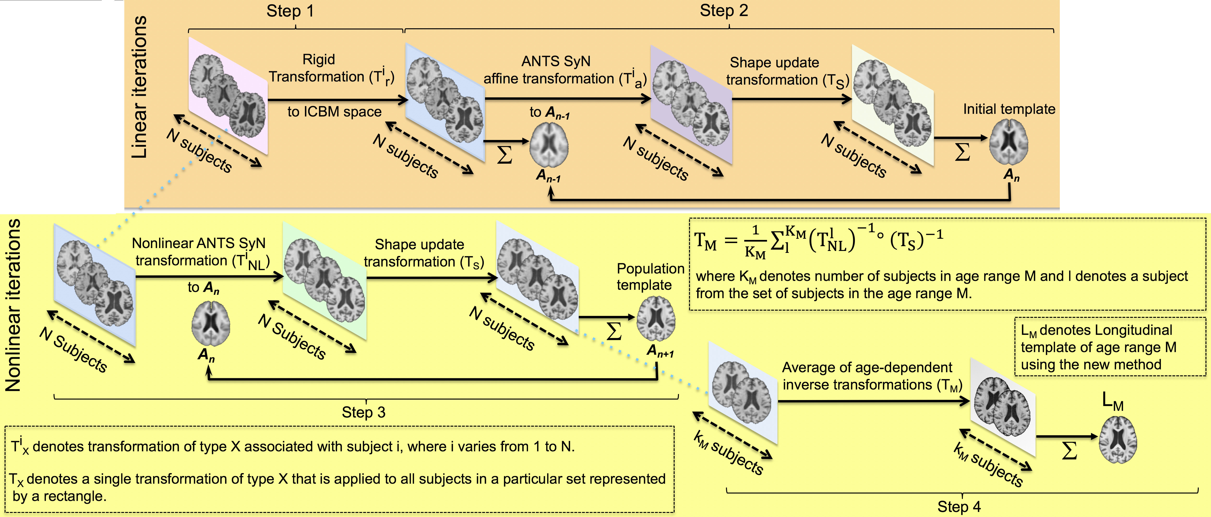
The first approach tested involved kernel regression, according to which the contribution of each cross-sectional dataset to a certain time-point of the longitudinal template is determined by the kernel weight2. The kernel used was 10 years wide with a constant weight, and was moved to cover eight partially overlapping age-ranges (65-75, 68-78, 71-81, 74-84, 77-87, 80-90, 86-95), each including 58 participants with male:female=1:1. The data within each age-range were spatially normalized (SyGN)3,4 and averaged, resulting into a longitudinal template with eight time-points: R65-75, R68-78, R71-81, R74-84, R77-87, R80-90, R83-93, R86-95 (Fig.1 steps 1,2,3).
Method 2 for longitudinal template construction
This was a variant of Method 1 and included an initial affine registration of individual datasets to the same initial template to limit group biases. This method resulted into a second longitudinal template with eight time-points: A65-75, A68-78, A71-81, A74-84, A77-87, A80-90, A83-93, A86-95.
Proposed method for longitudinal template construction
The proposed method first builds a common population template using data from all participants from 65-95 years of age using SyGN registration (Fig.1 steps 1,2,3). Next, the inverse of the non-linear deformations and the inverse of the shape-update deformation field for participants in each 10 years age-range were concatenated and averaged separately. Each resulting deformation was applied to data from the corresponding sub-group of participants and the data were averaged, resulting into the proposed longitudinal template with eight time-points: L65-75, L68-78, L71-81, L74-84, L77-87, L80-90, L83-93, L86-95.
Evaluation
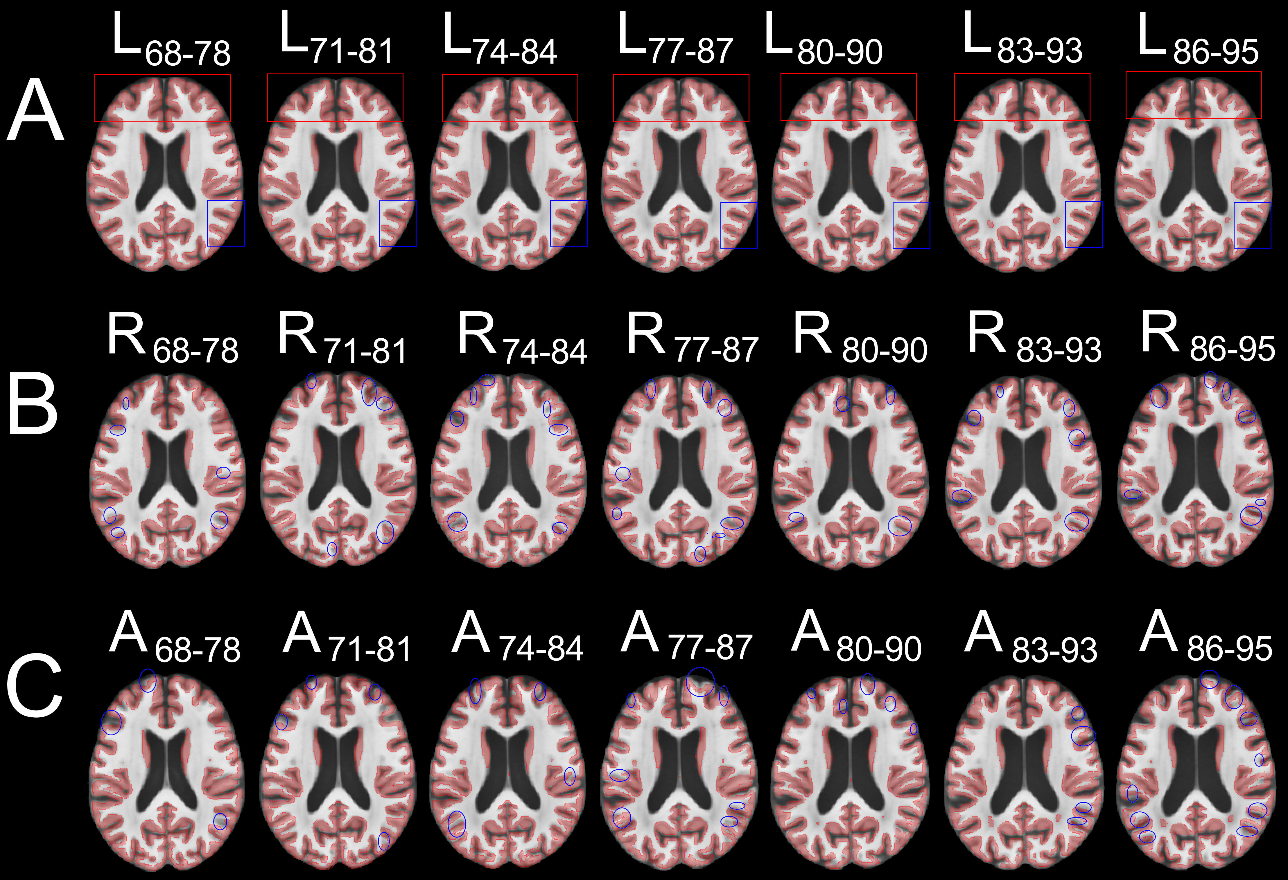
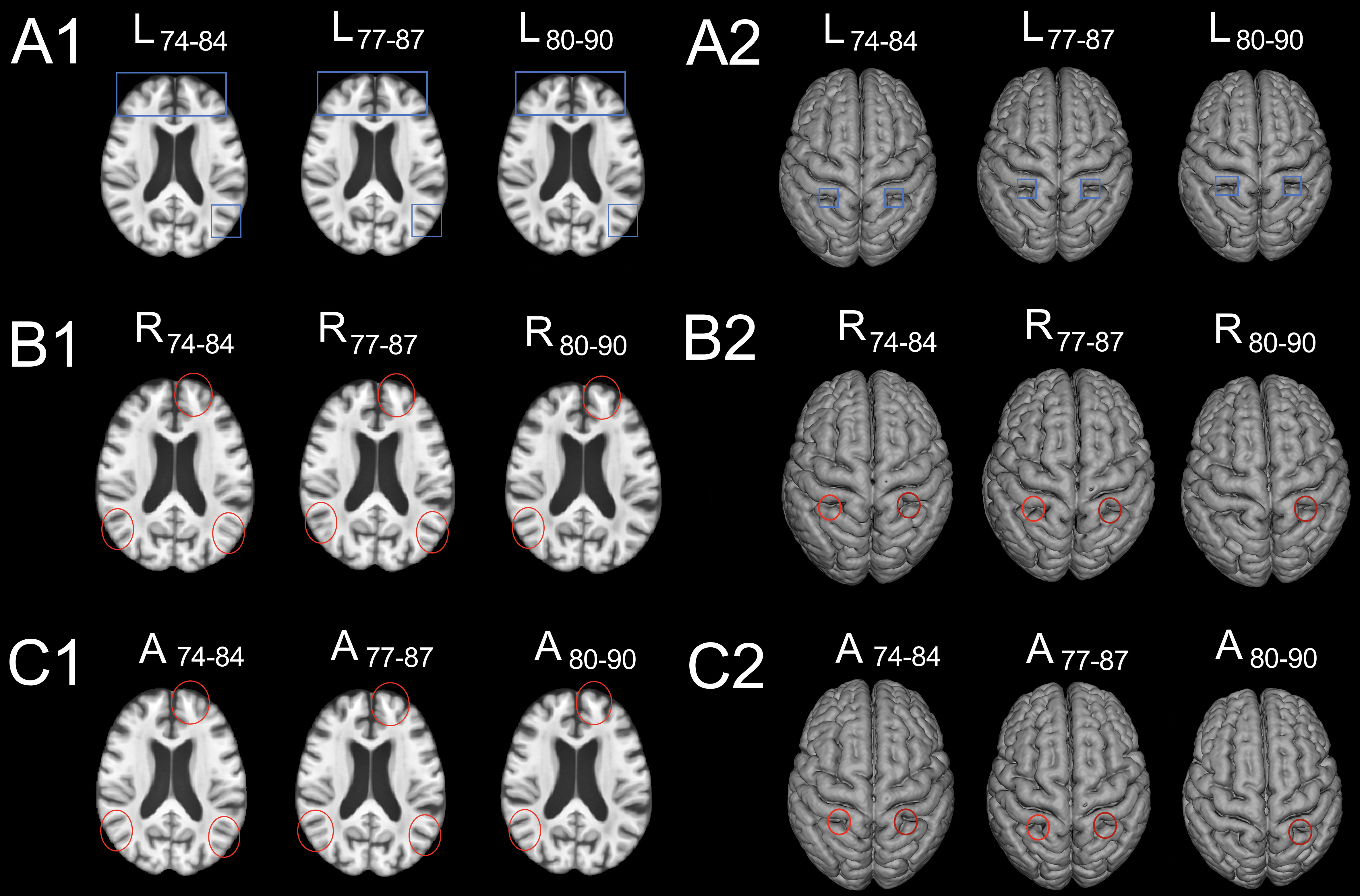
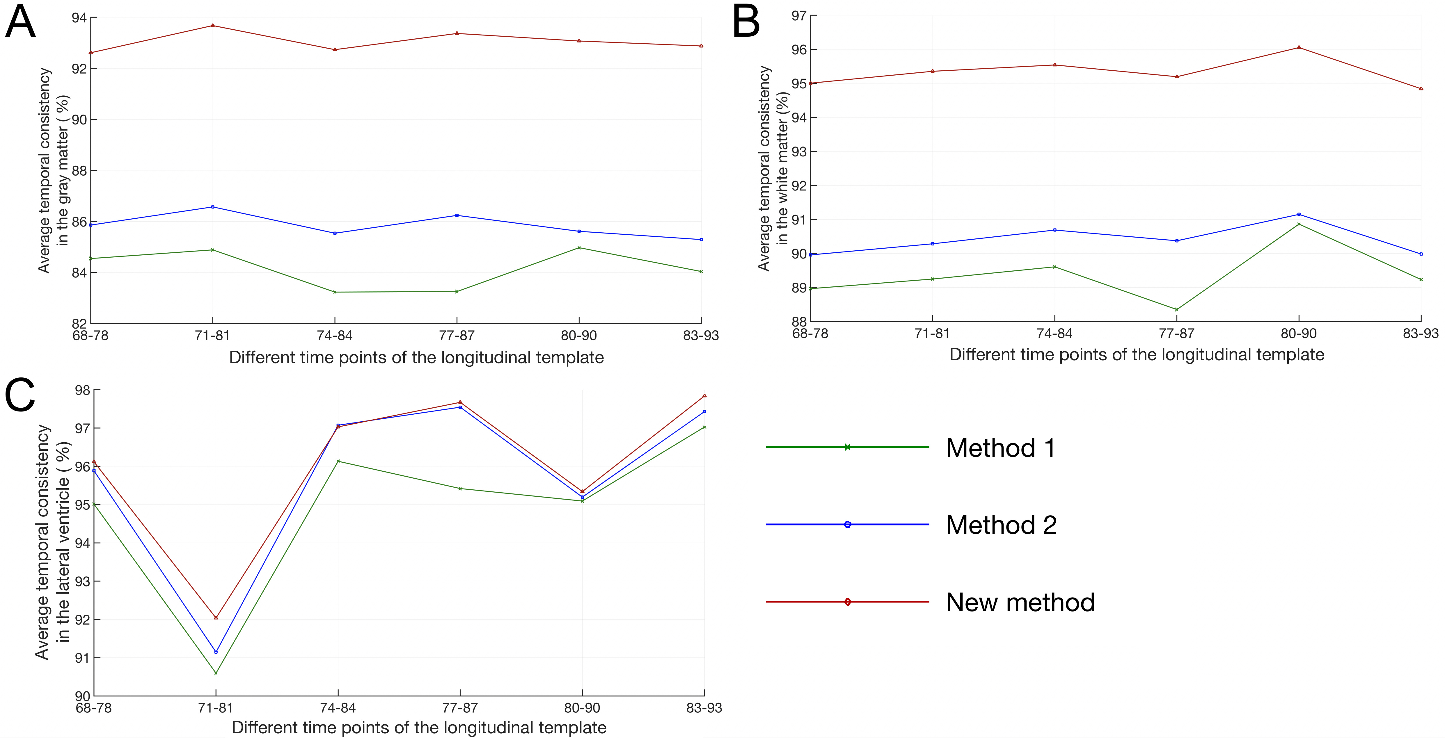
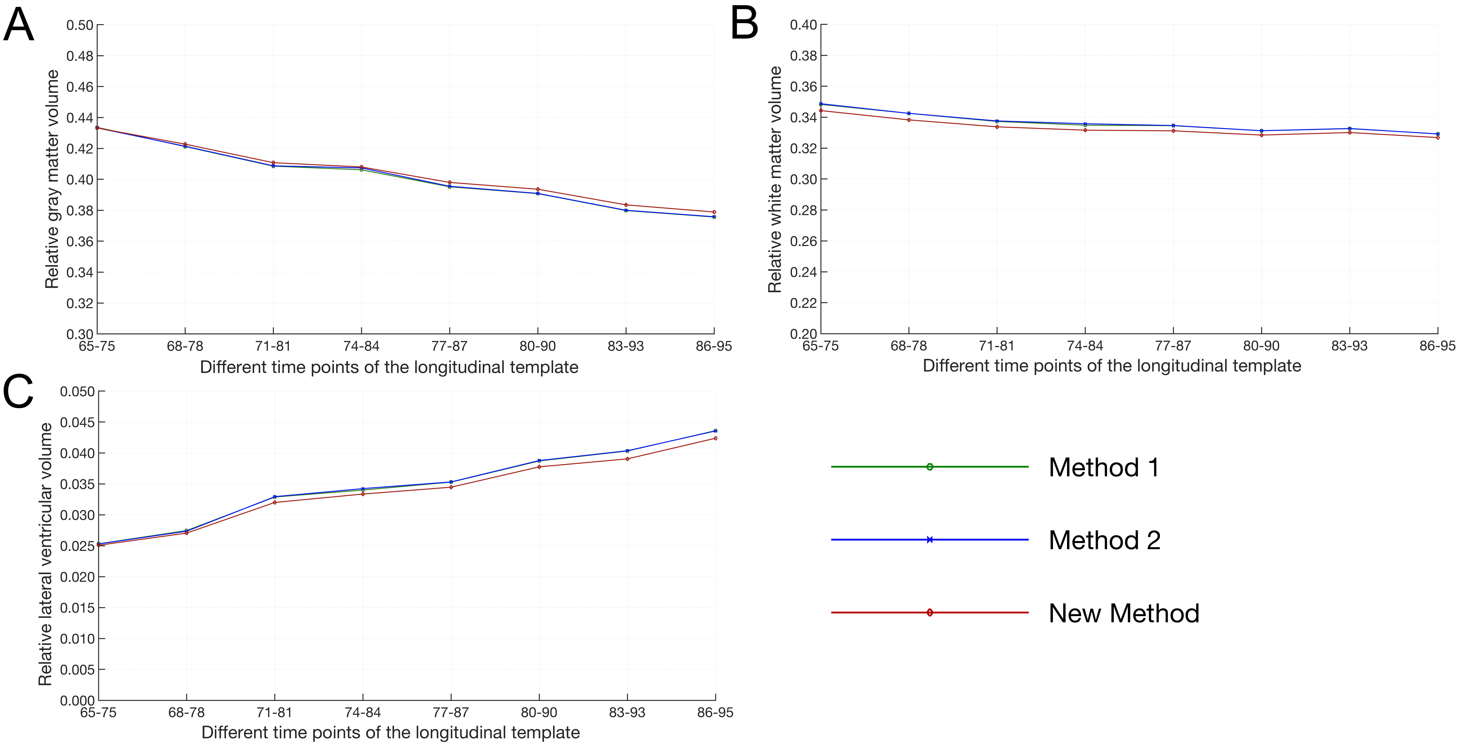
The three longitudinal templates were compared in terms of spatio-temporal consistency, image quality, and representativeness of age-related brain changes. Spatio-temporal consistency was assessed quantitatively using the metric average temporal consistency factor(TC)5, as well as by visual inspection (both in 2D and 3D) of how well cortical gray matter of a specific time-point overlapped with cortical gray matter of the previous time-point. Assessment of image quality included an evaluation of image sharpness and artifacts. Finally, the ability of longitudinal templates to accurately represent age-related structural brain changes was assessed by investigating the temporal progression of the relative gray matter volume, white matter volume, and lateral ventricular volume of the longitudinal templates.
Results and Discussion
The longitudinal template constructed using the proposed method resulted in the highest spatio-temporal consistency as illustrated both visually (Figs.2,3) and quantitatively for gray matter, white matter and the lateral ventricles (Fig. 4). Method 1 resulted in the lowest consistency as expected. Image sharpness was similar across methods (Fig.3). No image artifacts were detected. A steady decline in relative gray matter and white matter volumes and a steady increase in lateral ventricular volume was observed across time-points of the longitudinal templates constructed using all methods (Fig 5), in agreement with previous research on age-related structural brain changes6,7,8,9Conclusion
The present work introduced a new method for the development of a longitudinal structural brain template of the older adult brain with high spatio-temporal consistency. The template developed using the new method was based on high-quality data from a large cohort of non-demented older adults. The new template exhibited higher spatio-temporal consistency compared to two previously published methods also based on cross-sectional data, it was characterized by high image sharpness and no image artifacts, and accurately captured well-known age-related structural brain changes. It is expected that the new template will have a significant positive impact in longitudinal studies of structural brain MRI changes in aging, and investigations tracking progression of age-related diseases or treatment outcomes. The new longitudinal template is part of the MIITRA atlas (www.nitrc.org/projects/miitra).Acknowledgements
This study was supported by National Institutes of Health grant R01AG052200, P30AG010161, UH2NS100599, UH3NS100599, R01AG064233 and R01AG17917References
1. Bennett DA, Schneider JA, Buchman AS, Barnes LL, Boyle PA, Wilson RS. Overview and findings from the Rush Memory and Aging Project. Curr Alzheimer Res 2012;9:646–663.
2. Davis, Brad C., et al. Population shape regression from random design data. International journal of computer vision. 2010;90(2):255-266.
3. Avants, Brian B., et al. The optimal template effect in hippocampus studies of diseased populations. Neuroimage. 2010;49(3):2457-2466.
4. Avants, Brian B., et al. A reproducible evaluation of ANTs similarity metric performance in brain image registration. Neuroimage. 2011;54(3):2033-2044.
5. Xue, Zhong, Dinggang Shen, and Christos Davatzikos. CLASSIC: consistent longitudinal alignment and segmentation for serial image computing. NeuroImage. 2006;30(2):388-399.
6. Matsumae, Mitsunori, Ron Kikinis., et al. Age-related changes in intracranial compartment volumes in normal adults assessed by magnetic resonance imaging. Journal of neurosurgery 1996;84(6): 982-991.
7.Mortamet, Bénédicte, Donglin Zeng, Guido Gerig., et al. Effects of healthy aging measured by intracranial compartment volumes using a designed MR brain database. International Conference on Medical Image Computing and Computer-Assisted Intervention 2005:383-391.
8. Taki, Yasuyuki, Benjamin Thyreau., et al. Correlations among brain gray matter volumes, age, gender, and hemisphere in healthy individuals. PloS one. 2011;6(7): e22734.
9. Ge, Yulin, Robert I. Grossman., et al. Age-related total gray matter and white matter changes in normal adult brain. Part I: volumetric MR imaging analysis. American journal of neuroradiology. 2002;23(8): 1327-1333.
Figures