0185
High-Resolution Distortion-Free Whole-Brain MR Elastography using Multiband DIADEM (DIADEM-MRE)1Radiology, Mayo Clinic, Rochester, MN, United States
Synopsis
Introduction
MR elastography (MRE) has become a useful tool to assess brain stiffness change due to neurodegenerative diseases and normal aging.1 It can also reliably characterize tumor consistency and adherence, offering key information for surgery planning.2,3 The most common pulse sequence used for brain MRE is echo-planar-imaging (EPI), which, however, is prone to image distortion caused by local susceptibility‐induced B0 inhomogeneities. Recently, a distortion‐free imaging technique inspired by a point-spread-function mapping approach, termed DIADEM (Distortion-free Imaging: A Double Encoding Method),4 has been demonstrated for diffusion imaging. In this study, we implemented this approach to the standard EPI-MRE sequence to reduce geometric distortion, and demonstrated its feasibility for brain applications in healthy volunteers and patients with brain tumors.Methods
Sequence: DIADEM-MRE is based on the multi-band spin-echo EPI-MRE sequence5 with additional spin-warp phase-encoding gradient right before the EPI acquisition.6,7Phantom study: To understand how the geometric distortion may affect the stiffness calculation, a brain phantom with spherical inclusions was scanned on a high-performance compact 3T scanner8,9 with DIADEM-MRE. Given that both distorted (like EPI) and distortion-free images can be generated from the DIADEM dataset,10 a direct pixel-wise comparison of wave images and stiffness maps was performed.
Volunteer and patient study: With IRB approval and written informed consent, 3 healthy volunteers and 2 meningioma patients were scanned on the compact 3T scanner using a Nova Medical 32-channel receiver coil. The DIADEM-MRE data with 2-mm isotropic resolution was acquired with the imaging parameters of TR/TE=2100/65.8ms; FOV=21.6 cm; 108×108 matrix; 63 contiguous 2-mm-thick axial slices; 2x in-plane acceleration; 3x multiband (MB) acceleration, 316 μs echo spacing, 60-Hz mechanical vibrations; 6 motion encoding directions, 3 phase offsets; 9 shots for each DIADEM dataset and total acquisition time of 5:44 minutes.
Results
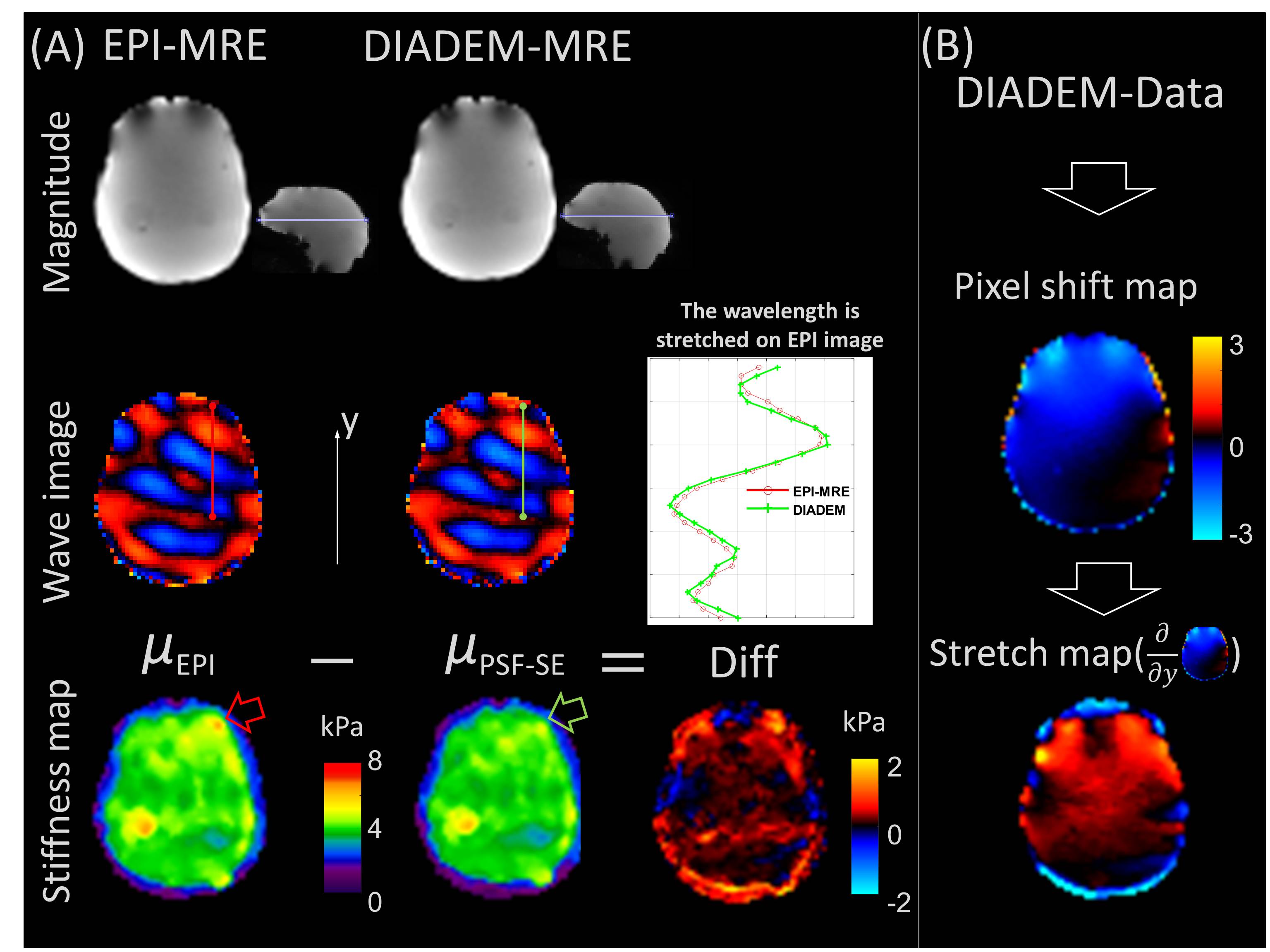
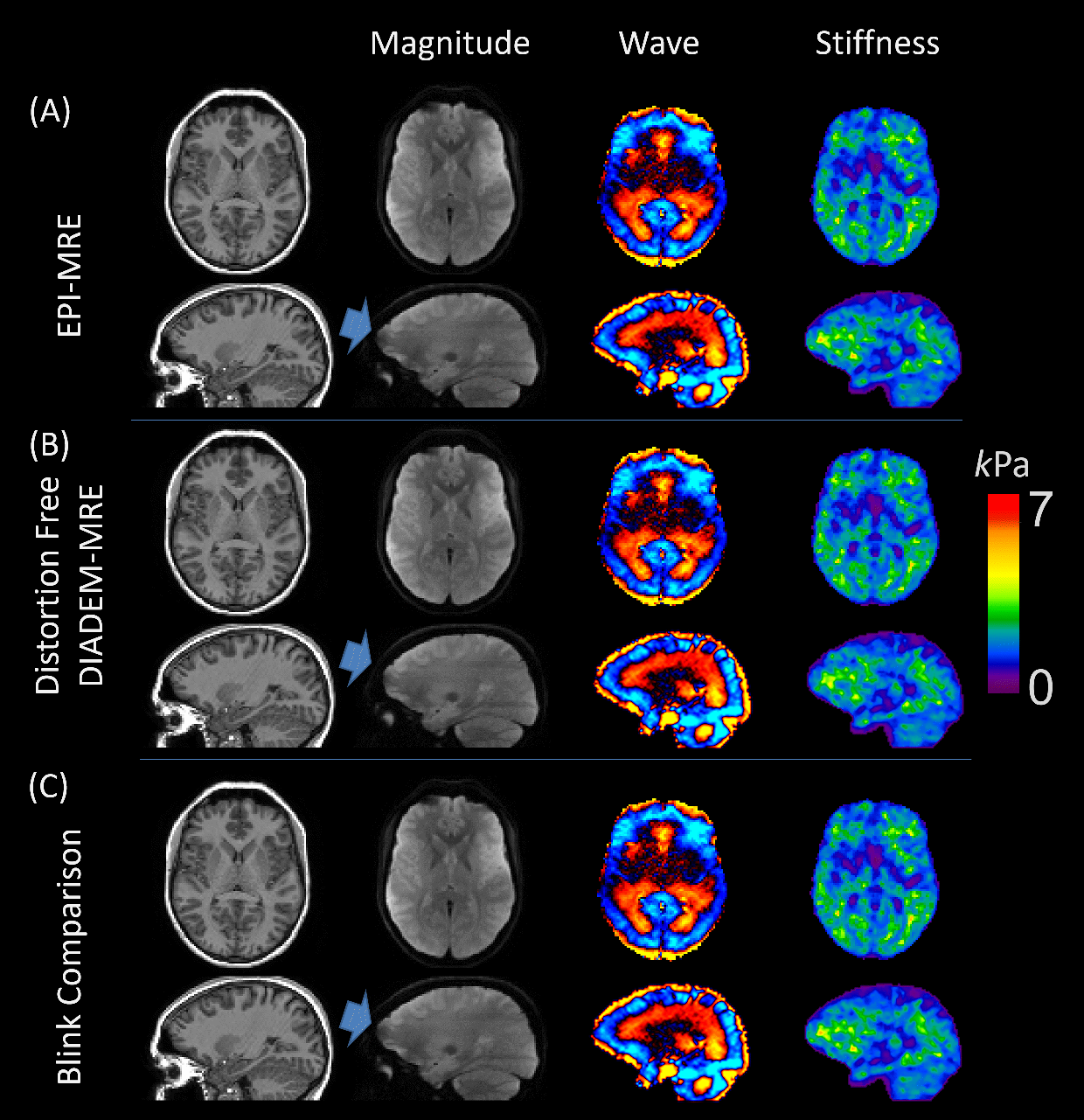
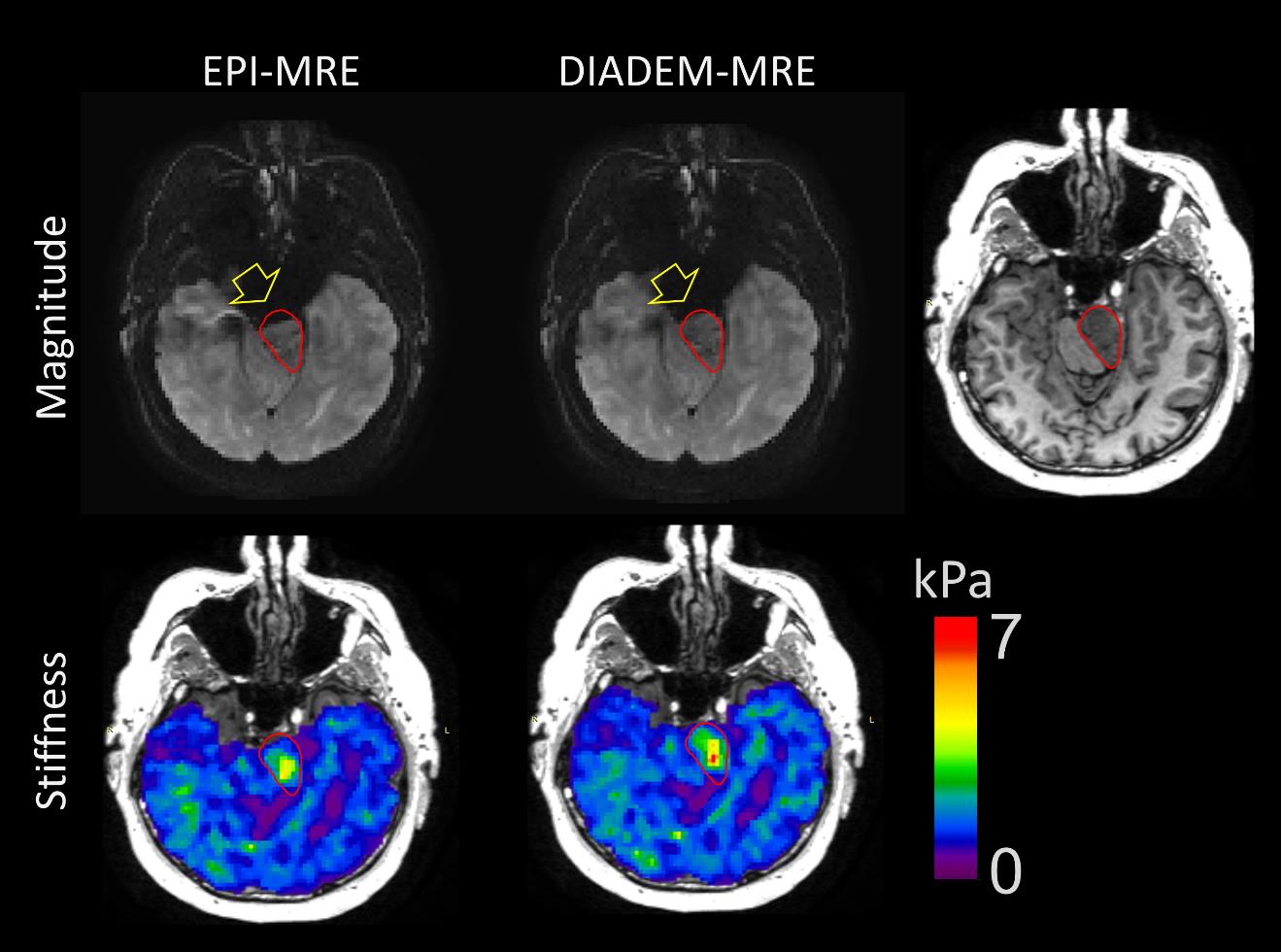
Fig.1A compares the magnitude, wave images and stiffness maps between the EPI-MRE and the distortion-free MB-DIADEM-MRE. The difference in stiffness was also computed. The wavelength was stretched on EPI images on the line profile (red and green lines), resulting in an over-estimated stiffness in that region. Fig.1B shows the pixel shift (or distortion) map calculated from DIADEM data and the stretch map generated by taking the derivative of pixel shift map along the y-direction. The stretch map indicates how much the wave is stretched (positive value) or compressed (negative value) on each location, which also corresponds to the stiffness difference map. Fig.2 shows the T1 anatomic images, as well as the magnitude, wave, and stiffness maps from the EPI-MRE (Fig.2A) and the distortion-free DIADEM-MRE (Fig.2B) in 3 healthy volunteers, respectively. The difference can be clearly appreciated in the animation that blinks between the two methods (Fig.2C). EPI- and DIADEM-MRE results in a meningioma patient are shown in Fig.3. The tumor ROI was first drawn on T1 images registered to the MRE space, and overlaid onto MRE magnitude images. A substantial amount of image distortion arising from strong susceptibility effects can be seen on the magnitude image for the EPI-MRE. However, no observable distortion is observed with DIADEM-MRE, where the ROI was well-matched with the anatomical data. The tumor stiffness was measured as 1.9±1.1 kPa and 2.3±1.1 kPa using EPI-MRE and DIADEM-MRE, respectively. The EPI distortion compressed the wavelength in the tumor region, resulting in an underestimate of tumor stiffness.Discussion and Conclusion
The distortion-free MRE images with 2-mm isotropic resolution can be achieved with the proposed multiband DIADEM-MRE as demonstrated in brain applications. It can also offer improvements for the understanding of brain stiffness in the regions of rapid susceptibility variation. The scan time was 5:44 minutes and can be further improved by reducing the number of shots for each DIADEM acquisition when combined with tilted-CAIPI method.11 These preliminary results demonstrate feasibility and motivate further exploration of DIADEM-MRE for expanded clinical applications.Acknowledgements
This work was supported by grants from the National Institute of Health (R01 EB001981, R01 EB010065, R01 NS113760, and NIH U01 EB024450)References
1 Yin, Z., Romano, A. J., Manduca, A., Ehman, R. L. & Huston, J. I. Stiffness and Beyond: What MR Elastography Can Tell Us About Brain Structure and Function Under Physiologic and Pathologic Conditions. Topics in Magnetic Resonance Imaging 27, 305-318, doi:10.1097/rmr.0000000000000178 (2018).
2 Murphy, M. C. et al. Preoperative assessment of meningioma stiffness using magnetic resonance elastography. Journal of Neurosurgery 118, 643-648, doi:10.3171/2012.9.Jns12519 (2013).
3 Yin, Z., Glaser, K. J., Manduca, A., Van Gompel, J. J., Link, M. J., Hughes, J. D., Romano, A., Ehman, R. L. & Huston, J., 3rd. Slip interface imaging predicts tumor-brain adhesion in vestibular schwannomas. Radiology 277, 507-517, doi:10.1148/radiol.2015151075 (2015).
4 In, M. H. et al. Distortion-free imaging: A double encoding method (DIADEM) combined with multiband imaging for rapid distortion-free high-resolution diffusion imaging on a compact 3T with high-performance gradients. J Magn Reson Imaging, doi:10.1002/jmri.26792 (2019).
5 Sui, Y., Yin, Z., Rossman, P. J., Murphy, M. C., Trzasko, J. D., Scott, J. M., Glaser, K. J., McGee, K. P., Bernstein, M. A., Ehman, R. L. & Huston, J., 3rd. Fast Brain MR Elastography Using a Simultaneous Multislice EPI Acquisition on a Compact 3T Scanner. ISMRM Proceeding, 3971 (2019).
6 Robson, M. D., Gore, J. C. & Constable, R. T. Measurement of the point spread function in MRI using constant time imaging. Magnetic resonance in medicine 38, 733-740 (1997).
7 Zeng, H. & Constable, R. T. Image distortion correction in EPI: comparison of field mapping with point spread function mapping. Magn Reson Med 48, 137-146, doi:10.1002/mrm.10200 (2002).
8 Weavers, P. T. et al. Technical Note: Compact three-tesla magnetic resonance imager with high-performance gradients passes ACR image quality and acoustic noise tests. Med Phys 43, 1259-1264, doi:10.1118/1.4941362 (2016).
9 Foo, T. K. F., Laskaris, E., Vermilyea, M., Xu, M., Thompson, P., Conte, G., Van Epps, C., Immer, C., Lee, S.-K., Tan, E. T., Graziani, D., Mathieu, J.-B., Hardy, C. J., Schenck, J. F., Fiveland, E., Stautner, W., Ricci, J., Piel, J., Park, K., Hua, Y., Bai, Y., Kagan, A., Stanley, D., Weavers, P. T., Gray, E., Shu, Y., Frick, M. A., Campeau, N. G., Trzasko, J., Huston III, J. & Bernstein, M. A. Lightweight, compact, and high-performance 3T MR system for imaging the brain and extremities. Magnetic Resonance in Medicine 80, 2232-2245, doi:10.1002/mrm.27175 (2018).
10 In, M. H., Posnansky, O. & Speck, O. High-resolution distortion-free diffusion imaging using hybrid spin-warp and echo-planar PSF-encoding approach. Neuroimage 148, 20-30, doi:10.1016/j.neuroimage.2017.01.008 (2017).
11 Dong, Z. et al. Tilted-CAIPI for highly accelerated distortion-free EPI with point spread function (PSF) encoding. Magn Reson Med 81, 377-392, doi:10.1002/mrm.27413 (2019).
Figures