0182
Protocol Optimization for in vivo Electrical Propertes Tomography of the Human Breast at 3T1C.J. Gorter Center for High Field MRI, dept. Radiology, Leiden University Medical Center, Leiden, Netherlands
Synopsis
This work demonstrates a clinically feasible protocol and reconstruction pipeline which is relatively straightforward to implement, and achieves reliable conductivity reconstructions of the human breast. We aim to establish a reliable MR protocol with a scan time of 6 minutes to further develop the clinical potential of this technique.
Introduction
Electrical Properties Tomography (EPT) is a promising technique which allows probing the electrical properties of tissues noninvasively via MRI.1,2 During the past decade a tremendous scientific effort has focused on developing robust reconstruction approaches, each involving specific assumptions and pre-requisites, but all sharing the need for high-quality input data. On the other hand, only a small number of clinical studies have demonstrated the added value of EPT in characterizing suspected lesions.3–5 In the particular case of breast cancer, measured conductivity values were demonstrated to yield additional information confirming the clinical grading of breast tumours, however a systematic comparison of MR sequences and reconstruction options has not been shown to date.In this study, we have evaluated different MR sequence considerations for performing EPT in the human breast at 3T. Three MR sequences have been compared and evaluated in terms of artefact level and SNR, both in vivo and in a multi-compartment phantom to validate the reconstructed values against the ground truth. With this study, we aim to establish a reliable protocol with a scan time of 6 minutes for EPT in the human breast to further develop the clinical potential of this technique.
Methods
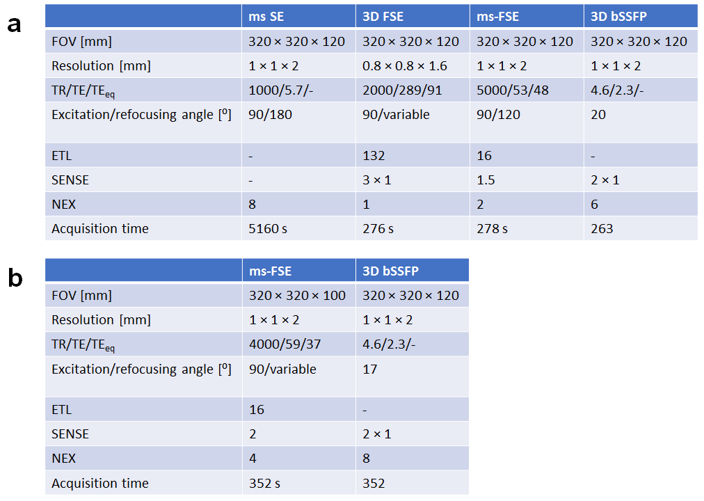
A cylindrical phantom with three cylindrical inserts of different electrical conductivity was constructed. Polyvinyl Pyrollidone (PVP10, Sigma Aldrich, the Netherlands) was used for permittivity adjustment and NaCl for conductivity adjustment to achieve conductivity values representative to fat, normal glandular tissue a high conductivity lesion. All solutions were gelled using 1.5% of agarose. In vivo data was acquired in healthy volunteers (N=3) after obtaining written informed consent. All imaging was performed on a Philips 3T system (Ingenia, Philips Healthcare, Best, The Netherlands) using either a 16-channel head coil in the phantom and a 7-channel breast coil in vivo.To lower the imaging requirements and improve the clinical potential, the conventional EPT approach based on the homogeneous Helmholtz equation was chosen which allows deriving conductivity reconstructions from basic MR phase data.1 Three MR sequences which have been applied in EPT before have been evaluated with the goal to establish an EPT protocol within a clinically feasible scan time of 6 minutes. Specifically, a 3D fast spin echo (FSE), multi-slice FSE and a balanced steady state free precession (bSSFP) sequence were evaluated.5,6 As a ground truth, a multi-slice spin echo (SE) sequence was acquired in the phantom only due to time limitations in vivo. Imaging parameters are listed in the table illustrated in Fig. 1.
Conductivity reconstructions were obtained using the noise-robust Laplacian kernel as proposed by van Lier et al.7 All reconstructions employed additional denoising with a restricted median filter as proposed by Stehning et al.,8 restricting the kernel of 5 mm radius to voxels that have a signal magnitude within 10% of the target voxel.
Results
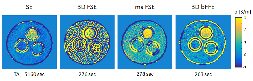
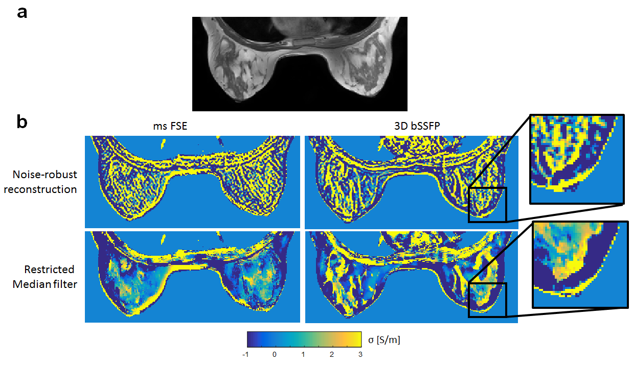
Figure 2 shows the reconstructed conductivity for the cylindrical phantom. The 3D FSE sequence shows to yield substantially worse reconstructions, compared to the ms FSE. The ms FSE and 3D bSSFP yield a comparable conductivity reconstruction, with the bSSFP reaching a slightly higher accuracy and precision compared to the ms FSE.Figure 3 shows reconstructions based on in vivo data acquired using the ms FSE and 3D bSSFP sequence, indicating that the bSSFP sequence yields the best overall reconstructed conductivity map. Also, the effect of median filtering is clear when compared to the noise-robust reconstruction, indicating that additional information on tissue compartments improves the reconstruction.
Discussion and Conclusion
This work demonstrates a clinically feasible protocol and reconstruction pipeline which is relatively straightforward to implement, and achieves reliable conductivity reconstructions of the human breast. In contrast to earlier work on EPT in the breast,5,6 both the multi-slice and 3D FSE sequences were not yielding satisfactory reconstructions and were outperformed by the SNR-efficient bSSFP sequence, which has been identified as a high performance sequence for EPT of the brain as well.8–10 Also, the restricted median filtering is an approach which is fairly straightforward, and reduces appearance of edge artefacts originating from the discrete Laplacian kernel.One of the disadvantages of the bSSFP sequence is the potential presence of stop-band artefacts, which have been described before in the context of EPT.10 In this study, second order shimming reached a reasonable B0 homogeneity across the breast, mitigating the occurrence of major stop-band artefacts within the breast despite the use of a modest TR (4.6 ms).
Acknowledgements
This work was supported by the Netherlands Organization for Scientific Research (NWO) through a VENI fellowship (016.Veni.188.040 and TTW.16820).References
1. Haacke, E. M., Petropoulos, L. S., Nilges, E. W. & Wu, D. H. Extraction of conductivity and permittivity using magnetic resonance imaging. Phys. Med. Biol. 36, 723–734 (1991).
2. Katscher, U. et al. Determination of electric conductivity and local SAR via B1 mapping. IEEE Trans. Med. Imaging 28, 1365–1374 (2009).
3. Balidemaj, E. et al. In vivo electric conductivity of cervical cancer patients based on $B_{1}^{+}$ maps at 3T MRI. Phys. Med. Biol. 61, 1596–1607 (2016). 4. Tha, K. K. et al. Noninvasive electrical conductivity measurement by MRI: a test of its validity and the electrical conductivity characteristics of glioma. Eur. Radiol. 28, 348–355 (2018).
5. Mori, N. et al. Diagnostic value of electric properties tomography (EPT) for differentiating benign from malignant breast lesions: comparison with standard dynamic contrast-enhanced MRI. Eur. Radiol. 29, 1778–1786 (2019).
6. Lee, J., Shin, J. & Kim, D.-H. MR-based conductivity imaging using multiple receiver coils. Magn. Reson. Med. 76, 530–9 (2016).
7. van Lier, A. L. H. M. W. et al. B1(+) phase mapping at 7 T and its application for in vivo electrical conductivity mapping. Magn. Reson. Med. 67, 552–561 (2012).
8. C, S., Voigt, T. & Katscher, U. Reproducibility study of 3D SSFP phase-based brain conductivity imaging. MAGMA 25, S197–S198 (2012).
9. Hampe, N., Katscher, U., Berg, C. A. T. van den, Tha, K. K. & Mandija, S. Deep learning brain conductivity mapping using a patch-based 3D U-net. (2019).
10. Gavazzi, S. et al. Transceive phase mapping using the PLANET method and its application for conductivity mapping in the brain. Magn. Reson. Med. 83, 590–607 (2020).
Figures