0176
Gadolinium-induced Marrow Signal Changes with Metabolic Correlation: Are Cell Lines Directly Affected?1UConn Health, Farmington, CT, United States, 2University of Iowa, Iowa City, IA, United States
Synopsis
In the BECOME trial, subjects experienced progressively increased T1W signal changes in deep grey matter nuclei. This retrospective analysis concluded these signal changes also can be seen in the diploic space bone marrow. While increasing trends in hypophosphatemia and leukopenia were also seen in the original study, this analysis has shown that these metabolic abnormalities are not associated with increased marrow signal and that low phosphate and low WBC were not associated with one another. This rejects our hypothesis that gadolinium deposition might interact with a common osteoclast-WBC progenitor cell to result in the metabolic abnormalities.
Background
In the prospectively-designed BECOME Trial, patients received monthly contrast-enhanced scans with off-label triple-dose gadopentetate dimeglumine (0.3 mmol/kg) to track MS lesions for up to two years. Our retrospective analysis of this data has previously shown signal increases within deep grey matter nuclei on T1W images. Blood work obtained prior to each monthly scan revealed a trend of several concurrent hematologic abnormalities, including leukopenia and hypophosphatemia. In the case of leukopenia, this occurred with a significantly higher incidence than could be explained solely by treatment side effects during the original BECOME trial. Hypophosphatemia was an entirely unexpected finding not associated with either drug under investigation. Both osteoclasts and white blood cells share a common progenitor hematopoietic stems cells within bone marrow. Therefore, we hypothesized that gadolinium deposition could manifest as similar signal increases in the bone marrow as seen in deep grey matter nuclei, and that any correlation between these cell-derived metabolic abnormalities could be the result of gadolinium deposition in the bone marrow.Materials and Methods
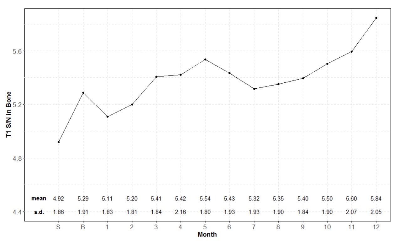
Subjects (N=67) received monthly triple-dose gadopentetate dimeglumine (0.3 mmol/kg) as part of a head-to-head MS treatment comparison beteween Betaseron and Copaxone for up to two years (up to 78 dose-equivalents). Manual segmentation of the cranial diploic space was performed using ITK-SNAP on a T2W sequence. Using an automated technique, this ROI was overlaid on the monthly T1W fat-saturated images to determine monthly changes in pixel intensity. S/N ratios established average SI change over the 12 months of the study, defined as S/N = Bone/Air. Linear mixed regression modeling with random intercept was used to test for significance of the signal intensity. Incidence of leukopenia and hypophosphatemia compared with monthly marrow signal changes was performed using the type 3 test of fixed effects. Chi square analysis was performed to find correlation between hypophosphatemia and leukopenia.Results
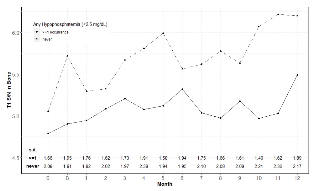
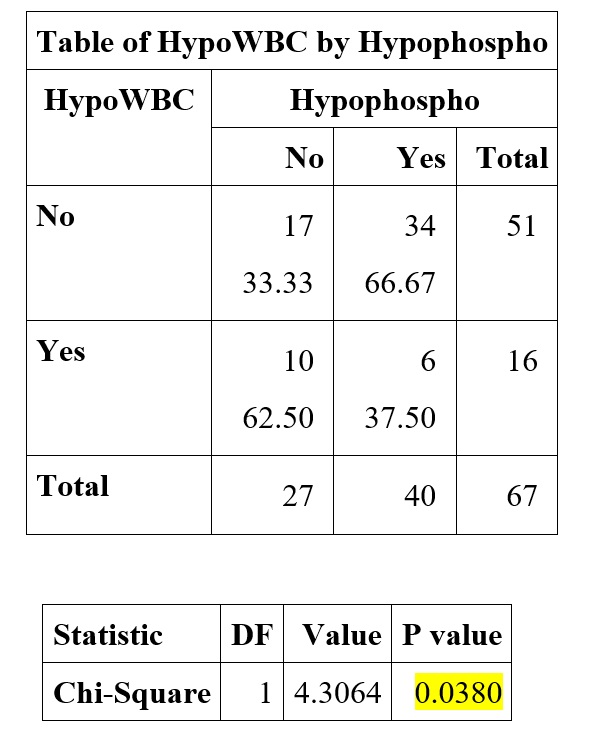
A statistically significant increased T1 fat-sat SI changes was seen in the cranial diploic space during the first 14 months of gadolinium administration: S/N=0.039 (S.E. 0.008; p<0.0001). Incidence of leukopenia increased significantly over the course of the trial with OR 1.103 (1.047, 1.162, p<0.001). Marrow signal increase was not different between leukopenic and normal subjects (p = 8.160). In the hypophosphatemia subgroup analysis, patients with consistently normal phosphate levels (n= 32) experienced a significantly higher rate of increasing T1 fat-sat S/N in bone than patients who experienced at least one episode of hypophosphatemia (n=35) during that time (mean difference in monthly S/N increase = 0.034; p=0.037). Among 16 patients with low WBC, 37.5% (6/16) patients demonstrated hypophosphatemia, while 66.7% patients with normal WBC demonstrated hypophosphatemia. Pearson’s Chi-square test showed that the difference is statistically significant (P=0.038).Conclusion
Our analysis of data from the BECOME trial has demonstrated evidence of progressive intracranial marrow signal increase on T1W fat-saturated images that may be attributable to gadolinium deposition in a study cohort receiving monthly triple-dose gadopentetate dimeglumine. However, while leukopenia and hypophosphatemia correlated with increased signal in deep grey matter nuclei, these findings did not correlate with increase in marrow signal. Furthermore, no correlation was revealed between hypophosphatemia and leukopenia.Acknowledgements
No acknowledgement found.References
1. Wolansky LJ, Haghighi MH, Sevdalis E, et al. Safety of serial monthly administration of triple-dose gadopentetate dimeglumine in multiple sclerosis patients: preliminary results of the BECOME trial. J Neuroimaging 2005;15:289-290.
2. Wolansky LJ, Cadavid D, Punia V, et al. Hypophosphatemia is associated with the serial administration of triple-dose gadolinium to patients for brain MRI. J Neuroimaging 2015;25:379-383.
3. Murata N, Murata K, Gonzalez-Cuyar LF, et al. Gadolinium tissue deposition in brian and bone. Magnetic Resonance Imaging 2016;34:1359-1365.
4. https://stemcells.nih.gov/info/2001report/chapter5.htm
Figures