0170
Accelerating DENSE MR elastography by including multi-axes motion encoding into the multiphase DENSE-MRE acquisition scheme1Department of Neurology, Medical University of Graz, Graz, Austria
Synopsis
In MR elastography, the propagation of three-dimensional wave motion is acquired to assess mechanical tissue properties. We here propose an accelerated approach of the multiphase DENSE-MRE acquisition scheme which additionally includes three-dimensional motion encoding besides the multiple phase offsets within one TR. In addition to phantom experiments, this multi-axes encoding concept was also investigated in the human brain in vivo. The gathered wave images and shear modulus maps are confirmed by three consecutive single-axes multiphase DENSE-MRE acquisitions for x-, y- and z-motion encoding direction. With this concept, the acquisition can be accelerated up to a factor of 3.
Introduction
In MR elastography, the propagation of three-dimensional wave motion is acquired to assess mechanical tissue properties.1,2 We recently presented a multiphase DENSE-MRE acquisition approach which uses stimulated echo motion encoding together with multiple phase offset sampling within each TR for reducing echo times especially at low vibration frequencies and for reducing total acquisition time.3 Though, all phase offsets are acquired within each TR, three sequence repetitions have to be performed to gather wave motion information along all three axes x, y and z.Purpose
We here propose an acceleration of the multiphase DENSE-MRE concept, which includes encoding of all three orthogonal motion directions simultaneously within each TR which allows to further reduce acquisition time.Methods
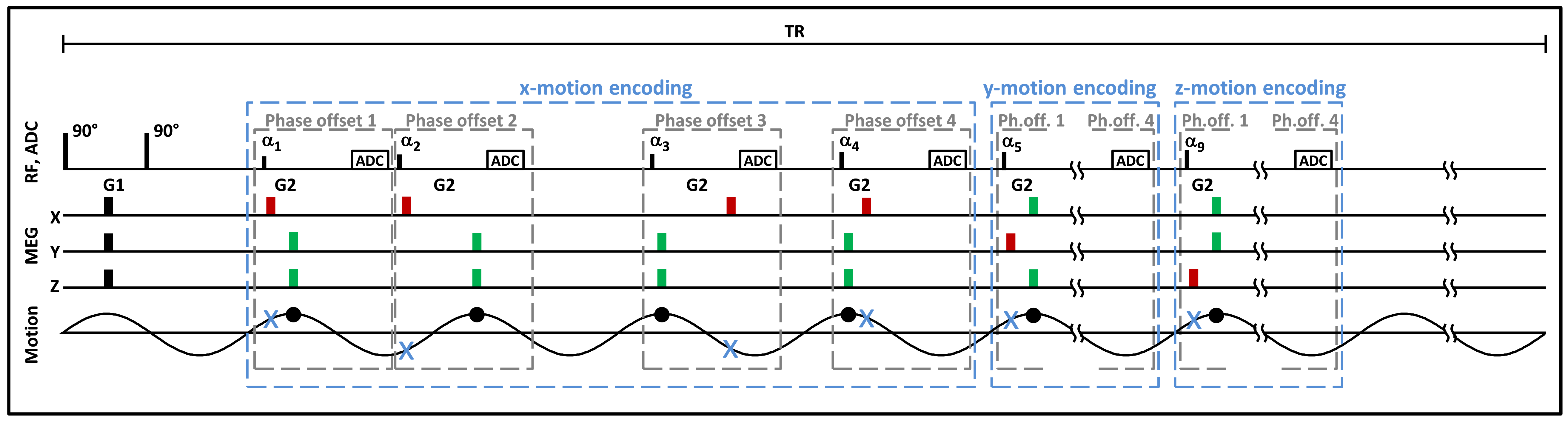
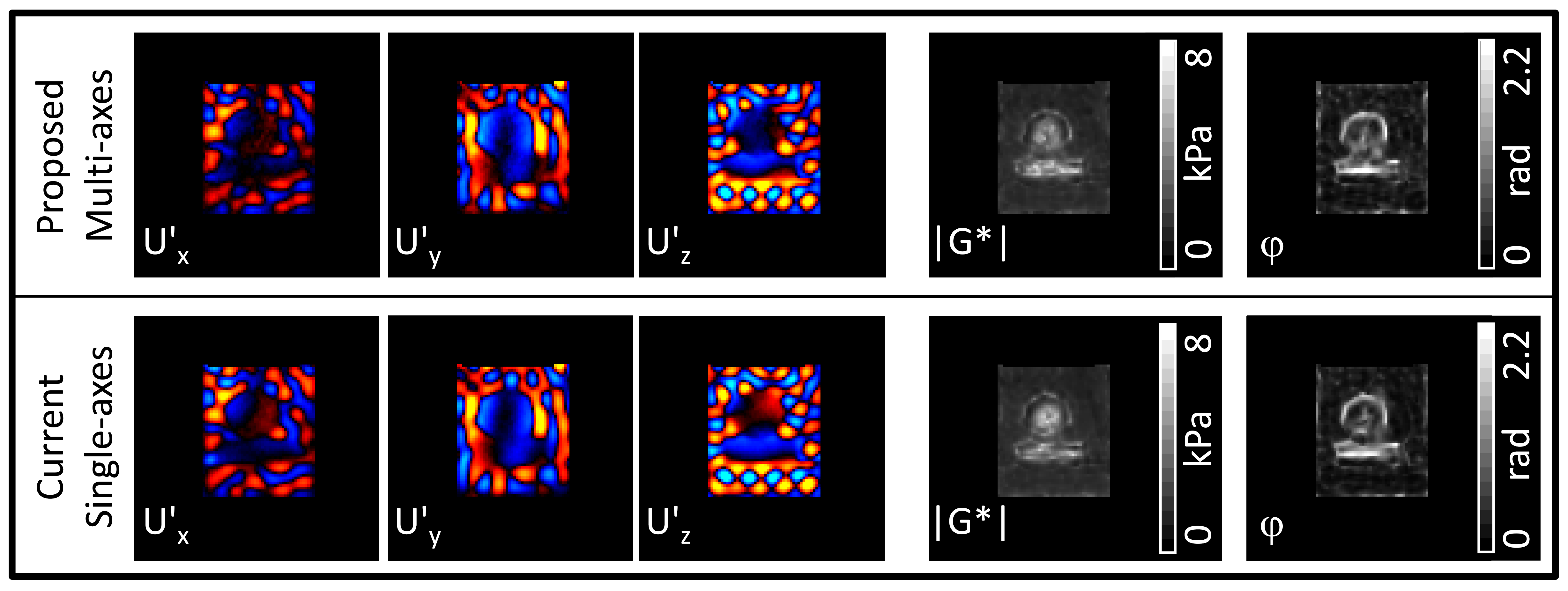
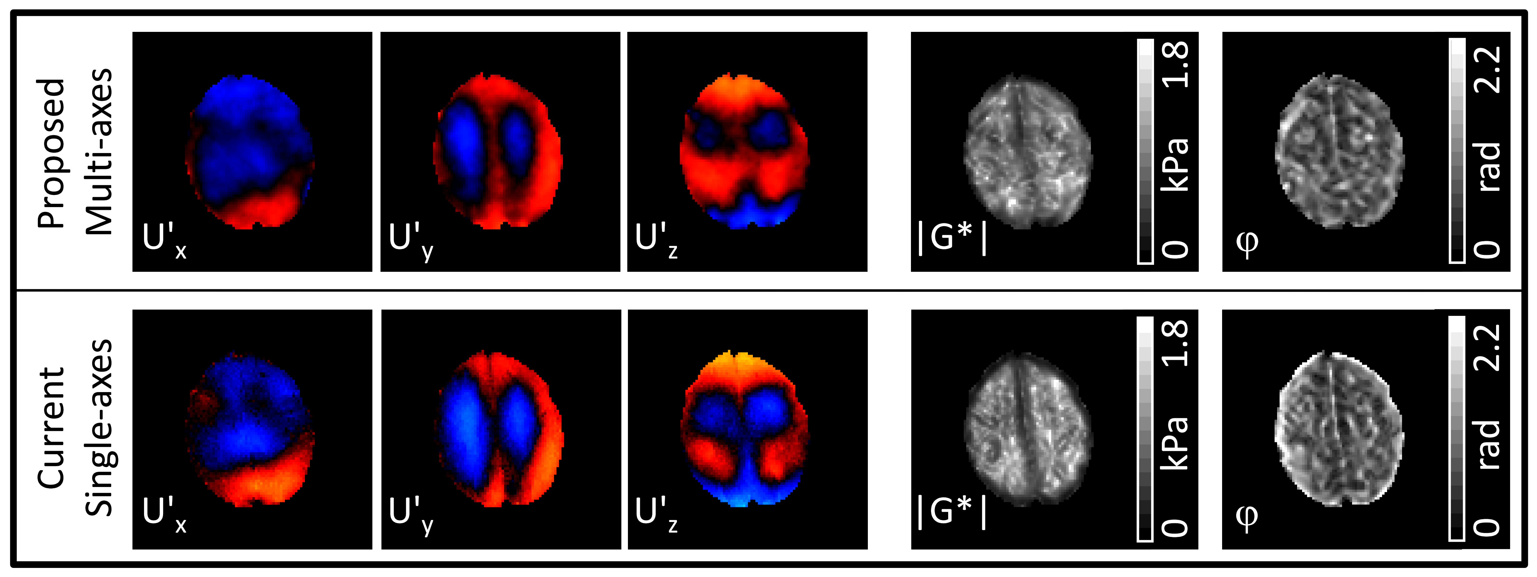
In multiphase DENSE-MRE, the motion encoding gradient consists of two monopolar gradients G1 and G2. Whereas G1 prepares the magnetization, G2 encodes the motion at the desired sampling points of the wave in a series of readout blocks during each TR. In the new multi-axes concept, the preparation is performed simultaneously in three dimensions with G1s on all three gradient axes. The set of readout blocks is divided into three subsets for encoding x-, y- and z-motion. In each subset, the timings of the G2s are adapted. For one axis, the G2s sample the wave at equidistant time points over the wave period like in multiphase DENSE-MRE. However on the remaining two axes, G2 every time captures the same sampling point of the wave, resulting in a constant background phase in the subset of images which can be eliminated during post-processing. Figure 1 illustrates this acquisition method schematically.This multi-axes concept was investigated in an agar phantom with two stiff inclusions (bar and cylinder) and in the brain of a healthy volunteer and compared to consecutive single-axes multiphase DENSE-MRE acquisitions. MRE experiments were performed on a 3T MRI scanner (Magnetom Prisma, Siemens, Erlangen, Germany) using a head cradle driven by a piezoelectric actuator. Geometric parameters for all sequences were FOV=300²mm², Matrix=128², slice thickness=4mm. Sequence parameters for the phantom study: 9 slices, TR=2520ms, readout-TE=11.8ms, 50Hz driving frequency; multi-axes: 12 mixing times between 8-230.5ms (4 phase offsets, 3 directions), TA=2min31s; single-axes: 4 mixing times between 8-53ms (4 phase offsets, 1 direction), TA=2min31s*3 (x-, y-, z-encoding). Sequence parameters for the in-vivo study: 5 slices, 20Hz driving frequency; multi-axes: TR=3250ms, readout-TE=23.0ms, 12 mixing times between 8-564.25ms (4 phase offsets, 3 directions), TA=3min15s; single-axes: TR=2500ms, readout-TE=3.8ms, 4 mixing times between 8-45.5ms (4 phase offsets, 1 direction), TA=2min30s*3 (x-, y-, z-encoding). Constant background phase was eliminated for each image subset in the first step of post-processing by mean value subtraction. Furthermore, butterworth filtering was applied prior to inversion. For the phantom a band-pass (cutoff=20-100 m-1) was used for the multi-axes and single-axes data to reduce low frequency waves and noise. In the brain only the multi-axis data was low-pass (cutoff=50m-1) filtered to reduce noise. A multifrequency dual elasto visco (MDEV) inversion algorithm was used to obtain maps of the magnitude |G*| and phase φ of the complex shear modulus G*.4
Results
The phantom images (Fig. 2) gathered with the proposed method clearly depict the wave patterns for all motion components and allowed inversion to G* maps. Comparable wave patterns and G* maps were obtained by consecutive single-axes acquisitions. G* calculation resulted in a global mean across all slices of |G*|=1.66kPa, φ=0.42° and |G*|=1.64kPa, φ=0.42° for the multi-axes and the single-axes acquisitions, respectively. In the brain (Fig. 3) also clear wave images and G* maps could be achieved by the multi-axes acquisition and confirmed by the single-axes investigation. Mean values in the brain across all slices were |G*|=0.69kPa, φ=0.66° and |G*|=0.73kPa, φ=0.83° for the multi-axes and the single-axes acquisitions, respectively.Discussion and conclusion
With the proposed multi-axes modification of the multiphase DENSE-MRE, wave images for all three-dimensional motion components could be acquired which allowed shear modulus estimation in the phantom and brain. Comparable results between the multi-axes and single-axes acquisitions confirmed the approach. Due to the higher number of readout blocks in the multi-axes scheme and prolonged readout-TE to allow G2 shifts between the axes, the SNR of the images is lower than in the single-axes acquisitions. Since the driving frequency in the phantom experiment was higher, the prolonged readout-TE had less influence than in the brain investigation with the lower frequency. However, this could be tackled by additional low-pass filtering prior to the inversion. In return, by acquiring all motion encoding directions simultaneously, the total acquisition time can be decreased by up to a factor of 3 which compensates for the SNR loss.Acknowledgements
References
1. Mariappan YK, Glaser KJ, Ehman RL. Magnetic resonance elastography: A review. Clin. Anat. 2010;23:497–511. doi: 10.1002/ca.21006.
2. Hiscox L V, Johnson CL, Barnhill E, McGarry MDJ, Huston J, van Beek EJR, Starr JM, Roberts N. Magnetic resonance elastography (MRE) of the human brain: technique, findings and clinical applications. Phys. Med. Biol. 2016;61:R401–R437. doi: 10.1088/0031-9155/61/24/R401.
3. Strasser J, Haindl MT, Stollberger R, Fazekas F, Ropele S. Magnetic resonance elastography of the human brain using a multiphase DENSE acquisition. Magn. Reson. Med. 2019;81:3578–3587. doi: 10.1002/mrm.27672.
4. Streitberger KJ, Reiss-Zimmermann M, Freimann FB, Bayerl S, Guo J, Arlt F, Wuerfel J, Braun J, Hoffmann KT, Sack I. High-resolution mechanical imaging of glioblastoma by multifrequency magnetic resonance elastography. PLoS One 2014;9. doi: 10.1371/journal.pone.0110588.
Figures