0122
MR-guided neuromodulation of visual networks in Rhesus Monkey at a 3T system
Xiaojing Long1, Yangzi Qiao1, Teng Ma1, Weibao Qiu1, Chao Zou1, Jo Lee1, Yang Liu1, Changjun Tie1, Ye Li1, Lijuan Zhang1, Qiang He2, Xin Liu1, and Hairong Zheng1
1Shenzhen Institutes of Advanced Technology, Chinese Academy of Sciences, Shenzhen, China, 2Shanghai United Imaging Healthcare Co., Ltd., Shanghai, China
1Shenzhen Institutes of Advanced Technology, Chinese Academy of Sciences, Shenzhen, China, 2Shanghai United Imaging Healthcare Co., Ltd., Shanghai, China
Synopsis
In this work, we applied BOLD fMRI in Rhesus monkey on a 3T MR system and investigated the functional effects induced by transcranial ultrasound stimulation (TUS) in both the target spot (the primary visual cortex) and the remote interconnected brain regions. We found that TUS can evoke BOLD reaction not only on the region-specific region but also the interconnected areas in the monkey brain. Additionally, our results demonstrated that the temporal features of BOLD time courses of TUS on the primary visual cortex and those of real visual stimulation have no significant difference in the regions of primary visual pathway.
INTRODUCTION
Transcranial ultrasound stimulation (TUS) is a noninvasive tool that can excite or inhibit neural activity in targeted brain regions by delivering pulsed ultrasonic waves. The combination of neuro-stimulation with functional MRI (fMRI) has received increasing attention over the past years as fMRI could provide a good illustration of brain responses to stimulation and their relation to cognition and perception, as well as a better understanding of the mechanisms underlying modulation effects. In this work, we applied BOLD fMRI in Rhesus monkeys on a 3T MR system, aiming to study the brain effects induced by TUS in both the target spot and the remote interconnected brain regions.METHODS
Experiments were conducted on a 3T MR scanner (uMR790, United Imaging Healthcare; Shanghai; China) with the simultaneous strength of 80 mT/m, slew rate of 200 mT/m/s, and a customized 8-channel surface coil. A customer-designed 300 kHz MR-compatible single element ultrasound transducer was used with a coupling cone filled with degassed ultrapure water, and fixed on the hair-removed scalp above the V1 area of the Rhesus monkey. The location of TUS target was confirmed using the MR-based acoustic radiation force imaging (ARFI). The stimulation experiment comprised two functional runs: (1) a visual stimulation and (2) a TUS run. The stimulation consisted of 8 blocks, starting with 12 seconds baseline followed by 30-second duration blocks of visual or TUS stimuli repeated for four times with 30-second baseline intervals in between. The visual stimulation was performed to the left eye of the monkey with a flashing LED. As for TUS, an acoustic pressure of 800kPa was used for stimulation. The whole-brain fMRI data was obtained using a single-shot gradient echo planar imaging (EPI) sequence with parameters of TR=2000ms, TE=30ms, flip angle 90°, 64×64 matrix, 32 slices (interleaved), 1.5×1.5×2.5 mm3 voxel size. All fMRI data analysis was performed off-line using SPM12 with the typical processing pipeline.RESULTS
Activation maps revealed that real visual stimulation activated primary visual cortex and lateral geniculate nuclei (LGN) which are in the primary visual pathway, while TUS not only activated the sonicated V1 area but also other brain regions including LGN, anterior cingulate cortex (ACC), parahippocampal gyrus, supramarginal gyrus, caudate and putamen. By comparing the temporal features of the event-related BOLD time courses, no significant difference was observed between visual stimulation and TUS on the peak amplitude and 50% peak latency of BOLD signals in the primary visual cortex and LGN.CONCLUSIONS
This work have shown that TUS can serve as a novel non-invasive stimulation technique to modulate region-specific and remote interconnected areas in the monkey brain, which suggested that TUS may provide a new mode for neuroscientific studies such as assessment of brain functions and their functional connectivity to different parts of the brain. In addition, TUS may also have the potentials for neurotherapeutics for remedying a certain pathological conditions associated with regional or network dysfunction.Acknowledgements
This work was supported by the National Natural Science Foundation of China (No.81527901).References
No reference found.Figures
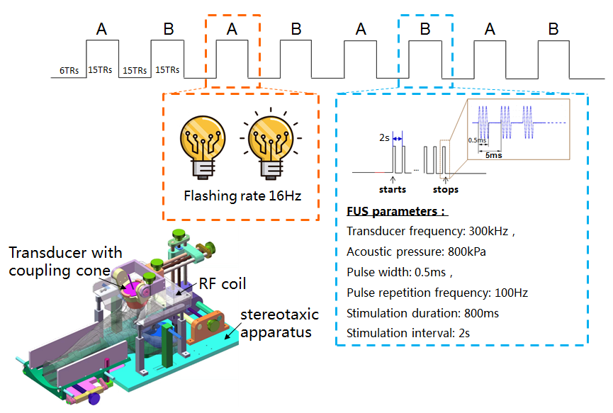
Figure 1. Experiment design and parameters.

Figure 2. BOLD activation maps of real visual stimulation (left) and TUS (right) on primary visual cortex.

Figure 3. Comparison of event-related BOLD time courses on selected regions.