0117
A Pilot Clinical Study of Quantitative Silicone Oxygen Sensors in Cervical Cancer1Koch Institute for Integrative Cancer Research, Massachusetts Institute of Technology, Cambridge, MA, United States, 2Radiology, Brigham and Women's Hospital, Boston, MA, United States, 3Radiation Oncology, Brigham and Women's Hospital, Boston, MA, United States, 4Materials Science and Engineering, Massachusetts Institute of Technology, Cambridge, MA, United States
Synopsis
We report on the first-in-human evaluation of a class of silicone oxygen sensors capable of both high sensitivity and repeated and long-term monitoring of tumor oxygen levels. We are evaluating the use of this sensor in patients receiving high dose rate brachytherapy for cervical cancer. This sensor is a direct and quantitative measurement of tumor oxygen. Low oxygen regions of tumors are more resistant to many common forms of treatment. Understanding tumor oxygen levels can enable personalized radiation and chemotherapy treatments to overcome resistance and improve outcomes for patients.
Background and Motivation
Tumor hypoxia, the extreme depletion of tumor oxygen, is a well-known adverse prognostic marker for poor clinical outcomes, including radiation and chemotherapy resistance and development of distant metastases.1–3 These trends are experienced in a number of cancers including cervical, prostate, and head and neck. Currently available clinical techniques for measuring hypoxia are qualitative, indirect, or limited by poor sensitivity. A compelling approach to combat hypoxia-induced radiation resistance is radiation dose escalation to hypoxic tumor sub-volumes. An ideal option for dose escalation is interstitial high dose rate brachytherapy (HDR-brachy) which is uniquely suited for hyper-localized dose escalation to hypoxic tumor sub volumes. HDR-brachy involves the placement of a series of catheters (2 mm diameter hollow tubes) throughout a tumor to serve as conduits for the temporary placement of radioactive seeds. Given the emerging importance of MRI guidance in brachytherapy catheter placement, an MRI-compatible quantitative oxygen sensor would be a valuable tool for tumor oxygen measurement during radiation treatment.Silicones, which have been shown to be safe in vivo, have longitudinal relaxation times that are dependent on oxygen concentration.4,5 The suitability of silicones for in vivo oxygen sensing applications is strongly dependent on formulation and materials design. Cross-linked/elastomeric silicones generally have lower oxygen sensitivity, but don’t clear from the tissue thus enabling repeated measurements at fixed locations, while silicone oils are generally more sensitive to oxygen but clear quickly from the tissue.4 The spatial and temporal heterogeneity of tumor hypoxia, and the extended duration of treatment, require repeated measurements throughout the course of treatment with high oxygen sensitivity. We report on the clinical translation of a silicone material, previously developed in our lab, that offers high sensitivity and locational stability (doesn’t clear). This is the first-in-human use of this oxygen sensing family of materials.
Methods
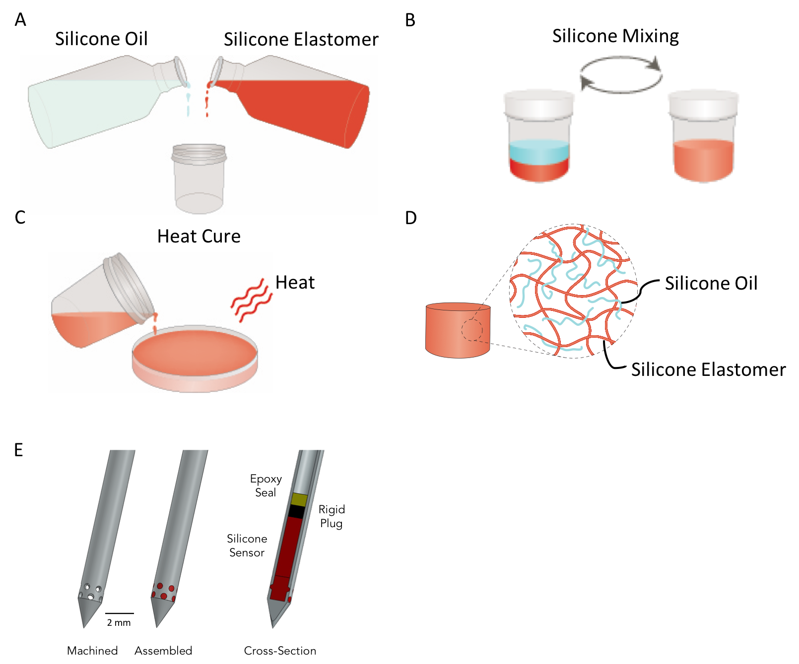
Silicone elastomer and silicone oil were mixed using dual asymmetric centrifugal mixing (Flacktek Inc.) (Figures 1A and 1B). Silicone mixtures were dispensed into molds and/or directly into the devices and heat cured to allow cross-linking of the elastomer (Figures 1C and 1D). Oxygen sensing catheters were fabricated by machining 12 holes that are each 0.5 mm in diameter at the distal tip of the catheter. The silicone was then sealed from the remainder of the inner lumen with acrylic epoxy to ensure that gas could only be exchanged with the silicone through the holes in the tip of the catheter (Figure 1E).The primary objective of this pilot study is to evaluate the feasibility of obtaining tumor oxygen measurements at the time of MR-guided interstitial brachytherapy with the temporary placement of an oxygen sensing catheter. Study eligibility required diagnosis of cervical cancer for which interstitial brachytherapy was planned. At the time of MR-guided brachytherapy, two oxygen sensor catheters are placed into the cervical tumor. Target accrual for this study is 10 patients. Two patients have been completed to date with active recruitment in the coming months and trial completion expected by March 2020. The primary endpoint of the study is the successful insertion, measurement, and removal of the oxygen sensing catheter which has been achieved in both patients.
All clinical catheter measurements were made using a 3T MRI scanner (Siemens Verio) with endorectal, spine, and body matrix receive coils. Longitudinal relaxation measurements are conducted using an inversion recovery turbo spin echo sequence. MRI data acquisition was conducted using the following parameters, inversion range: 80 to 2200 ms, repetition time: 3000 ms, echo time: 15 ms, slice thickness: 2 mm, matrix: 256 x 256, and field of view: 120 x 120 mm. Two averages were collected for each inversion time.
Results
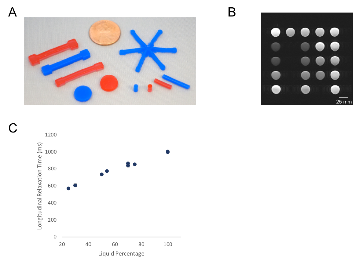
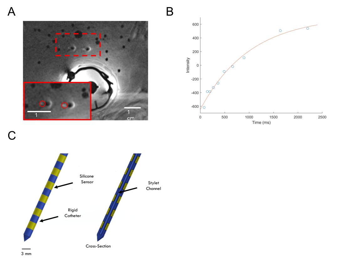
Oxygen sensors can be formed into a multitude of shapes, enabling easy integration with a wide range of clinical applications (Figure 2A). Cups of cured silicone oxygen sensors (Figure 2B) were evaluated to obtain the longitudinal relaxation times of a range of sensor formulations. Material performance of different formulations was used to inform device design (Figure 2C). Two oxygen sensing catheters were inserted into a patient’s cervical tumor. Catheter intensities were measured in a single slice (Figure 3A). These intensities were fit to the inversion recovery equation to obtain relaxation times (function of oxygen) for each catheter (Figure 3B).Discussion
Silicone oxygen sensor performance is influenced by formulation (ratio of oil and elastomer) and the necessary sensor performance (both mechanical properties and material sensitivity) should be considered when deciding on the appropriate formulation. Incorporation of the silicone sensor in the tip of a modified brachytherapy catheter serves to enable validation of this material family clinically with minimal disruption to the clinical workflow. A design (Figure 3C) that shifts the silicone to the outer surface of the catheter, thus freeing the inner conduit, will enable both oxygen sensing and radiation dose delivery capabilities in a single catheter.Conclusion
Silicone oxygen sensors measured using MRI offer a method highly compatible with the current clinical workflow and address a pressing and unmet medical need in radiation oncology. Incorporation of the sensor in a high dose rate brachytherapy catheter allows quantitative mapping of tumor oxygen levels, enabling oxygen-guided dose escalation to hypoxic tumor sub-volumes with the goal of improving clinical outcomes.Acknowledgements
This work is supported by a Bridge Project Expansion Grant (The Koch Institute for Integrative Cancer Research/Dana Farber Cancer Center), Frontier Research Grant (The Koch Institute for Integrative Cancer Research), and Lemelson Vest Grant. J. Tokuda is supported by the Image Guided Therapy Center (NIH P41EB015898). G. Ekchian is supported by The Koch Institute Quinquennial Cancer Research Fellowship and the Kavanaugh Translational Innovation Fellowship.References
1. Höckel, M. et al. Association between tumor hypoxia and malignant progression in advanced cancer of the uterine cervix. Cancer Res. 56, 4509–4515 (1996).
2. Nordsmark, M. & Overgaard, J. A confirmatory prognostic study on oxygenation status and loco-regional control in advanced head and neck squamous cell carcinoma treated by radiation therapy. Radiother. Oncol. 57, 39–43 (2000).
3. Knocke, T. H., Weitmann, H. D., Feldmann, H. J., Selzer, E. & Pötter, R. Intratumoral pO2-measurements as predictive assay in the treatment of carcinoma of the uterine cervix. Radiother. Oncol. 53, 99–104 (1999).
4. Kodibagkar, V. D., Cui, W., Merritt, M. E. & Mason, R. P. Novel 1H NMR approach to quantitative tissue oximetry using hexamethyldisiloxane. Magn. Reson. Med. 55, 743–748 (2006).
5. Liu, V. H., Vassiliou, C. C., Imaad, S. M. & Cima, M. J. Solid MRI contrast agents for long-term, quantitative in vivo oxygen sensing. Proc. Natl. Acad. Sci. U. S. A. 111, 6588–6593 (2014).
Figures