0048
A Multi-Inversion-Recovery (mIR) Myelin Water Mapping (MWF)-MRF Sequence1Diagnostic Radiology, The University of Hong Kong, Hong Kong, China, 2Philips Healthcare, Hong Kong, China
Synopsis
We proposed a new multi-inversion-recovery (mIR) myelin water mapping (MWF)-MRF sequence that allows 24 s/slice scan speed for four-compartment (myelin water, cerebrospinal fluid, gray matter and white matter) brain mapping on clinical 3T MRI.
Purpose
Myelin or myelin water mapping by MRI is highly desirable for monitoring the demyelination and remyelination processes. Such myelin measurement by MRI was conventionally based on the measurement of 16 to 32 echoes to depict the T2 differences between tissue compartments, namely free water and myelin water 1–3. The myelin water has a significantly shorter T1 and T2 than free water, which allows the myelin water to be distinguished by multi-compartment analysis on MRF data 1–3. The multi-compartment analysis is a recent key breakthrough in MRF methodology 4. However, in the previous study, the measurement was based on the combination of two separated MRF scans, one without delays after repetitions and one with 5s-delays 4. It is desirable to further simplify and integrate the two scans into one. This study aims to develop an MRF sequence sensitized for myelin water fraction measurement (MWF-MRF).Methods
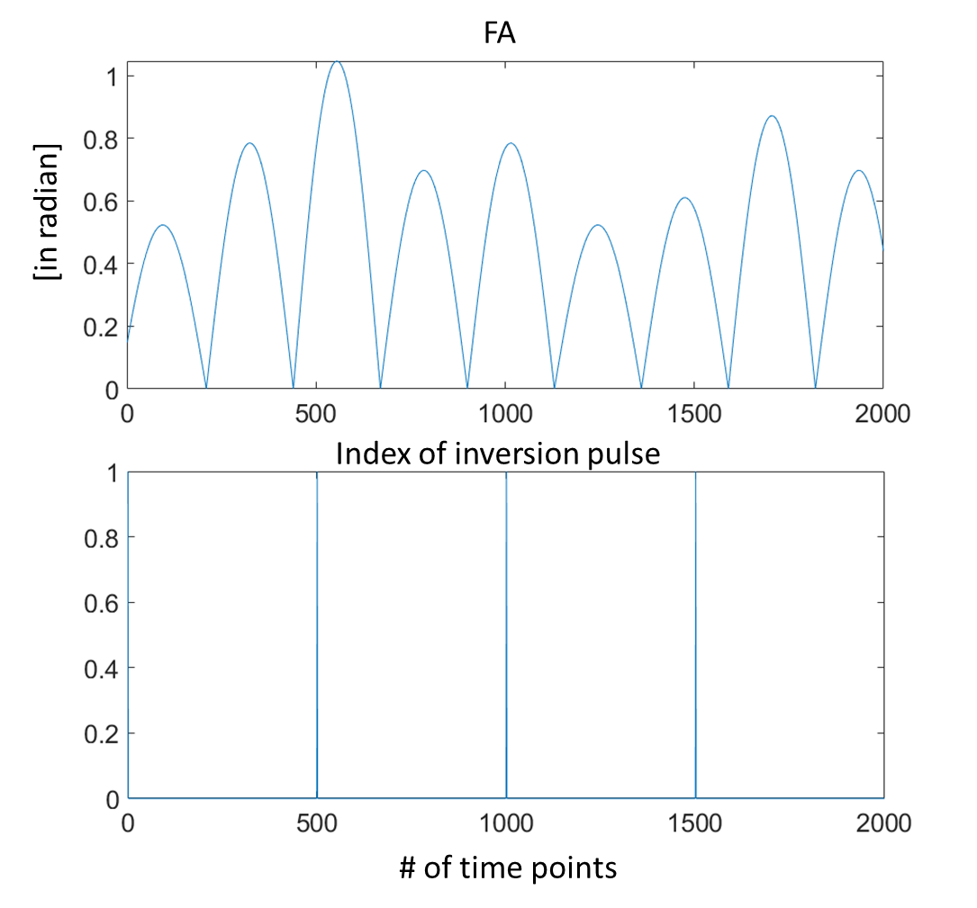
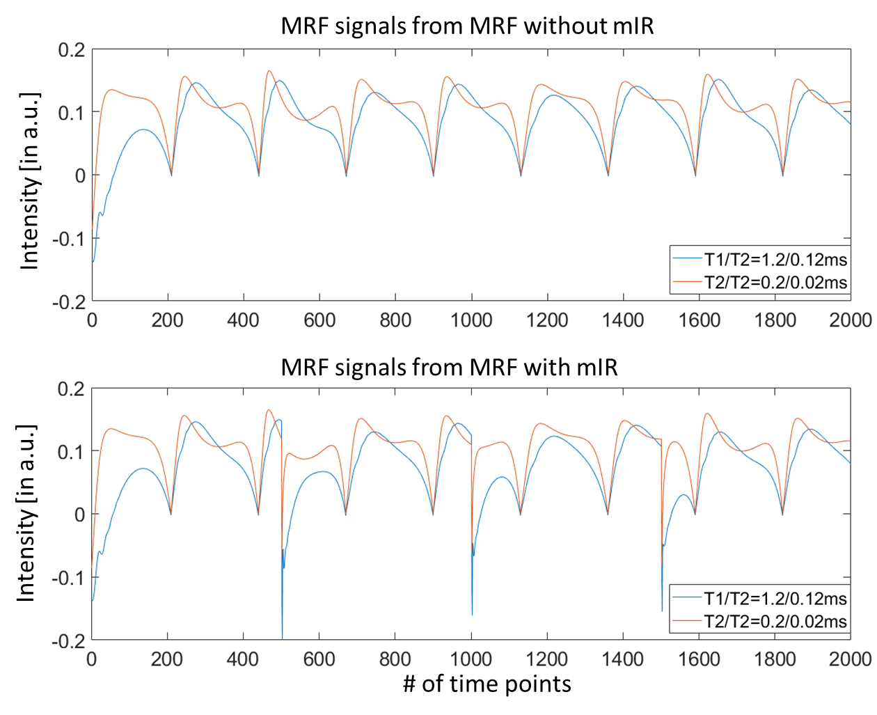
We used a 3T human MRI scanner (Achieva TX, Philips Healthcare) with an 8-channel brain coil for signal reception in the brain. The MRF was based on an inversion recovery steady-state precession (IR-FISP) sequence with variable flip angles (FA) and repetition times (TR) 5. A constant speed spiral readout trajectory with an acquisition window of 8.4 ms and acquisition factor = 58.4% was used. The trajectory was rotated by 222.5° after each dynamic. A spiral-in/out trajectory was used for increasing sensitivity while reducing the susceptibility effect 6. This new multi-IR MRF sequence (in Figs. 1 and 2) is sensitive enough to probe myelin content in the brain. The magnetization obtained after repeatedly applying the IR pulse is more specific to the myelin water signals due to the inversion and prolonged recovery of long T1 signal. Therefore, we propose to use this new MRF sequence for rapid measurement of brain myelin water content in vivo. Our preliminary in vivo experiment used the following imaging parameters: four IR pulses applied at the 1, 501, 1001, and 1501 TR, TR = 12.1 to 14.1 ms, FA = 0 to 60°, field of view = 300 × 300 mm2, acquisition matrix = 256 × 256, image resolution = 1.17 × 1.17 mm2, and slice thickness = 5 mm. The quantification was based on an in-house implementation of the non-negative least-square (NNLS) algorithm with a reweighting iteration for updating the joint distribution of the T1/T2 parameter across the slice/volume 4.Results
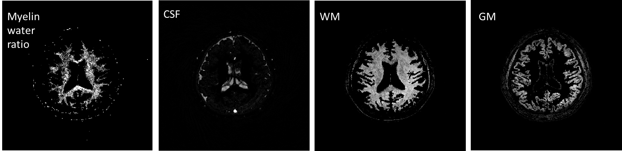
Figure 3 shows the multi-compartment myelin water mapping of a healthy volunteer using our proposed multi-IR MRF. The four-compartment brain maps were obtained from a pixel-wise separation. More importantly, the myelin water could be reliably and rapidly measured using the proposed multi-IR MRF with a scan time of 24 s/slice. The T1 and T2 ranges of the four compartments were 202 ms < T1 < 363 ms and 14 ms < T2 < 37 ms for myelin water, 1200 ms < T1 < 2630 ms and 28 ms < T2 < 300 ms for gray matter, 628 ms < T1 < 1211 ms and 34 ms < T2 < 73 ms for white matter, and 1200 ms < T1 < 2630 ms and 28 ms < T2 < 300 ms for myelin water, and 557 ms < T1 < 3768 ms and 856 ms < T2 < 1820 ms for cerebrospinal fluid. The method achieved maximal spatial separation of compartments as determined empirically from the MRF data in vivo. Figure 4 compared the MWF-MRF with or without mIR insertions. The mIR obviously improved the detection of myelin water content.Discussion
The proposed multi-IR scheme can repeatedly invert the signal of the long T1 compartment, which enhances the detection of myelin water. In this project, the mIR MWF-MRF and multi-compartment analysis successfully estimated the volume fraction of myelin water, gray matter, white matter, and cerebrospinal fluid by four-compartment separation and quantification.Conclusion
We have successfully developed a new mIR MWF-MRF sequence. The myelin water content could be reliably and rapidly measured using the proposed multi-IR MRF with a scan time of 24 s/slice.Acknowledgements
This work is supported by HKU URC seed fund.References
1. Laule, C. et al. Water content and myelin water fraction in multiple sclerosis: A T 2 relaxation study. J. Neurol. (2004). doi:10.1007/s00415-004-0306-6
2. Du, Y. P. et al. Fast multislice mapping of the myelin water fraction using multicompartment analysis of T2* decay at 3T: A preliminary postmortem study. Magn. Reson. Med. (2007). doi:10.1002/mrm.21409
3. Laule, C. et al. Myelin water imaging of multiple sclerosis at 7 T: Correlations with histopathology. Neuroimage (2008). doi:10.1016/j.neuroimage.2007.12.008
4. Nagtegaal, M., Koken, P., Amthor, T. & Doneva, M. Fast multi‐component analysis using a joint sparsity constraint for MR fingerprinting. Magn. Reson. Med. 1–14 (2019). doi:10.1002/mrm.27947
5. Jiang, Y., Ma, D., Seiberlich, N., Gulani, V. & Griswold, M. A. MR fingerprinting using fast imaging with steady state precession (FISP) with spiral readout. Magn. Reson. Med. 74, 1621–1631 (2015).
6. Glover, G. H. & Law, C. S. Spiral-in/out BOLD fMRI for increased SNR and reduced susceptibility artifacts. Magn. Reson. Med. 46, 515–522 (2001).
Figures