0031
Improved accuracy of blood-brain barrier (BBB) assessment with water-extraction-with-phase-contrast-arterial-spin-tagging (WEPCAST) MRI1Department of Radiology, Johns Hopkins University, Baltimore, MD, United States
Synopsis
A new scheme of water-extraction-with-phase-contrast-arterial-spin-tagging (WEPCAST) MRI was proposed for non-invasive assessment of blood-brain-barrier (BBB) permeability to water. In this scheme, venous bolus-arrival-time was measured first by Look-Locker WEPCAST and then applied to single-delay long-labeling-duration WEPCAST scan to estimate water extraction fraction. The results showed an improved accuracy for estimation of BBB permeability.
INTRODUCTION
Integrity of blood-brain-barrier (BBB) is traditionally assessed by its permeability to relatively large molecules, such as CSF/serum albumin ratio for non-imaging approaches1 and Gd-based MRI for imaging approaches2. Recently, there is a growing interest to measure BBB permeability to smaller molecules such as water3-5, with the promise of early detection or increased sensitivity to BBB breakdown. Water-extraction-with-phase-contrast-arterial-spin-tagging (WEPCAST) is a new, non-contrast method to measure BBB permeability to water6. This method works by selective quantification of arterially labeled spins on the venous side, providing a measurement of venous bolus-arrival-time (vBAT) and water extraction fraction (E). In the original report, vBAT and E were estimated simultaneously. This is, however, less optimal because sequence requirements for the measurements of these two parameters are different. Here we proposed a new scheme of WEPCAST technique in which two separate scans are performed, one specifically measuring vBAT and the other specifically focusing on quantification of venous signal.METHODS
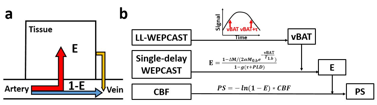
TheoryWEPCAST measures arterially labeled signal on the venous side (Figure 1a), which can be written as $$ΔM=ΔM_a+ΔM_b$$ where $$ΔM_a=2\alpha(1-E)M_{0,blood}e^{-\delta_v/T_{1,blood}}c(t)$$ denotes the non-extracted labeled spins and $$ΔM_b=2\alpha f/\lambda M_{0,blood}e^{-\delta_v/T_{1,blood}}c(t)\ast[r(t)m(t)]$$ represents the extracted spins that are re-exchanged into vessel. $$$\delta_v$$$ is vBAT, $$$\lambda$$$ is blood-brain-partition-coefficient, $$$\alpha$$$ is labeling efficiency, $$$c(t)=\{\begin{array}{l}1,if\delta_v<t<\delta_v+\tau\\0,otherwise\end{array}$$$ is arterial-input-function, $$$r(t)=e^{-ft/\lambda}$$$ is residue function and $$$m(t)=e^{-t/T_{1,tissue}}$$$ is T1 relaxation. Here, $$$\delta_v$$$ and E are the only unknowns. One way to estimate them, as proposed in the original report, is to measure ΔM at multiple post-labeling-delay times (PLD) and fit for $$$\delta_v$$$ and E simultaneously. However, to reliably estimate $$$\delta_v$$$, the labeling duration needs to be short (e.g. 2s) so that the bolus wash-in/wash-out curve is clear. On the other hand, short labeling duration may not allow an accurate estimation of ΔM due to bolus dispersion. This study therefore proposes to measure $$$\delta_v$$$ and E through a two-step procedure (Figure 1b): 1) use a short-labeling-duration Look-Locker (LL) WEPCAST sequence to determine $$$\delta_v$$$; 2) apply $$$\delta_v$$$ value to another single-delay WEPCAST scan with long labeling-duration to estimate E.
Simulation
One set of simulations were performed to show that short-labeling-duration LL-WEPCAST can estimate $$$\delta_v$$$ reliably but its quantification of ΔM can be affected by large number of background-suppression pulses. Another set of simulations were performed to show that long labeling duration is necessary to alleviate the confounding effect of bolus dispersion on ΔM measurement.
MRI Experiment
Ten healthy volunteers (29.3±5.2years, 5F/5M) were scanned on a 3T system. WEPCAST with a labeling duration of 2, 3, 4, 5s and a fixed PLD of 2.5s was performed in mid-sagittal plane, followed by a M0 scan for normalization. A conventional single-delay WEPCAST sequence with labeling duration of 2s and PLD of 3.5s6 was performed for comparison. In a subset of seven volunteers (28.4±5.9years, 3F/4M), LL-WEPCAST (labeling duration=2s, 6 PLDs) was also acquired in coronal view.
Data processing
WEPCAST processing followed Lin et al6. Briefly, phase-contrast complex-difference images were collected for control and label conditions and a subtraction between them gives WEPCAST difference images. For LL-WEPCAST, superior-sagittal-sinus (SSS) signals at different PLDs were fit for the kinetic model to estimate vBAT. For single-delay WEPCAST, ROI signals for anterior, middle and posterior SSS were quantified. E was estimated by applying vBAT from LL-WEPCAST to single-delay scan results. BBB permeability-surface-area-product (PS) was then quantified using the relationship $$$PS=-ln(1-E)· CBF$$$, where CBF is cerebral-blood-flow, assumed to be 50 mL/100g/min for male and 60 mL/100g/min for female.
RESULTS AND DISCUSSION
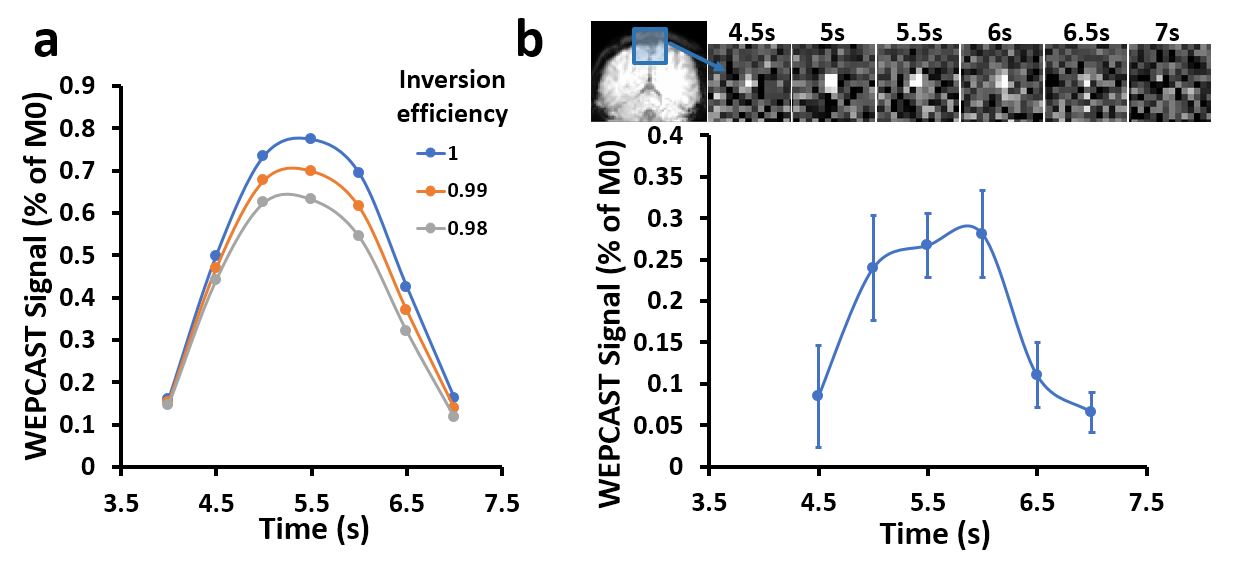
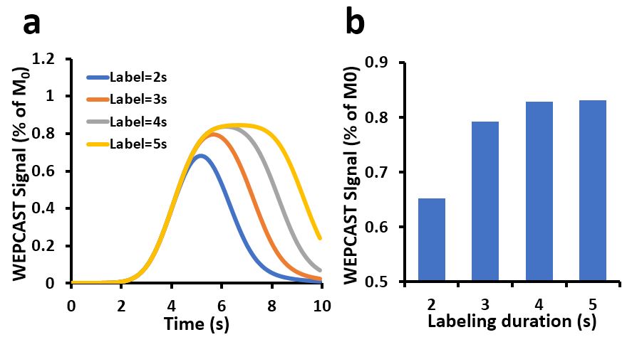
Figure 2a showed simulated LL-WEPCAST signal time course as a function of inversion efficiency of the background-suppression pulses. The signal intensity ΔM deviated from theoretical value (blue curve) when the inversion efficiency deteriorates, especially in later time points. However, $$$\delta_v$$$ was not affected. Figure 2b showed experimental LL-WEPCAST images as well as signal time course. The averaged vBAT was found to be 4.5±0.2s.Figure 3a showed simulated WEPCAST signal ΔM as a function of time, at various labeling durations. As can be seen, bolus dispersion caused an underestimation of in short-labeling-duration scans. Figure 3b showed simulation data of ΔM as a function of labeling duration, at a fixed PLD of 2.5s.
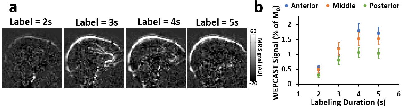
Figure 4a showed representative single-delay WEPCAST images of different labeling duration for one participant. Clear signal was presented at SSS and appeared brighter when labeling duration increased. Quantitative ROI results were shown in Figure 4b. As labeling duration increased, all ROI signals first increased and then reached plateau. No significant difference was found between labeling duration of 4s and 5s (p>0.05 for all locations). Considering both signal accuracy and scan duration, we recommend a WEPCAST protocol of 4s labeling duration.
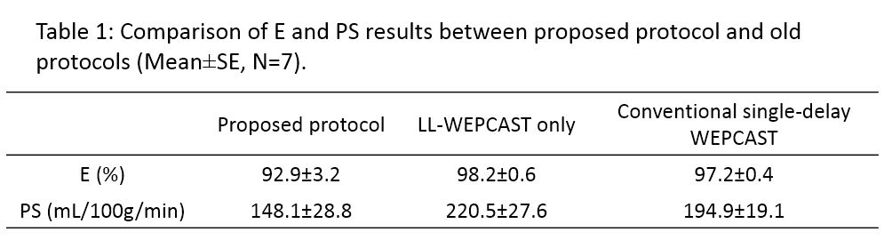
When applying the vBAT values estimated from LL-WEPCAST scan to single-delay, 4s-labeling-duration WEPCAST, E and PS were found to be 92.9±3.2% and 148.1±28.8mL/100g/min. Table 1 also summarized the results using approaches proposed in the original WEPCAST report, i.e. using LL-WEPCAST only or conventional single-delay WEPCAST (in the same subjects). Compared with current results, a clear overestimation of E and PS using old approaches can be seen.
CONCLUSION
A new scheme of WEPCAST technique was proposed to improve the accuracy of BBB permeability measurement by estimating venous BAT and water extraction with LL-WEPCAST and single-delay long-labeling-duration WEPCAST in a two-step procedure.Acknowledgements
No acknowledgement found.References
1. Alafuzoff I, Adolfsson R, Bucht G, Winblad B. Albumin and immunoglobulin in plasma and cerebrospinal fluid, and blood-cerebrospinal fluid barrier function in patients with dementia of Alzheimer type and multi-infarct dementia. J Neurol Sci 1983;60:465-472.
2. Montagne A, Barnes SR, Sweeney MD et al. Blood-brain barrier breakdown in the aging human hippocampus. Neuron 2015;85:296-302.
3. Shao X, Ma SJ, Casey M, D'Orazio L, Ringman JM, Wang DJJ. Mapping water exchange across the blood-brain barrier using 3D diffusion-prepared arterial spin labeled perfusion MRI. Magn Reson Med 2019;81:3065-3079.
4. He X, Wengler K, Schweitzer ME. Diffusion sensitivity of 3D-GRASE in arterial spin labeling perfusion. Magn Reson Med 2018;80:736-747.
5. Ohene Y, Harrison IF, Nahavandi P, Ismail O, Bird EV, Ottersen OP, Nagelhus EA, Thomas DL, Lythgoe MF, Wells JA. Non-invasive MRI of brain clearance pathways using multiple echo time arterial spin labelling: an aquaporin-4 study. Neuroimage 2019;188:515-523.
6. Lin Z, Li Y, Su P, Mao D, Wei Z, Pillai JJ, Moghekar A, van Osch M, Ge Y, Lu H. Non-contrast MR imaging of blood-brain barrier permeability to water. Magn Reson Med 2018;80:1507-1520.
Figures