0021
Super-Resolution Multi-band ASL using Slice Dithered Enhanced Resolution (SLIDER) Technique1University of Southern California, Los Angeles, CA, United States
Synopsis
Arterial Spin Labelling (ASL) is a noninvasive imaging technique that can quantitatively measure Cerebral Blood Flow (CBF). However, existing ASL techniques generally have a low spatial resolution due to a relative low Signal-to-noise ratio (SNR). In this study, we develop a super-resolution ASL method by combining the Slice Dithered Enhanced Resolution (SLIDER) with multi-band ASL with optimized slice-dependent background suppression to enhance both the resolution and SNR. The reconstructed images achieve a resolution of isotropic 2x2x2 mm3, and show increased spatial and temporal SNR compared to standard high-resolution ASL images.
Introduction
As an entirely noninvasive and quantitative imaging method, arterial spin labeled (ASL) perfusion MRI is increasingly being used for clinical applications. However, existing ASL techniques generally have a coarse spatial resolution, due to a low signal-to-noise ratio (SNR). Simultaneous Multi-Slice (SMS) or Multiband (MB) is a new accelerated imaging technology that simultaneously excites multiple slices and recovers each slice with parallel imaging techniques1. Preliminary studies combining MB with ASL showed that MB can reduce T1 relaxation of the label2, improve spatial coverage and resolution compared to those of standard 2D ASL. Super-resolution imaging techniques are ideal for high-resolution ASL by increasing both resolution and SNR efficiency. It utilizes sub-voxel spatial shifts in the slice direction to achieve a √n gain (n is the number of spatially shifted thick slices) in SNR efficiency compared to that of a standard acquisition of thin slices. The goal of this project is to develop and evaluate cutting-edge MB pCASL protocols and apply super-resolution reconstruction to offer a perfusion image with a high spatial resolution of isotropic 2mm.Methods
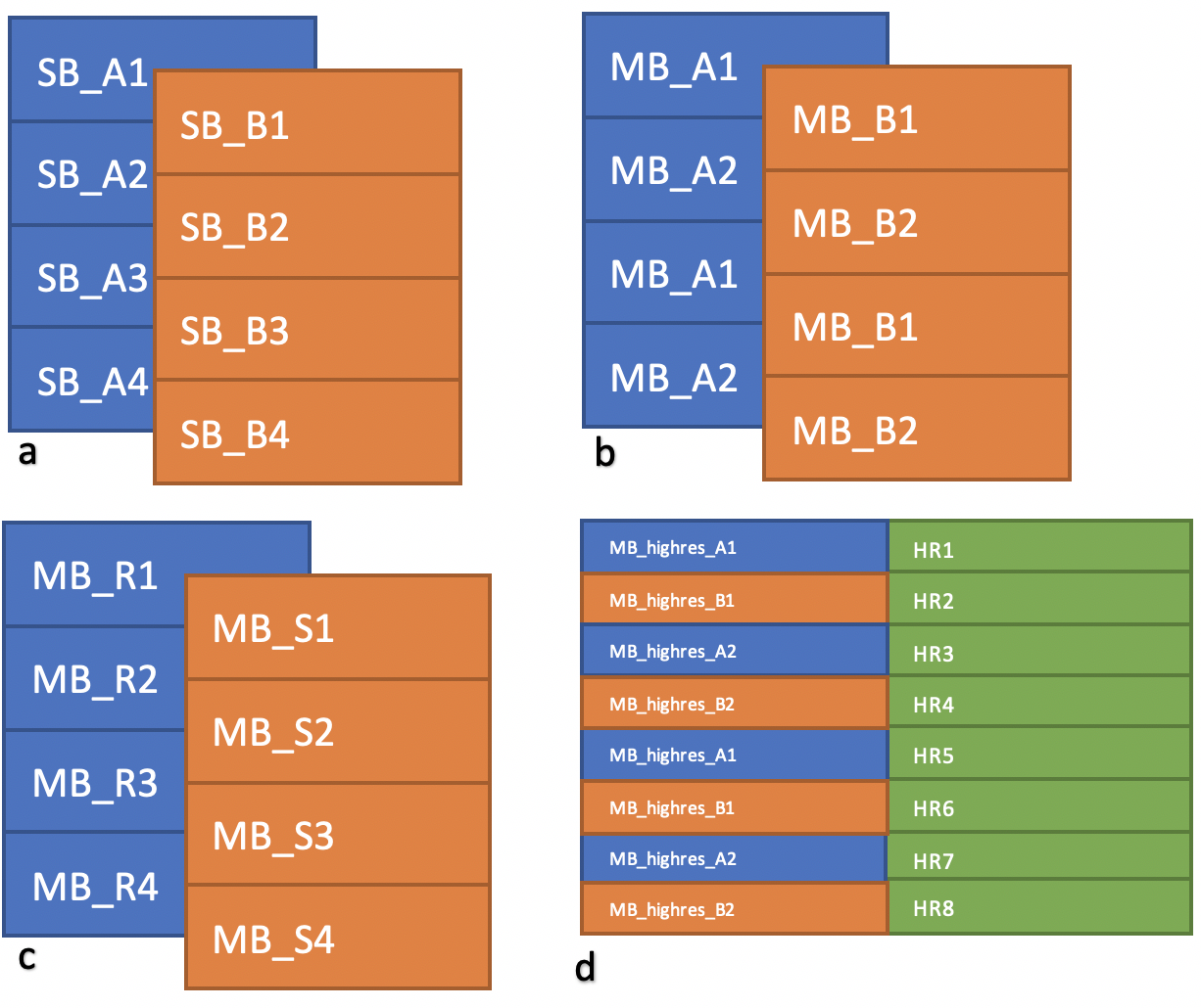
1. SMS pCASL ProtocolTo get the high-resolution perfusion image, low-resolution multi-band images were acquired, as well as their single-band reference images, which were used to reconstruct the unaliased images. The sequence used was a constrained slice-dependent background suppressed simultaneous multi-slice3 pseudo-Continuous Arterial Spin Labeling(pCASL). Imaging parameters for this SMS-pCASL sequence were: FOV=192mm, resolution=2x2x4mm3, TR=5000ms, TE=20ms, Post Labeling Delay (PLD)=1800ms. The multi-band acceleration factor was chosen to be four, so as to reduce the T1 relaxation effect of the label. The two multi-band acquisitions had a slice-shift of 2 mm (half of the slice thickness), so that they can be used for super-resolution reconstruction. Besides the low-resolution multi-band images, standard high-resolution multi-band images, with resolution of 2x2x2 mm3, were also acquired for comparison with the reconstructed super-resolution results. When acquiring high-resolution images, two interleaved scans were obtained (as shown in Figure 1) to be comparable to the super-resolution images.
2. Super-Resolution Method
we used the super-resolution algorithm called the SLIce Dithered Enhanced Resolution (SLIDER) technique4, which has been successfully applied for SMS diffusion MRI and BOLD fMRI to achieve sub-millimeter spatial resolutions. In this study, we integrated and adapted the SLIDER technique for 2D MB pCASL to achieve a high resolution of iso-2mm, without significant loss in SNR. The basic idea of this algorithm was to use low-resolution images as sub-samples of high-resolution images to be reconstructed. When calculating the inverse model, the Toeplitz matrix was used as the forward model and Tikhonov regularization with a tuning parameter λ applied instead of direct pseudo inverse for denoising purpose. λ values were chosen to be ranging from 10 to 50% of the largest eigenvalue of the Toeplitz matrix.
3. Results evaluation
To evaluate the results of super-resolution images, spatial and temporal SNR in the grey matter areas were calculated. Spatial SNR was defined by mean signal in the sum of even and odd perfusion images divided by the standard deviation of difference between even and odd perfusion images2. We used this different definition from the traditional one because of the inhomogeneous noise distribution of the images. Temporal SNR was defined by mean signal of all the time points of perfusion images divided by the standard deviation in signals between different time points.
Results and Discussion
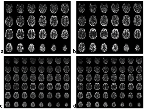
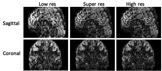
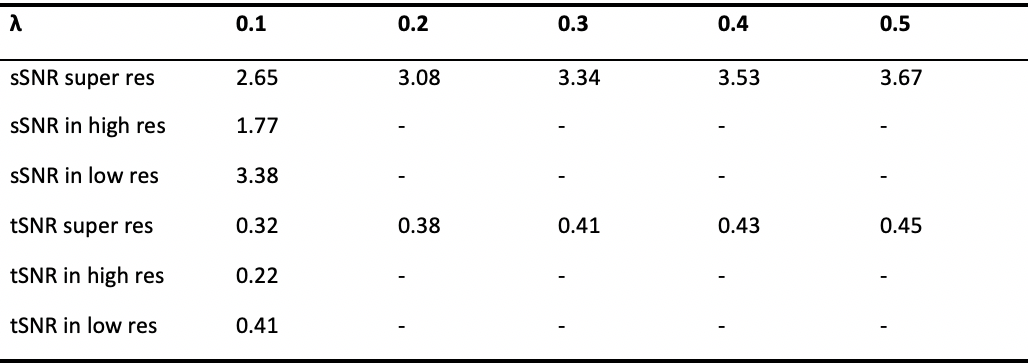
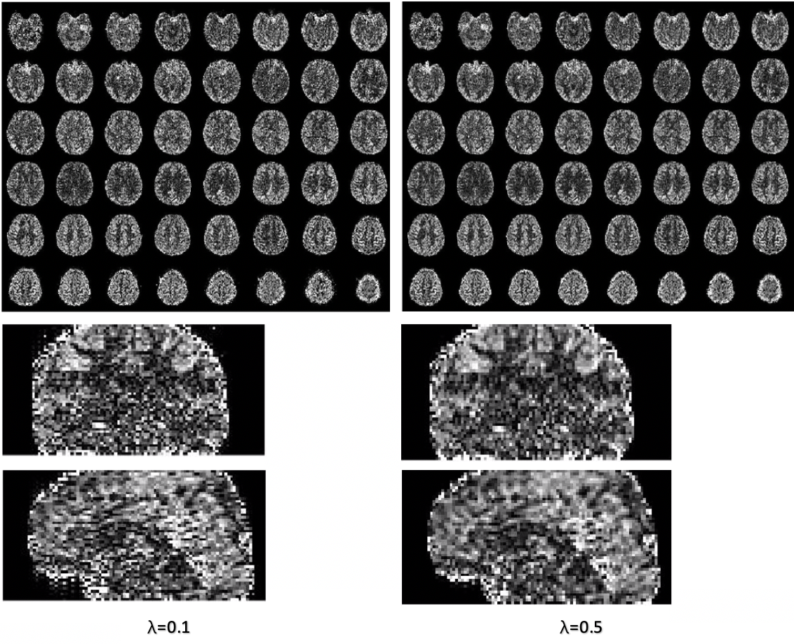
Figure 2 and figure 3 show the reconstructed super-resolution images. Qualitatively, super-resolution images are comparable to the original high-resolution images with the same spatial resolution of 2x2x2 mm3 without apparent blurring. Figure 4 shows the spatial and temporal resolution of the reconstructed super-resolution images compared to that of the low-resolution and high-resolution images. When the regularization term λ is 0.1, the sSNR and tSNR of the super-resolution are 41.6% and 45.5% higher than that of the standard high resolution, which is similar to the theoretical gain. Figure 5 shows that as λ becomes larger, though the SNR of super-resolution improves, there appears to be some blurring effects in the images, as a result of over regularization.Conclusion
With a super resolution method and an optimized SMS pCASL protocol, we can acquire high-resolution perfusion images with a higher SNR efficiency, compared to current existing ASL techniques. This technique may be particularly useful to differentiate effects of brain structural and functional changes.Acknowledgements
No acknowledgement found.References
1. Setsompop K, Gagoski BA, Polimeni JR, Witzel T, Wedeen VJ, Wald LL. Blipped-controlled aliasing in parallel imaging for simultaneous multislice echo planar imaging with reduced g-factor penalty. Magn Reson Med 2012; 67:1210-1224.
2. Wang Y, Moeller S, Li X, et al. Simultaneous multi-slice Turbo-FLASH imaging with CAIPIRINHA for whole brain distortion-free pseudo-continuous arterial spin labeling at 3 and 7 T. Neuroimage 2015;113:279-288.
3. Shao X, Wang Y, Moeller S, et al. A constrained slice‐dependent background suppression scheme for simultaneous multislice pseudo‐continuous arterial spin labeling[J]. Magnetic resonance in medicine, 2018, 79(1): 394-400.
4. Vu A T, Beckett A, Setsompop K, et al. Evaluation of SLIce Dithered Enhanced Resolution Simultaneous MultiSlice (SLIDER-SMS) for human fMRI[J]. Neuroimage, 2018, 164: 164-171.
Figures