Motion of the Heart with the Respiratory & Cardiac Cycles: Advanced Motion Correction Techniques
1King's College London
Synopsis
MRI acquisition is slow compared to physiological motion, thus the extensive cardiac- and respiratory- induced motion of the heart during the acquisition period can degrade image quality by introducing ghosting and/or blurring like motion artefacts. Several cardiac and respiratory motion compensation techniques have been proposed to overcome this problem. These techniques are based on minimizing, correcting or resolving the motion during the acquisition. This talk will include examples of recently introduced advanced methods to deal with cardiac and respiratory motion, discussing their strengths and limitations.
Introduction
MRI acquisition is slow compared to physiological motion, thus the extensive cardiac- and respiratory- induced motion of the heart during the acquisition period can degrade image quality by introducing ghosting and/or blurring like motion artefacts. Several cardiac and respiratory motion compensation techniques have been proposed to overcome this problem. These techniques are based on minimizing, correcting or resolving the motion during the acquisition. This talk will include examples of recently introduced advanced methods to deal with cardiac and respiratory motion, discussing their strengths and limitations.Cardiac motion
External electrocardiogram (ECG) devices are usually employed to synchronize the acquisition with the cardiac cycle. To minimize motion within the cardiac cycle the acquisition is targeted to periods with minimal cardiac motion, usually end-systole or in mid-diastole. Conventional ECG-triggered acquisitions provide a static visualization of the heart. So-called free-running acquisitions image the heart at different points of the cardiac cycle. With these approaches, data acquisition is performed continuously throughout the entire cardiac cycle and retrospective ECG-gating is used to assign the data to different cardiac phases. However this approach is usually limited to 2D scans. More recently whole-heart free-running approaches have been propose to enable functional assessment and whole-heart anatomy visualization from a single free-running scan [1-2]. Non-rigid motion compensation algorithm have been also proposed to align the different cardiac phases of a free-running acquisition to a reference phase, correcting rather than resolving the motion [3].
Respiratory-induced motion of the heart
A simple approach to minimize the respiratory-induced motion of the heart is to perform the acquisition under one or multiple breath-holds. However, this approach is incompatible with high-resolution 2D or 3D acquisitions due to the long scan time required to satisfy spatial resolution and volumetric coverage requirements. Moreover breath-holding can be difficult especially for paediatric and geriatric patients.
Respiratory motion monitoring can be used to enable free breathing scans, combining data from multiple breathing cycles acquired at a similar respiratory position. One-dimensional (1D) diaphragmatic navigator echoes are widely used in clinical practice to monitor the respiratory motion of the dome of the diaphragm. This signal is used to gate the acquisition, that is, data are accepted/acquired only when the respiratory signal is within a predefined narrow acceptance window (3–5 mm) of the breathing cycle (typically end-expiration) with all other data being rejected. Conversely, data falling outside the acceptance window are rejected and need to be re-acquired at subsequent cardiac cycles. Diaphragmatic navigators can be also used to correct for residual motion within the gating window, usually referred to as tracking. This method assumes that the heart motion is dominated by translation in the superior-inferior (SI) direction, and that this displacement is proportional to that of the diaphragm. However 1D diaphragmatic navigator approaches lead to prolonged and unpredictable scan times since only a portion of the data (typically 30–50%) is accepted for reconstruction (referred to as scan efficiency). Moreover, the diaphragmatic navigator method is limited because it infers (rather than measures) the motion of the heart, does not account for the multidimensional non-linear motion of the heart or hysteresis effects between inspiration and expiration.
Several technical developments have been proposed to overcome some of these drawbacks and achieve 100% scan efficiency (thus shorter scan times) with none or minimal data rejection. Self-navigation (SN) methods [4-5] have been proposed to derivate the respiratory-induced motion of the heart from the acquired data itself without the need of either a 1D diaphragmatic navigator or a heart-diaphragm tracking factor. Respiratory-induced displacements of the heart can be directly estimated from the repetitive acquisition of the central k-space point or the central k-space line. SI translational respiratory motion compensation, typically to end-expiration, is performed directly in k-space by applying a linear phase shift before image reconstruction. However, respiratory SN techniques account for translational motion only and inaccuracies in motion estimation can be introduced from the contribution of the static tissues (such as the chest wall) present in the zero- or one-dimensional projection of the entire imaging volume. In order to address this hurdle, the so-called image navigator (iNAV) methods have been proposed [6-7]. Such approaches aim at spatially isolating the moving heart from the surrounding tissues by acquiring a low resolution 2D image or 3D volume prior to data collection or as part of the acquired data itself. This improves the quality of motion detection not only by eliminating the contribution from the chest wall, but also by enabling multiple degrees of freedom for motion correction. iNAVs have been used to extract information on the translational motion of the heart in 2D and 3D.
In several approaches, the SI translational respiratory motion of the heart—estimated either via SN or iNAV strategies—is used to bin the imaging data at different respiratory stages or bins. Such bin images can be used for the estimation of inter-bin non-rigid motion fields, thus allowing for the reconstruction of a single, non-rigid motion-corrected, 3D whole-heart volume composed of all the acquired k-space data [8].
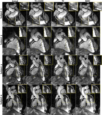
Additionally, the image quality of each individual bin can be augmented by exploiting sparsity along the respiratory dimension, obtaining respiratory motion-resolved images. Recently, approaches aimed at resolving the motion of the heart along both the respiratory and the cardiac dimensions have been introduced, allowing for respiratory-resolved images of the heart throughout the entire cardiac cycle [9].
Acknowledgements
No acknowledgement found.References
1. J. Pang et al., “ECG and navigator-free four-dimensional whole-heart coronary MRA for simultaneous visualization of cardiac anatomy and function,” Magn Reson Med, vol. 72, no. 5, pp. 1208–1217, Nov 2014. 20.
2. S. Coppo et al., “Free-running 4D whole-heart self-navigated golden angle MRI: initial results,” Magn Reson Med, vol. 74, no. 5, pp. 1306–1316, Nov 2015. 21.
3. J. Pang et al., “High efficiency coronary MR angiography with nonrigid cardiac motion correction,” Magn Reson Med, vol. 76, no. 5, pp. 1345–1353, Nov 2016.
4. C. Stehning, P. Bornert, K. Nehrke, H. Eggers, and M. Stuber, “Free-breathing whole-heart coronary MRA with 3D radial SSFP and self-navigated image reconstruction,” Magn Reson Med, vol. 54, no. 2, pp. 476–480, Aug 2005. 31.
5. D. Piccini, A. Littmann, S. Nielles-Vallespin, and M. O. Zenge, “Respiratory self-navigation for wholeheart bright-blood coronary MRI: methods for robust isolation and automatic segmentation of the blood pool,” Magn Reson Med, vol. 68, no. 2, pp. 571–579, Aug 2012.
6. M. Henningsson, P. Koken, C. Stehning, R. Razavi, C. Prieto, and R. M. Botnar, “Whole-heart coronary MR angiography with 2D self-navigated image reconstruction,” Magn Reson Med, vol. 67, no. 2, pp. 437–445, Feb 2012.
7. J. Luo et al., “Nonrigid Motion Correction With 3D Image-Based Navigators for Coronary MR Angiography,” Magn Reson Med, vol. 77, no. 5, pp. 1884–1893, May 2017.
8. G. Cruz, D. Atkinson, M. Henningsson, R. M. Botnar, and C. Prieto, “Highly efficient nonrigid motion-corrected 3D whole-heart coronary vessel wall imaging,” Magn Reson Med, vol. 77, no. 5, pp. 1894–1908, May 2017
9. L. Feng et al., “5D whole-heart sparse MRI,” Magn Reson Med, vol. 79, no. 2, pp. 826–838, Feb 2018.
Figures
