g-Ratio Mapping: Challenges & Validation
1McGill University, Montreal, QC, Canada, 2University of Calgary, Calgary, AB, Canada
Synopsis
The fiber g-ratio is the ratio of the inner to the outer diameter of the myelin sheath of a myelinated axon. In healthy neural tissue, it is optimized for speed of signal conduction, cellular energetics, and spatial constraints. The g-ratio is a fundamental contributor to functionally relevant neuronal properties such as conduction velocity. g-Ratio imaging has the potential to allow us to investigate in vivo g-ratio changes in health and disease. This lecture details the methodology for imaging the g-ratio with MRI, describes the known challenges in doing so, presents validation work to date, and discusses challenges in validating the technique.
Lecture Overview
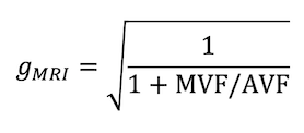
The g-ratio has been investigated with MRI in the context of healthy white matter in the adult brain {Stikov and Campbell et al., 2015; Mohammadi et al., 2015; Thapaliya et al., 2017; Jung et al., 2018} and spinal cord {Duval et al., 2017}, age and gender variation {Cercignani et al., 2016; Berman et al., 2017}, neurodevelopment {Dean et al., 2016; Melbourne et al., 2016}, Multiple Sclerosis {Cercignani et al., 2015; Hagiwara et al., 2017}, and connectomics {Mancini et al., 2018}. The original MRI-based g-ratio formulation {Stikov et al., 2011; Stikov and Campbell et al., 2015} computes a voxel-aggregate g-ratio using one or more MR contrasts sensitive to the axon volume fraction (AVF) and the myelin volume fraction (MVF) (Eq. 1), meaning it is determined entirely by the ratio MVF/AVF. Most implementations thus far have been multi-modal, with diffusion MRI providing sensitivity to the AVF, while providing insignificant signal from the myelin compartment. Complementary myelin-sensitive contrast has been used to estimate the MVF. This poses several challenges: first, image registration is critical for such a voxelwise computation {Mohammadi et al., 2015}. Second, the fact that the axon signal fraction estimate comes from an acquisition that has insignificant signal from myelin means that the absolute AVF depends on the MVF estimate, and this creates a coupling between gMRI and the total fiber volume fraction if the MVF is miscalibrated {Campbell et al., 2018}. This may be less of a confound for an alternative gradient-echo based approach, where the ratio of the axon to myelin signal fractions can be estimated directly and then scaled to obtain a volume fraction ratio {Thapaliya et al., 2017}.
The g-ratio estimate is fundamentally limited by the estimates of the AVF and MVF, which have their own sets of limitations. Diffusion modeling suffers from inaccuracy due to assumptions on parameter values, discrete and continuous degeneracy, T2 relaxation effects, and other details {Jelescu et al, 2017; Lampinen et al., 2019}. Myelin imaging suffers from non-specificity and inaccuracy {West et al., 2018}.
A fundamental limitation of the g-ratio formulation above is that it provides one aggregate g-ratio per imaging voxel. The g-ratio is defined on a per axon basis, and is known to vary on the scale of micrometers, therefore it would be ideal to estimate the g-ratio distribution instead of an aggregate value. It may be possible to estimate at least an aggregate g-ratio for each major tract system in a voxel, if one can estimate a tract-specific MVF {de Santis et al., 2016; Andrews et al., 2019} and AVF. This would effectively be a quantized version of multidimensional correlation spectroscopy {de Almeida Martins et al., 2016; Benjamini et al., 2018}, which could possibly provide insight into the g-ratio distribution.
Validation is critical for g-ratio imaging to become a useful tool in neuroscience research. The accuracy and reproducibility of the gMRI metric are dependent on the accuracy and reproducibility of the AVF and MVF estimates. Validation can be done using numerical phantoms, physical phantoms, healthy biological tissue, disease models, and postmortem human tissue. Postmortem human tissue is most valuable when it includes the range of pathology we wish to characterize in vivo. Validation of diffusion MRI models is challenging but ongoing {Jelescu et al., 2017; Dyrby et al., 2018}, as is validation of myelin sensitive contrasts {Laule et al., 2007}. Some recent studies have investigated both of these types of contrast in one setting using histology {Duval et al., 2016; West et al., 2017} and scan-rescan analysis {Duval et al., 2018}. One major confound of validation using ex vivo tissue is that the assumptions used in microstructural imaging in vivo may not hold ex vivo.
In summary, g-ratio imaging is fundamentally limited by our ability to estimate axon and myelin volume fractions with MRI, which is the topic of ongoing research. The aggregate g-ratio is limited to contexts where one summary metric per voxel is useful. This is an ill-posed scenario, because different types of pathology or plastic change could result in the same change to the aggregate g-ratio. However, the g-ratio formulation is an intuitive way to interpret multi-modal quantitative MRI data in terms of a parameter with functional ramifications. Given its current limitations, gMRI is best interpreted as a g-ratio weighted metric.
Acknowledgements
No acknowledgement found.References
Andrews, D. A., Campbell, J. S. W., Leppert, I. R., Park , D. J., Pike, G. B., Polimeni, J. R., Tardif, C. L. (2019). Efficient whole-brain tract-specific T1 mapping with slice-shuffled inversion-recovery diffusion-weighted imaging at 3T. In ISMRM 2019.
Benjamini, D., & Basser, P. J. (2018). Towards clinically feasible relaxation-diffusion correlation MRI using MADCO. Microporous and Mesoporous Materials : The Official Journal of the International Zeolite Association, 269, 93–96.
Berman, S., West, K. L., Does, M. D., Yeatman, J. D., & Mezer, A. A. (2017). Evaluating g-ratio weighted changes in the corpus callosum as a function of age and sex. NeuroImage.
Campbell, J. S. W., Leppert, I. R., Narayanan, S., Boudreau, M., Duval, T., Cohen-Adad, J., … Stikov, N. (2018). Promise and pitfalls of g-ratio estimation with MRI. NeuroImage, 182, 80–96.
Cercignani, M., Giulietti, G., Spanò, B., & Bozzali, M. (2015). Mapping the g-Ratio within MS lesions. In ISMRM 2015 (p. 1402).
Cercignani, M., Giulietti, G., Dowell, N. G., Gabel, M., Broad, R., Leigh, P. N., … Bozzali, M. (2016). Characterizing axonal myelination within the healthy population: a tract-by-tract mapping of effects of age and gender on the fiber g-ratio. Neurobiology of Aging.
de Almeida Martins, J. P., & Topgaard, D. (2018). Multidimensional correlation of nuclear relaxation rates and diffusion tensors for model-free investigations of heterogeneous anisotropic porous materials. Scientific Reports, 8(1), 2488.
Dean, D. C., O’Muircheartaigh, J., Dirks, H., Travers, B. G., Adluru, N., Alexander, A. L., & Deoni, S. C. L. (2016). Mapping an index of the myelin g-ratio in infants using magnetic resonance imaging. NeuroImage, 132, 225–237.
Duval, T., Levy, S., Stikov, N., Campbell, J., Mezer, A., Witzel, T., … Cohen-Adad, J. (2017). g-Ratio weighted imaging of the human spinal cord in vivo. NeuroImage, 145(Pt A), 11–23.
Duval, T., Perraud, B., Vuong, M.-T., Rios, N. L., Stikov, N., & Cohen-Adad, J. (2016). Validation of quantitative MRI metrics using full slice histology with automatic axon segmentation. In ISMRM 2016 (p. 396).
Duval, T., Smith, V., Stikov, N., Klawiter, E. C., & Cohen-Adad, J. (2018). Scan-rescan of axcaliber, macromolecular tissue volume, and g-ratio in the spinal cord. Magnetic Resonance in Medicine, 79(5), 2759–2765.
Dyrby, T. B., Innocenti, G. M., Bech, M., & Lundell, H. (2018). Validation strategies for the interpretation of microstructure imaging using diffusion MRI.
Hagiwara, A., Hori, M., Yokoyama, K., Nakazawa, M., Ueda, R., Horita, M., … Aoki, S. (2017). Analysis of white matter damage in patients with multiple sclerosis via a novel in vivo MR method for measuring myelin, axons, and g-ratio. AJNR. American Journal of Neuroradiology, 38(10), 1934–1940.
Jelescu, I. O., & Budde, M. D. (2017). Design and Validation of Diffusion MRI Models of White Matter. Frontiers in Physics, 5.
Jung, W., Lee, J., Shin, H.-G. G., Nam, Y., Zhang, H., Oh, S.-H. H., & Lee, J. (2018). Whole brain g-ratio mapping using myelin water imaging (MWI) and neurite orientation dispersion and density imaging (NODDI). NeuroImage, 182, 379–388.
Lampinen, B., Szczepankiewicz, F., Novén, M., van Westen, D., Hansson, O., Englund, E., … Nilsson, M. (2019). Searching for the neurite density with diffusion MRI: Challenges for biophysical modeling. Human Brain Mapping, 1–17.
Laule, C., Vavasour, I., Kolind, S., Li, D., Traboulsee, T., Moore, & MacKay, A. (2007). Magnetic resonance imaging of myelin. Neurotherapeutics : The Journal of the American Society for Experimental NeuroTherapeutics, 4(3), 460–484.
Mancini, M., Giulietti, G., Dowell, N., Spanò, B., Harrison, N., Bozzali, M., & Cercignani, M. (2018). Introducing axonal myelination in connectomics: A preliminary analysis of g-ratio distribution in healthy subjects. NeuroImage, 182, 351–359.
Melbourne, A., Eaton-Rosen, Z., Orasanu, E., Price, D., Bainbridge, A., Cardoso, M. J., … Ourselin, S. (2016). Longitudinal development in the preterm thalamus and posterior white matter: MRI correlations between diffusion weighted imaging and T2 relaxometry. Human Brain Mapping, 37(7), 2479–2492.
Mohammadi, S., Carey, D., Dick, F., Diedrichsen, J., Sereno, M. I., Reisert, M., … Weiskopf, N. (2015). Whole-Brain In-vivo Measurements of the Axonal G-Ratio in a Group of 37 Healthy Volunteers.
Stikov, N., Perry, L. M., Mezer, A., Rykhlevskaia, E., Wandell, B. A., Pauly, J. M., & Dougherty, R. F. (2011). Bound pool fractions complement diffusion measures to describe white matter micro and macrostructure. NeuroImage, 54(2), 1112–1121.
Stikov, N., Campbell, J. S. W., Stroh, T., Lavelée, M., Frey, S., Novek, J., … Pike, G. B. (2015). In vivo histology of the myelin g-ratio with magnetic resonance imaging. NeuroImage, 118, 397–405.
Thapaliya, K., Vegh, V., Bollmann, S., & Barth, M. (2017). Assessment of microstructural signal compartments across the corpus callosum using multi-echo gradient recalled echo at 7 T. NeuroImage.
West, K. L., Kelm, N. D., Carson, R. P., Gochberg, D. F., Ess, K. C., & Does, M. D. (2018). Myelin volume fraction imaging with MRI. NeuroImage, 182, 511–521. https://doi.org/10.1016/j.neuroimage.2016.12.067
West, K. L., Kelm, N. D., Carson, R. P., Alexander, D. C., Gochberg, D. F., & Does, M. D. (2017). Experimental studies of g-ratio MRI in ex vivo mouse brain.
Figures