Clinical Applications of Synthetic MRI
1Children's Hospital of Philadelphia, United States
Synopsis
Synthetic MRI is now available for clinical use. The ability to automatically perform brain segmentation, volume estimages and myelin measurements offer potential for quantitative neurological studies. The measured pathological changes can be applies to surgical planning and image guided interventions.
Abstract and Learning Objectives
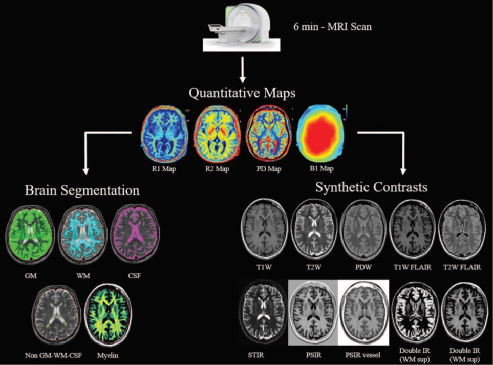
Synthetic magnetic resonance imaging is a novel imaging based technique that allows generating multiple contrast weighted images based on relaxivity (R1, R2 and PD) measurements of tissues in a single acquisition using a multi-echo, multi-delay saturation recovery turbo spin-echo sequence. In its current 2D version, the scan time of the acquisition sequence is around 6 minutes, with automatic post-processing completed in less than 10 seconds. From the quantitative maps, it is possible to create multiple conventional contrast images called synthetic MR images, which can be beneficial to reduce scan time and improve patient throughput. An advantage with this approach is that the synthesized TE, TR and TI can be customized for a given pathology, patient age, or radiologist preference without the need to repeat acquisition. The quantitative maps are generated on an absolute scale and the resulting measurements can therefore be compared between patients, even across scanner platforms. Using the relaxometry maps, Synthetic MRI can also perform brain tissue volumetrics, segmentation and myelin quantification without additional scan time. The quantitative analysis may have implications for understanding and monitoring of the evolution of the maturation process. Especially, the myelination process is vitally important to central nervous system functioning. Accurate invivo myelin measurements could be valuable for differential diagnostics of white matter changes, and to study treatment response in demyelinating and neurogenerative disorders.
Acknowledgements
No acknowledgement found.References
1. Warntjes JBM. Novel method for rapid, simultaneous T1, T2, and proton density quantification. Magn. Reson. Med. 2007.
2. Betts AM. Brain imaging with synthetic MR in children: clinical quality assessment. Neuroradiology 2016.
3. West H. Clinical validation of synthetic brain MRI in children: initial experience. Neuroradiology 2017.
4. Goncalves FG. Synthetic brain MRI: review of current concepts and future directions. Topics in Mag. Reson. Imag. 2018.
5. McAllister A. Quantitative synthetic MRI in children: normative intracranial tissue segmentation values during development. Am. J. Neuroradiol. 2017.
Figures